Practice Essentials
Slipped capital femoral epiphysis (SCFE) is the most common hip abnormality presenting during adolescence and is a primary cause of early osteoarthritis. SCFE usually occurs in children 8-15 years of age. [1] It is primarily associated with obesity and growth surges but occasionally can occur in patients with endocrine disorders such as hypothyroidism and hypogonadism. [2] Presentation before their 10th birthday or after their 16th birthday and height below the 10th percentile are reliable risk factors for identifying children with an SCFE who are at heightened risk for an underlying endocrinopathy and can be used to guide selective laboratory testing. Patients with unilateral SCFE presentations with an underlying endocrinopathy are at heightened risk for contralateral slip development and should be treated with prophylactic fixation. [3]
Unfortunately, SCFE is frequently misdiagnosed, and it has symptoms that can be misleading. [4] Early diagnosis of SCFE is essential, as a delay in diagnosis regularly leads to an increase in slippage with increased risk of subsequent damage to the blood vessels, which can result in irreversible damage to the joint. Symptoms and findings are frequently subtle and nonspecific, often leading to delay in diagnosis and treatment and, consequently, to the manifestation of massive deformities. [5] Pathology is localized to the proximal aspect of the femur, but symptoms may occur in the knee or the thigh, thereby leading to misdiagnosis. [2, 6]
(See the image below.)
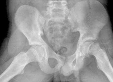
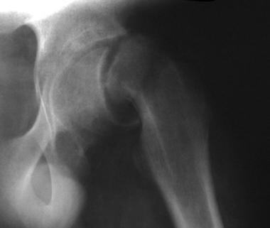 Slipped capital femoral epiphysis. This child had undergone radiation treatment for a pelvic malignancy. On this anteroposterior hip image, pathologic slippage is not subtle.
Classification and grade
SCFE is not frequently encountered during routine practice, and diagnosis and treatment are often delayed. It is important to understand symptoms and imaging features to avoid delayed diagnosis. After the diagnosis is made, correct classification of the disease is required. Classification should be based on physeal stability to enable selection of safe and effective treatment. However, surgeons should bear in mind that assessment is challenging and actual physeal stability is not always consistent with the stability predicted by a clinical classification method. [7]
SCFE is commonly classified as (1) acute or chronic, and (2) stable or unstable. The slip is classified as acute if it is present for less than 3 weeks, and as chronic if present for 3 weeks or longer. Under the second classification, SCFE is considered stable if the patient can bear weight on the extremity without a crutch or a walker; if the patient is unable to bear weight, SCFE is considered unstable. [1, 2]
Slip progression after in situ fixation of SCFE has been reported as occurring in up to 20% of patients. [8] The Wilson method or the Southwick method can be used to measure the grade of severity of the slip in SCFE. With the Wilson method, a mild slip is described as epiphysis displacement that is less than one third the width of the metaphysis; a moderate slip as between one third and one half the width; and a severe slip as more than one half the width. The Southwick method uses the epiphyseal shaft angle on the frog-leg lateral radiograph, with the angle calculated by subtracting the epiphyseal shaft angle on the uninvolved side from that on the SCFE side. A mild slip is considered less than 30 degrees; moderate, between 30 and 50 degrees; and severe, greater than 50 degrees. [1]
Imaging modalities
Children with SCFE are often seen by an array of medical professionals prior to diagnosis. Patients with mild slips, slips with knee pain, or bilateral slips can occasionally present a diagnostic challenge that increases the risk of a delay in diagnosis and associated complications. [9]
Research has shown that when a nonorthopedic provider evaluates this patient population, there can be a significant delay in appropriate treatment, which may have serious consequences for the prognosis of the patient. These delays are often caused by the practitioner's inability to put the clinical picture into focus with regard to how these patients typically present. [10]
Radiographs are the easiest images to obtain and provide an excellent screening examination for hip pain in any patient. For patients with SCFE, advanced stages of the disease are easy to identify; however, subtle changes early in the disease course are more difficult to detect. Before the femoral epiphysis actually has become displaced, only slight widening of the affected physis may be evident. [1]
Diagnosis is made via anteroposterior (AP) pelvic and lateral frog-leg radiographs. [1, 2, 11, 12] Magnetic resonance imaging (MRI) or computed tomography (CT) may be used to detect SCFE in early stages. CT scanning is a sensitive method of measuring the degree of tilt and detecting early disease, but it is rarely needed. CT may be performed with low doses, and reconstructions may allow viewing of the relationship of the femoral head to the metaphysis in 3 planes. MRI depicts slippage earliest and can show early marrow edema and slippage. MRI may be helpful in follow-up studies of the contralateral hip. [13, 14, 15, 16]
Metaphyseal blanch is seen as an increase in density in the proximal metaphysis. It is presumed that metaphyseal blanch represents an attempt at healing that occurs before displacement of the epiphysis is visible.
CT scanning is a sensitive method used for measuring degree of tilt and for detecting disease, but it is rarely needed. Usually, CT scanning is performed only at the request of the treating physician for documenting the severity of the tilt.
The earliest way to detect SCFE is by using magnetic resonance imaging (MRI). With MRI, early marrow edema and slippage can be detected. These are demonstrated with increased signal on T2-weighted and water-sensitive images. MRI can be considered in patients for whom the clinical suspicion of SCFE is high and for whom radiographs appear normal. MRI can also be considered for follow-up imaging of the contralateral hip. [14]
Ultrasonographic findings are rarely specific, and the sensitivity of sonography is unknown. Hip effusions of blood have often been reported and are suggestive of fracture.
Marrow edema is a nonspecific finding; although it can indicate early bone changes in SCFE, marrow edema has numerous other causes such as infection or even tumor. These diagnoses are rarely considered with proper clinical evaluation.
Radiographic imaging is the primary imaging modality, whereas MRI, CT, and ultrasound are helpful in surgical planning and prognostic evaluation. Postoperatively, they provide information on short-term and long-term complications. [5]
The radionuclide bone scan is sometimes used during workup but prior to diagnosis. Accumulation of bone scanning agents can be decreased after fixation and in patients with an acute slip and significant displacement. This decrease is usually limited to the epiphysis. Decreased accumulation is associated with an increased incidence of chondrolysis.
Radiography
Diagnosis is made on the basis of AP pelvic and lateral frog-leg radiographs. Abduction of the femur for the frog-leg view may result in increased slippage and should be performed with caution. Radiologic signs include the Steel sign, which reveals double density at the metaphysis on anteroposterior radiography; widening of the physis compared with the uninvolved sign; and the epiphysis falling below the Klein line (a line drawn along the superior edge of the femoral neck that should cross the epiphysis, but in SCFE, the epiphysis falls below the Klein line). [1, 2, 11, 12, 17]
(See the images below.)
Slipped capital femoral epiphysis. This child had undergone radiation treatment for a pelvic malignancy. On this anteroposterior hip image, pathologic slippage is not subtle.
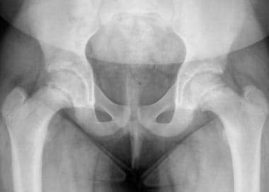 Slipped capital femoral epiphysis. A 13-year-old female adolescent with acute-onset right hip pain. She had presented to the emergency department 1 week prior with right knee pain. On this anteroposterior pelvic view, note increased opacity of her right metaphysis and subtle widening of the physis.
 Slipped capital femoral epiphysis. A 13-year-old female adolescent with acute-onset right hip pain had presented to the emergency department 1 week prior with right knee pain (detailed view, same patient as in previous image). Blanching of the metaphysis and only subtle widening of the physis are evident. The epiphysis is yet to be displaced.
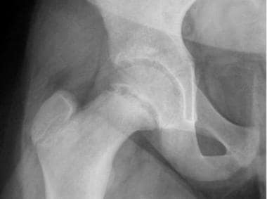
 Slipped capital femoral epiphysis. Image in a 14-year-old male adolescent who came to the emergency department with reports of thigh and knee pain. A relatively subtle medial slip is seen here.
 Slipped capital femoral epiphysis. Image of a 14-year-old male adolescent who came to the emergency department with reports of thigh and knee pain (same patient as in previous image). A more obvious posterior slip is noted on this frog-leg lateral view.
On AP radiographs, close attention should be paid to the physis. Early in SCFE, the physis may widen. Increased opacity in the metaphysis, described as blanching, may occur as an early healing response, and the epiphysis may appear smaller because it is tilted dorsally. [1]
The lateral radiograph demonstrates slippage earliest because slippage begins with posterior displacement and progresses with medial rotation.
The Southwick method can be applied by creating an axis for the femoral neck and determining whether the epiphysis is tilted.
(See the image below.)
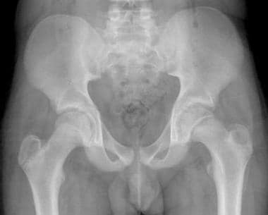
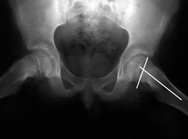 Slipped capital femoral epiphysis. Frog-leg pelvic image in a child with bilateral slipped capital femoral epiphysis. A line perpendicular to the epiphyseal axis and another along the axis of the femoral neck demonstrate the degree of tilt via the Southwick method.
An additional method of diagnosis consists of drawing a line along the lateral aspect of the femoral neck on the AP view; this line, known as the Klein line, should intersect a portion of the femoral head. [17]
Degree of confidence in radiographic findings of SCFE is high. When the aforementioned constellation of findings is present, false-positive and false-negative findings do not occur.
Treatment
Slipped capital femoral epiphysis (SCFE) is a frequent chronic and often bilateral atraumatic slippage of the epiphysis relative to the femoral neck that occurs during adolescence. The success of treatment depends on the extent of slippage and possible complications. [5]
In situ pinning of SCFE is a safe and effective treatment modality but often results in residual deformity leading to femoroacetabular impingement, which may limit patient activities and predispose to early-onset arthritis. [18]
Proximal femoral shortening osteotomies are becoming the treatment of choice for severe SCFE to reduce the risk of femoroacetabular impingement. Reported rates of complications seem reasonable. Although anterior subcapital shortening osteotomy (ASSO) is a technically demanding procedure, it is reliable and reproducible. The main risk factor for developing avascular necrosis (AVN) remains the unstable nature of SCFE—not the surgeon's level of experience. [19]
Treatment of patients with SCFE, especially in moderate and severe forms, remains controversial. Dunn osteotomy performed with the Ganz approach has become very popular, although it is a complicated and challenging surgical procedure with risk of AVN. A systematic review of the Dunn procedure modified by Ganz performed by an experienced surgeon for SCFE reveals that this procedure allows for anatomic reduction of moderate or severe SCFE with a low incidence of AVN. The few papers with long-term follow-up reported no progression of hip osteoarthritis; however, given that patients are adolescents at the time of surgery, longer follow-up studies are needed to validate this statement. It is still debated whether better results are obtained in stable or unstable SCFE. The indication for this procedure in mild SCFE remains controversial. [20]
-
Slipped capital femoral epiphysis. This child had undergone radiation treatment for a pelvic malignancy. On this anteroposterior hip image, pathologic slippage is not subtle.
-
Slipped capital femoral epiphysis. A 13-year-old female adolescent with acute-onset right hip pain. She had presented to the emergency department 1 week prior with right knee pain. On this anteroposterior pelvic view, note increased opacity of her right metaphysis and subtle widening of the physis.
-
Slipped capital femoral epiphysis. A 13-year-old female adolescent with acute-onset right hip pain had presented to the emergency department 1 week prior with right knee pain (detailed view, same patient as in previous image). Blanching of the metaphysis and only subtle widening of the physis are evident. The epiphysis is yet to be displaced.
-
Slipped capital femoral epiphysis. Frog-leg pelvic image in a child with bilateral slipped capital femoral epiphysis. A line perpendicular to the epiphyseal axis and another along the axis of the femoral neck demonstrate the degree of tilt via the Southwick method.
-
Slipped capital femoral epiphysis. Image in a 14-year-old male adolescent who came to the emergency department with reports of thigh and knee pain. A relatively subtle medial slip is seen here.
-
Slipped capital femoral epiphysis. Image of a 14-year-old male adolescent who came to the emergency department with reports of thigh and knee pain (same patient as in previous image). A more obvious posterior slip is noted on this frog-leg lateral view.


