Practice Essentials
Urolithiasis, kidney stones, renal stones, and renal calculi are interchangeably used to refer to the accretion of hard, solid, nonmetallic minerals in the urinary tract (see the image below). [1, 2] Nephrocalcinosis is a term that refers to increased calcium content in the parenchyma of the kidney.
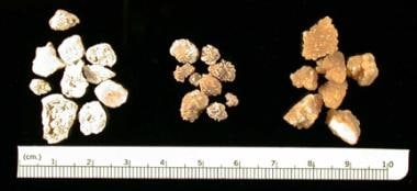 Three groups of kidney stones are shown. Groups at left and center contain varying concentrations of calcium, phosphate, and oxalate. The group of stones on the right is composed of cysteine.
Patient and parent education regarding risk factors for additional stone formation and diet and medication complications is very important.
Signs and symptoms
Presentation usually depends on age; symptoms such as flank pain and hematuria are more common in older children. Nonspecific symptoms (eg, irritability, vomiting) are common in very young children.
The following are five fairly typical presentations of stone disease in children:
-
Intense pain that suddenly occurs in the back and radiates downward and centrally toward the lower abdomen or groin
-
Hematuria, usually gross, occurring with or without pain
-
Infection leading to radiologic imaging in which a stone is identified
-
Asymptomatic stones, which are sometimes identified when abdominal imaging is performed for another reason
-
Persistent microscopic hematuria, which consists of 5 or more red blood cells per high-power field in 3 of 3 consecutive centrifuged urine specimens obtained at least 1 week apart
See Presentation for more detail.
Diagnosis
In children, laboratory studies provide only suggestive evidence of a kidney stone; however, certain laboratory studies (eg, calcium or uric acid excretion) may be very helpful in identifying risk factors for additional stone formation. Imaging studies are valuable. For example, renal ultrasonography is highly effective for identifying stones in the urinary tract. Direct assessment of stones is vital.
See Workup for more detail.
Management
The overall goals of care are as follows:
-
To prevent additional renal damage, which may lead to loss of renal parenchyma
-
To manage pain associated with current stone(s)
-
To expedite passage or removal of any stones present
-
To prevent new stones from forming.
Management may include medical approaches, surgical interventions, and dietary modification.
See Treatment and Medication for more detail.
For more information, see the Medscape Reference topic Nephrolithiasis.
Pathophysiology
Renal, urologic, endocrine, and metabolic disorders may lead to the development of crystallized material in the urinary system. Stones are most often classified into groups based on their chemical components.
Materials that produce stones in the urinary tract of children include the following:
-
Calcium with phosphate or oxalate
-
Purine derivatives
-
Magnesium ammonium phosphate (struvite)
-
Cysteine
-
Combinations of the preceding items
-
Drugs or their metabolites (eg, phenytoin, triamterene)
-
Melamine-contaminated milk powder consumption
In fluids contained within the urinary system, interaction between factors that promote and factors that inhibit crystallization is continuous. When solutes in solution are at concentrations below their solubility product (subsaturation of stone-forming compounds in the urine), added crystals dissolve (undersaturated region). Spontaneous precipitation can occur when concentration of constituents is above the formation product. The metastable region lies between solubility product and formation product. Existing crystals can grow, but spontaneous precipitation does not occur.
Etiology
Renal stones occur as a result of the following 3 factors:
-
Supersaturation of stone-forming compounds in urine
-
Presence of chemical or physical stimuli in urine that promote stone formation
-
Inadequate amount of compounds in urine that inhibit stone formation (eg, magnesium, citrate)
Contributing factors
Numerous dietary items may contribute to renal stone production. A high oxalate intake may contribute to calcium oxalate stone production. Excessive purine intake may contribute to the production of stones containing uric acid and uric acid plus calcium components. A ketogenic diet, prescribed to reduce seizures, places children at risk for both uric acid and calcium stone formation.
In general, urinary calcium increases with dietary calcium intake (see Hypercalciuria). Urinary calcium increases in patients with high sodium chloride intake. Dietary phosphate restriction, if severe, increases urine calcium excretion. A diet high in protein from animal sources, glucose or sucrose increases urinary calcium and, in some cases, may contribute to stone formation. [3] Vitamins A and D can contribute to calcium urolithiasis when taken in excessive amounts. Fructose consumption is also associated with an increased risk of kidney stones. [4]
Drug intake may contribute to stone formation in 3 basic ways (see the table below).
Table 1. Stone Formation (Open Table in a new window)
Mechanism of Stone Formation |
Drug |
Primary Stone Composition |
Crystallization of highly excreted, poorly soluble drug or metabolite causes stone formation. |
Phenytoin, triamterene, sulfonamides, felbamate, ceftriaxone, indinavir, ciprofloxacin, guaifenesin/ephedrine |
Drug or its metabolites |
Drug may increase the concentration of stone-forming minerals. |
1. Anti-cancer drugs 2. Glucocorticoid 3. Allopurinol (if used in tumor lysis) 4. Loop diuretics 5. Calcium and vitamin D |
1. Uric acid 2. Calcium 3. Xanthine 4. Calcium oxalate 5. Calcium |
Drug inhibits activity of carbonic anhydrase enzymes in the kidney, causing metabolic acidosis, hypocitraturia, and elevated urine pH. |
Topiramate, zonisamide, acetazolamide |
Calcium phosphate |
First, the drug or its metabolites may precipitate as stones (eg, phenytoin, triamterene, sulfadiazine, felbamate, ceftriaxone [5] ).
Second, the drug may increase the concentration of stone-forming minerals by increasing the filtered load or decreasing the tubular reabsorption. For example, anticancer agents increase the filtered load of uric acid and glucocorticoids increase the filtered load of calcium. Allopurinol increases the filtered load of xanthine in patients with tumor lysis to produce xanthinuria. Furosemide decreases tubular calcium reabsorption, leading to increased urine calcium concentration.
Third, the drug may alter urine pH, decreasing the solubility of a stone-forming agent. In children with distal renal tubular acidosis, bicarbonate probably contributes to stone formation by further alkalinizing the urine.
Fluid intake is important quantitatively and qualitatively. A low fluid intake leads to concentrated urine and increases the risk of stone formation. Water may have a high mineral content in some areas. Milk contains significant calcium and vitamin D. Orange juice may be supplemented with calcium. Tea contains oxalate and often sucrose. Many drinks (eg, sports drinks) contain sodium chloride and sucrose.
Some diseases, or the medications used to treat them, increase stone formation risk. Examples include distal renal tubular acidosis, short-gut syndrome, inflammatory bowel disease, intractable seizures, and cystic fibrosis. Urolithiasis is not uncommon in pediatric patients who have undergone a kidney transplant. [6] Factors associated with post-kidney transplant urolithiasis include retention of suture material, recurrent urinary tract infection, hypercalciuria, and urinary stasis. Gastrostomy tube–fed children are at higher risk of urolithiasis that may be related to concomitant topiramate administration, urinary tract infection, and shorter G-tube duration possibly affecting subclinical chronic dehydration. [7]
A population-based, case–control study by Tasian et al that included 25,981 patients with nephrolithiasis reported that oral antibiotics were associated with increased odds of nephrolithiasis. The adjusted odds ratio (95% confidence interval) was 2.33 (2.19 to 2.48) for sulfas, 1.88 (1.75 to 2.01) for cephalosporins, 1.67 (1.54 to 1.81) for fluoroquinolones, 1.70 (1.55 to 1.88) for nitrofurantoin/methenamine, and 1.27 (1.18 to 1.36) for broad-spectrum penicillins. The study also reported that the greatest odds for nephrolithiasis were recent exposure and exposure at younger age. [8]
Risk factors
Risk factors for pediatric urolithiasis include the following:
-
Habitually low urine volume
-
High urine excretion of calcium
-
High urine excretion of uric acid
-
High urine excretion of oxalate
-
Low urine pH: Uric acid and cysteine are less soluble in acid urine.
-
High urine pH: Struvite and calcium phosphate are less soluble in alkaline urine.
-
Nidus for crystal precipitation: A nidus for crystal precipitation (eg, uroepithelial surface properties that affect crystal retention) occurs when the crystalline lattice structure of one crystal is similar to another crystal and the second crystal grows on the first.
In children, hypercalciuria and a reduction in the gap between calcium phosphate upper limit of metastability and supersaturation are significant risk factors for stones. [9] Other factors such as developmental abnormalities of the urinary tract, urinary obstruction, urinary stasis, and infection with urea-splitting microorganisms may also be important.
Epidemiology
Frequency of urolithiasis in children has not been studied in a systematic population-based fashion. Institutional and case reports indicate regional variation. A significant increase in the number of children diagnosed with and treated for urolithiasis has occurred in the past decade. [10]
United States statistics
Approximately 11% of the US population has nephrolithiasis; however, it is less common in children, with an estimated prevalence of 1%. [1] The incidence of nephrolithiasis is rising among children in the United States. [2, 11] Population-based studies estimate that the incidence of pediatric urolithiasis was 65/100,000 person-years during 2005-2016, an increase from the 1999 estimate of 18/100,000 person-years. [1]
Children with stones now account for 1 in 685 pediatric hospitalizations in the United States. Surprisingly, more than half the patients are younger than 13 years at hospitalization. [12] Urolithiasis is relatively uncommon in the United States compared with some other areas of the world, possibly because of diet or public health measures. Endemic bladder stones (uric acid and ammonium acid urate) are rare in the United States, though common in developing countries.
The southeast region of the United States has a higher frequency of kidney stone formation in adults than do other regions of the United States. The reason for a higher incidence of stone formation in the southern United States is unknown. Suggested factors include climate, diet, genetics, state of hydration, and bacterial colonization.
A study by Tasian et al examined trends in the annual incidence and cumulative risk of nephrolithiasis among children and adults living in South Carolina over a 16-year period. The study reported that the incidence of kidney stones has increased among young patients (26% increase in 5 years for those 15-19 years of age). Incidence also increased 15% per 5 years among females and 15% per 5 years among African Americans. [13]
Urinary tract stones in children are a relatively infrequent problem. Regional rates of stone formation in children have not been reported.
International statistics
Stones are more common in certain areas. In Europe, kidney stones occur in 1-2 children per million population per year. In underdeveloped countries, children more frequently have endemic bladder stones than renal stones. Endemic bladder calculi are common in developing countries where dietary protein is mostly derived from plant sources rather than meat. These areas include Eastern Europe, Southeast Asia, India, and the Middle East. Upper urinary calculi associated with urease-producing bacterial infection occur in England and Europe.
A study by Pong et al, that investigated the incidence for pediatric urolithiasis in Taiwan from 1998-2007, reported that the trend of annual newly diagnosed incidences for boys, girls, and all children declined. This also coincided with a decline in medical costs and annual medical care visits for pediatric urolithiasis during this period. [14]
Age-, sex-, and race-related demographics
Peak presentation for adults is middle age. Adults are most often afflicted with calcium oxalate or calcium phosphate stones. In some cases, the primary cause of stone formation cannot be identified.
Children can present with stones at any age (eg, premature newborn to teenager). In children, calcium stones are most common. The approximate frequency of kidney stone types in the pediatric age group is calcium with phosphate or oxalate (57%), struvite (24%), uric acid (8%), cystine (6%), endemic (2%), mixed (2%), and other types (1%). With children, particularly younger children, the primary cause of stone formation (eg, hypercalciuria, hyperuricosuria) can usually be identified with a through evaluation.
Although stones are more frequent in men than in women (4:1), the boy-to-girl ratio (3:2) is closer to equal.
No population-based studies have been performed, but institutional reports indicate that, in the United States, white children develop urolithiasis more frequently than children who are black, Asian American, or Latin American.
Prognosis
Generally, the prognosis for children with kidney stones is good; most children do well. However, mortality and significant morbidity sometimes occur. Urolithiasis during childhood can have important lifelong implications for some patients.
Kidney stones are not usually fatal, although some primary conditions that produce kidney stones (eg, Lesch-Nyhan syndrome, oxalosis) can lead to death from problems associated with the primary disease or complications of renal failure. Infected stones may lead to urosepsis and death. Complete untreated renal outflow obstruction causes renal failure.
Acute renal colic may be very painful. Infected stones may produce pain as well as sepsis. Children who develop frequent painful stones or stones that require painful treatment such as urologic stone removal or extracorporeal shockwave lithotripsy (ESWL) may experience considerable morbidity.
Complications
The primary complications of urolithiasis include obstruction of the urinary tract, renal parenchymal damage, infection, and adverse effects of medication or diet.
-
Three groups of kidney stones are shown. Groups at left and center contain varying concentrations of calcium, phosphate, and oxalate. The group of stones on the right is composed of cysteine.
Tables
Mechanism of Stone Formation |
Drug |
Primary Stone Composition |
Crystallization of highly excreted, poorly soluble drug or metabolite causes stone formation. |
Phenytoin, triamterene, sulfonamides, felbamate, ceftriaxone, indinavir, ciprofloxacin, guaifenesin/ephedrine |
Drug or its metabolites |
Drug may increase the concentration of stone-forming minerals. |
1. Anti-cancer drugs 2. Glucocorticoid 3. Allopurinol (if used in tumor lysis) 4. Loop diuretics 5. Calcium and vitamin D |
1. Uric acid 2. Calcium 3. Xanthine 4. Calcium oxalate 5. Calcium |
Drug inhibits activity of carbonic anhydrase enzymes in the kidney, causing metabolic acidosis, hypocitraturia, and elevated urine pH. |
Topiramate, zonisamide, acetazolamide |
Calcium phosphate |


