Background
Sporotrichosis is caused by the acquisition of Sporothrix schenckii, a dimorphic, saprophytic, and geophilic fungus. Although this fungal infection has been reported in semiarid environments, it is most typically found in warmer temperatures and tropical climates around the world. [1] S schenckii usually grows amidst decaying vegetable matter and in the soil as a saprophyte. Saprophyte can also be commonly found in soil, straw, wheat grain, fruits, tree bark, wood, shrubs, thorns, rose shrubs, decaying vegetation and timber. However, it is most acquired by humans when a form of traumatic inoculation occurs after in contact with animals. The most common animals known to have spread Sporotrichosis are cats, dogs and horses. Sporotrichosis spreading from human to human is quite rare (international Journal of Dermatology).
Because sporotrichosis may be difficult to initially diagnose, a differential diagnosis should always be generated during the clinical evaluation (see Differentials).
Benjamin Schenck first noted Sporotrichosis in 1896. In the year 1900, Hekton and Perkins officially named the pathogenic fungus Sporothrix Schenkii after its original founder (international journal of dermatology). The association between sporotrichosis and acquired immunodeficiency syndrome (AIDS) was first reported in 1985. Although it is not considered an AIDS marker, it is included in a list of AIDS-related conditions. In immunocompromised patients, especially those with an impaired cell-mediated immunity, it can become an opportunistic infection with a possible life-threatening course.
 Sporotrichosis with cutaneous necrosis and lymphangitic (sporotrichoid) spread. A 28-year-old white man presented for evaluation of a poorly healing, asymptomatic, round plaque acquired on the dorsum of his left hand. The lesion had been present for approximately 3 weeks.
 Glucose-peptone agar culture plates revealing colony growth of Sporothrix schenckii. The left plate reveals older colonies as dark brown or black, and the right plate reveals younger white colonies with a brown center, characteristic of this fungus.
 Microscopic examination of a blue dye preparation from the colony surface reveals elongated septate hyphae with groups of microconidia in a flowerlike arrangement.
 A well-circumscribed, moderately elevated, erythematous plaque with central ulceration is found on the dorsum of this patient's left hand. Potassium chloride (KOH) stain was negative for fungal elements.
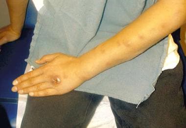
 A 2 X 2 cm, dome-shaped, well-circumscribed, erythematous plaque is shown proximal to the left ring finger. The lesion was draining a serosanguineous fluid. No purulence was noted.
 Biopsy rarely reveals the 6-mcg cigar-shaped yeast within tissue macrophages as shown in this histologic section. This is the morphology that Sporothrix schenckii assumes at 37°C.
 Moist cream-colored colonies with a central, dark, leathery, and wrinkled surface growing at 25°C is highly suggestive of Sporothrix schenckii.
 A fresh agar slant of Sporothrix schenckii reveals moist, white-to-cream–colored, yeastlike colonies.
 Cutaneous, ulcerating, painless nodule on the hand and a classic sporotrichoid lymphangitic pattern spreading proximally up the arm.
Pathophysiology
The fungus is most commonly acquired by traumatic implantation into the skin, causing a local pustule or ulcer with nodules developing proximally along the draining lymphatics. Once implanted, this saprophytic fungus can grow in human tissues.
When recognized by the immune system, an inflammatory response occurs. Physical signs and symptoms related to the location and degree of inflammation then ensues. Primary organ systems involved in sporotrichosis include the skin and subcutaneous tissues. Dissemination to osteoarticular structures, viscera, and the CNS is uncommon unless the fungus is inhaled or acquired by a patient who is immunocompromised. [2, 3] Inhalation may affect the lungs and cause a granulomatous pneumonitis. In a host who is immunocompromised, disseminated infection can occur from skin involvement or from primary pulmonary infection. For instance, one case has been reported of laryngeal and respiratory tract sporotrichosis after steroid inhaler use. [4]
Clinically, sporotrichosis may manifest as either an acute or a chronic subcutaneous mycotic infection. Although the acute phase is most common, chronic nodular lymphangitis also reportedly develops in some cases. A minor puncture wound or splinter is sufficient to inoculate the fungus into the tissue. Infection can also be related to zoonotic spread from infected cats or scratches from digging animals, such as armadillos. [2] Among animals that can spread sporotrichosis, cats have been found to be the most common. Cats most often transfer sporotrichosis through bites and scratches (Molecular Epidemiology of Feline Sporotrichosis).
Epidemiology
Frequency
United States
Sporotrichosis is an uncommon fungal infection of unknown frequency.
International
Distribution is global, but incidence is unknown. Deep mycoses have mainly been reported in the tropics and subtropics. A recent endemic of lymphocutaneous sporotrichosis were reported in Peru [5] where the epidemic has been occurring for three decades. [6] Brazil has also had a high outcome of sporotrichosis, and it was found to be due to zoonotic transmission most commonly as well through cats. It was found to have stemmed from the Rio de Janeiro [7] . A cluster of sporotrichosis cases occurred in Western Australia; a commercial hay supplier was the source of the outbreak. [8] Jilin Province, Northeast China, has recently been reported as a serious endemic region, as well as in the tropics and subtropics of Malaysia. [9]
Mortality/Morbidity
Sporotrichosis is usually associated with minimal morbidity. Increased morbidity and, rarely, mortality may occur if the diagnosis is delayed, if the fungus infects patients who are immunologically compromised, or if inadequate or inappropriate therapy is rendered.
Race
Sporotrichosis has no racial predilection.
Sex
Sporotrichosis occurs in men, women, and children. Men have a higher risk of acquiring this fungus because they have greater environmental exposure from outdoor occupations.
Age
Sporotrichosis may occur in people of any age. Yet, in children, sporotrichosis tends to present more frequently with a solitary ulcerative nodule. This is in contrast to adults in which a classic lymphocutaneous form is more common.
-
Sporotrichosis with cutaneous necrosis and lymphangitic (sporotrichoid) spread. A 28-year-old white man presented for evaluation of a poorly healing, asymptomatic, round plaque acquired on the dorsum of his left hand. The lesion had been present for approximately 3 weeks.
-
Glucose-peptone agar culture plates revealing colony growth of Sporothrix schenckii. The left plate reveals older colonies as dark brown or black, and the right plate reveals younger white colonies with a brown center, characteristic of this fungus.
-
Microscopic examination of a blue dye preparation from the colony surface reveals elongated septate hyphae with groups of microconidia in a flowerlike arrangement.
-
A well-circumscribed, moderately elevated, erythematous plaque with central ulceration is found on the dorsum of this patient's left hand. Potassium chloride (KOH) stain was negative for fungal elements.
-
A 2 X 2 cm, dome-shaped, well-circumscribed, erythematous plaque is shown proximal to the left ring finger. The lesion was draining a serosanguineous fluid. No purulence was noted.
-
Biopsy rarely reveals the 6-mcg cigar-shaped yeast within tissue macrophages as shown in this histologic section. This is the morphology that Sporothrix schenckii assumes at 37°C.
-
Moist cream-colored colonies with a central, dark, leathery, and wrinkled surface growing at 25°C is highly suggestive of Sporothrix schenckii.
-
A fresh agar slant of Sporothrix schenckii reveals moist, white-to-cream–colored, yeastlike colonies.
-
Cutaneous, ulcerating, painless nodule on the hand and a classic sporotrichoid lymphangitic pattern spreading proximally up the arm.


