Background
Rickettsiae comprise a group of microorganisms that phylogenetically occupy a position between bacteria and viruses. The genus Rickettsia is included in the bacterial tribe Rickettsiae, family Rickettsiaceae, and order Rickettsiales. They are obligate intracellular gram-negative coccobacillary forms that multiply within eukaryotic cells. Rickettsiae do not stain well with Gram stain, but they take on a characteristic red color when stained by the Giemsa or Gimenez stain. They have typical gram-negative cell walls and lack flagella. Their genome is very small, composed of 1-1.5 million bases. [1, 2]
Rickettsiae are a rather diverse collection of organisms with several differences; this prohibits their description as a single homogenous group. A general characteristic of rickettsiae is that mammals and arthropods are natural hosts. Rickettsioses are usually transmitted to humans by arthropods. Q fever, traditionally described among the rickettsial illnesses until recently, is primarily acquired by inhalation of contaminated airborne droplets. [3, 4, 5]
The epidemiology of human diseases caused by rickettsiae is intimately related to the biology of the vector that transmits it. Rickettsial diseases widely vary in severity from self-limited mild illnesses to fulminating life-threatening infections. [4]
Rickettsial illnesses, caused by organisms within the genus of rickettsiae, are recognized and can be divided into the following 3 biogroups: [2, 6]
Spotted fever biogroup (15 rickettsioses)
Included in this biogroup are the following:
-
Rocky Mountain spotted fever (RMSF), caused by Rickettsia rickettsii
-
Rickettsialpox, caused by Rickettsia akari
-
Boutonneuse fever (ie, Kenya tick-bite fever, African tick typhus, Mediterranean spotted fever, Israeli spotted fever, Indian tick typhus, Marseilles fever)
Typhus group
These are similar diseases that differ epidemiologically. The causative organisms (Rickettsia prowazekii and Rickettsia typhi) are similar to those of the spotted fever group but are antigenically distinct.
-
Louse-borne (epidemic) typhus
-
Brill-Zinsser disease (ie, relapsing louse-borne typhus)
-
Murine (endemic or flea-borne) typhus
Scrub typhus biogroup (Tsutsugamushi disease)
The rickettsial agents of scrub typhus have a single taxonomic name: Orientia tsutsugamushi. However, these organisms represent a heterogeneous group that strikingly differs from Rickettsial species of the spotted fever and typhus groups. The 3 major serotypes are Karp, Gilliam, and Kato.
Other rickettsioses and closely related illnesses
New or reemerging rickettsioses have been described in the last few decades, including tickborne lymphadenopathy (TIBOLA) and Dermacentor -borne-necrosis-eschar-lymphadenopathy (DEBONEL) related to Rickettsia slovaca infection, as well as lymphangitis-associated rickettsiosis attributed to Rickettsia sibricia infection. [1] Recently, a new Rickettsia species, 364D, that causes an eschar-associated illness was identified in California. [7, 8]
Ehrlichia organisms (the cause of human monocytic ehrlichiosis and Ehrlichia ewingii infection), Anaplasmaphagocytophilum (the cause of human granulocytic anaplasmosis), and Bartonella species (the cause of Catscratch disease, relapsing fever, and Trench fever) are organisms related to the rickettsiae. They are discussed in separate articles.
Q fever is a disease caused by Coxiella burnetii, which has recently been removed from the Rickettsiales. [5] The disease is described here for comparison with other rickettsioses.
Potential as biological weapons
The environmental stability, small size, aerosol transmission, persistence in infected hosts, low infectious dose, and high associated morbidity and mortality have made pathogenic rickettsiae desirable bioterrorism agents. In fact, R prowazekii and C burnetii have been weaponized. However, developing rickettsial pathogens as biological weapons has many drawbacks, such as the lack of direct host-to-host transmission and availability of therapeutic countermeasures against them. [9]
Pathophysiology
Rickettsiae microorganisms appear to exert their pathologic effects by adhering to and then invading the endothelial lining of the vasculature within the various organs affected. The adhesins appear to be outer membrane proteins that allow the rickettsia to be phagocytosed into the host cell. Once inside, the rickettsial organisms either multiply and accumulate in large numbers before lysing the host cell (typhus group) or they escape from the cell, damaging its membrane and causing the influx of water (spotted fever group). [4]
Rickettsiae rely on the cytosol of the host cells for growth. To avoid phagocytosis within the cells, they secrete phospholipase D and hemolysin C, which disrupt the phagosomal membrane, allowing for rapid escape.
The most important pathophysiologic effect is increased vascular permeability with consequent edema, loss of blood volume, hypoalbuminemia, decreased osmotic pressure, and hypotension. On the other hand, disseminated intravascular coagulation is rare and does not seem to contribute to the pathophysiology of rickettsiae.
Studies of murine models have demonstrated that rickettsiae are cleared by cytotoxic CD8 cells and by cytokine-activated rickettsicidal nitrogen and oxygen species. In fact, antibodies do not play an important role in immunity against pathogenic rickettsia upon fist exposure. Walker provided an excellent review of this topic. [1]
-
RMSF: In RMSF, rickettsiae multiply within the endothelial cells of small blood vessels and then gain access to the bloodstream after skin inoculation. Focal areas of endothelial proliferation and perivascular mononuclear cell infiltration cause leakage of intravascular fluid into tissue space. These vascular lesions can affect all organs; however, they most readily are found in the skin and adrenals. In the central nervous system and heart, a damaging host response (primarily cell-mediated) accompanies the vasculitis. The liver is usually affected with portal triaditis. Vascular wall destruction consumes platelets, causing thrombocytopenia. Multiple factors lead to hypoalbuminemia (eg, renal loss, decreased intake, hepatic involvement) and hyponatremia (eg, renal loss, extracellular fluid shifts, cellular exchange of sodium for potassium).
-
Rickettsialpox: The organism that causes this illness is known to cause angiitis similar to other rickettsiae. Biopsies, which are rarely needed to establish the diagnosis of rickettsialpox, show evidence of thrombosis and necrosis of capillaries, as well as perivascular mononuclear cell infiltration.
-
Boutonneuse fever: Features of this illness are related to involvement of the vascular structures of the dermis in a manner similar to that observed in RMSF. Endothelial cells of the capillaries, venules, and arterioles (ie, small-to-medium sized vessels) in various organs may also become involved as the organism disseminates. [10] Additionally, a few cases of leukocytoclastic vasculitis have been reported with this infection.
-
Louse-borne (epidemic) typhus: The pathology is similar to that described for the spotted fever group of rickettsial diseases. However, typhus group rickettsiae do not stimulate actin-based mobility and rather extensively multiply and accumulate intracellularly until they burst the endothelial cell and disseminate into the bloodstream.
-
Brill-Zinsser disease (ie, relapsing louse-borne typhus): The pathology is similar to that described for the spotted fever group of rickettsial diseases. However, the organisms appear to lie dormant, most likely in the cells of the reticuloendothelial system, until they are reactivated by an unknown stressor, multiply and cause another acute but milder infection.
-
Murine (endemic or flea-borne) typhus: Pathology is similar to that described for epidemic typhus.
-
Tsutsugamushi disease (ie, scrub typhus): After invading the host cell and replicating in its cytoplasm, the Orientia tsutsugamushi exits by budding enveloped by part of the host cell membrane as it invades adjacent cells. Perivasculitis of small blood vessels occurs similarly to other rickettsial diseases. Usually, a necrotic inflammatory skin lesion occurs at the mite bite site, and regional and generalized lymphadenopathy is associated with this infection.
-
Q fever: In Q fever, the Coxiella organism directly causes disease in various organs. It has been demonstrated in macrophages in the lungs and in vegetations of the heart valves. Host-mediated pathogenic mechanisms also appear to play an important role in disease pathogenesis; the disease causes granulomatous changes in reticuloendothelial organs (granulomatous hepatitis).
Etiology
RMSF
This disease is caused by R rickettsii. [1, 6]
Tick vectors of RMSF include the Rocky Mountain wood tick (Dermacentor andersoni) in the Western United States and Canada; the American dog tick (Dermacentor variabilis) in the Eastern United States, along the US Pacific coast, and in the central United States; and the Lone Star tick (Amblyomma americanum) in some southern areas. [3] Examples of the ticks are shown in the images below.
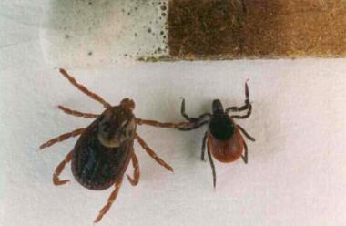 This photo shows the relative sizes of the adult forms of Ixodes scapularis (right) and Dermacentor variabilis (left). These ticks are shown next to a common match for scale. I scapularis is also referred to as Ixodes dammini. Photo by Darlyne Murawski; reproduced with permission.
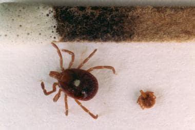 This photo is of an adult female, Amblyomma americanum, and a nymphal form of the same species (shown next to a common match for scale). Photo by Darlyne Murawski; reproduced with permission.
From 2002-2004, cases of RMSF reported from rural Arizona by Demma and colleagues were attributed to exposure to the common brown dog tick (Rhipicephalus sanguineus). [11] This represents a change from the typical vectors for this disease.
Expansions in tick populations can introduce rickettsial agents to new geographic areas and previously unrecognized rickettsiae -vector-human host relationships continue to evolve and be described. [12] Research from Germany identified a possible role for migratory birds in the distribution of emerging tick-borne pathogens including Rickettsiae. [13]
Rickettsiae multiply within ticks and pass to the next generation transovarially.
Rickettsiae are transmitted to a vertebrate host through saliva while a tick is feeding. It usually takes several hours of attachment and feeding before the rickettsiae are transmitted to the host. The risk of exposure to a tick carrying R rickettsii is low. Generally, about 1-3% of the tick population carries R rickettsii, even in areas where most human cases are reported.
Recognized or potential tick-borne spotted fever group rickettsial pathogens in the United States, other than R rickettsii include R akari, Rickettsia felis, Rickettsia parkeri, Rickettsia amblyomii, Rickettsia rhipicephali, and various unnamed serotypes (eg, Tillmook, 364-D). [14]
Rickettsialpox
It is caused by R akari, a member of the spotted fever group of Rickettsiae.
The disease is distinguishable from other rickettsial infections by the presence of an eschar at the site of the mouse mite (Liponyssoides sanguineus) bite, a vesiculopustular eruption, and the absence of Weil-Felix agglutinins.
The house mouse (Mus musculus) is the natural host of the mite transmitting rickettsialpox in the United States. Other rodents have been associated with the disease in other parts of the world. [6]
Boutonneuse fever
This disease is a tick-borne infection caused by various subspecies of Rickettsia conorii complex (R conorii conorii is the cause of Mediterranean spotted fever; R conorii israelensis is the cause of Israeli spotted fever; R conorii caspica is the cause of Astrakhan spotted fever; and R conorii indica is the cause of Indian tick typhus) , Rickettsia africae (the cause of African tick–bite fever) , or R slovaca, which are obligate intracellular organisms transmitted to humans by various ticks, depending on the geographical location. [10]
Contact with dogs carrying infected ticks appears to be the important risk factor for human infection.
Louse-borne (epidemic) typhus
This disease is caused by R prowazekii.
It is transmitted to humans by lice (ie, Pediculus humanus). Humans are the primary reservoir for R prowazekii.
Brill-Zinsser disease (ie, relapsing louse-borne typhus)
The rickettsial cause is the same but is related to the reactivation of the organism from a poorly defined latent state.
Murine (endemic or flea-borne) typhus
This disease is primarily caused by R typhi (Rickettsia mooseri) and R felis, which share a large antigenic moiety with R prowazekii.
It is transmitted from rat-to-rat by a rat flea (X cheopis) and accidentally to humans by the feces of infected fleas.
The cat flea (C felis) can also transmit the disease. [15]
Tsutsugamushi disease (ie, scrub typhus)
This disease is caused by O tsutsugamushi, which has a remarkable antigenic heterogeneity.
It is transmitted to humans by the larval form of trombiculid mites (ie, chiggers) that live and breed in the soil and scrub vegetation. The mite is both the reservoir and the vector that passes the bacteria transovarially. Rodents are also reservoirs. Humans are accidentally infected.
Q fever
The name derives from "Query Fever," given in 1935 following an outbreak of febrile illness in an abattoir in Australia. The disease is caused by C burnetii, a short, Gram negative, strictly intracellular bacterium.
Originally classified in the order Rickettsiales, C burnetii has since been placed (with Legionella and Francisella) into the gamma subdivision of the Proteobacteria on the basis of sequences of the 16SrDNA encoding genes. [5]
Unlike human rickettsial infections, it is a zoonosis transmitted from diseased animals to humans by the aerosol route or ingestion of raw milk.
Animals commonly infected include domestic livestock, especially cattle, sheep, and goats, as well as rodents, marsupials (in Australia), and cats (in Canada).
Ticks play a very minor role, if any, in transmission of the disease to humans; however, they transmit the disease to rodents and domestic animals.
C burnetii is a resilient organism that remains latent in infected hosts (eg, domestic livestock) until it is activated by a physiologic stressor, such as parturition. It then multiplies and contaminates the animals' surroundings, where it remains a potential source of infection for months. It is considered by the Center for Disease Control and Prevention (CDC) a potential agent of bioterrorism (class B). [6, 5]
Epidemiology
United States statistics
An analysis of insurance claims data showed that 14,830 patients in the United States received a diagnosis of rickettsial disease during 2005 to 2017. Of those patients, 7517 (50.7%) had spotted fever rickettsiosis, 4571 (30.8%) had ehrlichiosis, 1362 (9.2%) had typhus group rickettsiosis, and 1193 (8.0%) had other rickettsial diseases. [16]
RMSF
RMSF is caused by R rickettsii. Now reported in all geographic areas of the United States, RMSF was first recognized in and thought to be limited to the Rocky Mountain area. Its incidence sharply declined with the introduction of broad-spectrum antibiotics in the 1950s; the incidence soared again in the 1960s and peaked in 1981. Incidence has declined since that time. The average annual incidence from 1997-2002, based on passive surveillance, was 2.2 cases per million population. Incidence increased from 1.7 cases per million person-years in 2000 to 14.3 cases per million person-years in 2012. [17] The major endemic areas in the United States today include North Carolina, Oklahoma, South Carolina, Tennessee and Arkansas. More than 90% of patients with RMSF are infected from April through September. Individuals with frequent exposure to dogs and who reside near wooded areas or areas with high grass are at an increased risk of infection. [3, 18, 19]
Rickettsialpox
In the United States, rickettsialpox most commonly occurs in the Northeast, especially in New York City. Despite sporadic periodic outbreaks, incidence appears to be declining. The natural host is the common house mouse (Mus musculus).
Boutonneuse fever
This disease is very rare outside a limited geographical area in the Mediterranean, Africa, and India. With increased travel and ecotourism in endemic areas, more imported cases of the disease are described in travelers returning to the United States from Mediterranean regions. [10]
Louse-borne (epidemic) typhus
This is rare in the United States, but sporadic cases have been reported. The presumed source of infection is the southern flying squirrel.
Brill-Zinsser disease (ie, relapsing louse-borne typhus)
The distribution of this disease is analogous to louse-borne epidemic typhus. Recently, it has been rarely reported in the United States, and all cases had acquired the primary infection elsewhere. [20, 2, 21]
Endemic murine (flea-borne) typhus
This type of typhus is prevalent in urban cities and costal ports where rats are abundant. This is because it is transmitted rat-to-rat by a rat flea (Xenopsylla cheopis) and transmitted accidentally to humans by the feces of infected fleas. The cat flea (Ctenocephalides felis) may also serve as a vector for transmission of this disease to humans. These may be important vectors in Texas and southern California. Incidence has declined coincident with increased use of insecticides. Cases occur throughout the year, with peak prevalence from April through June in Texas and during the warm months of summer and early fall elsewhere. [20, 22]
Tsutsugamushi disease (ie, scrub typhus)
This is extremely rare outside the southwest Pacific and Southeast Asia. [23]
Q fever
Outbreaks are most common in slaughterhouses, research facilities, and plants, where handling of animals or their birth products is a source of exposure. Prevalence of Q fever in the United States is underestimated. Since Q fever became a nationally reportable disease in 1999 in the United States, a linear increase has been observed in the number of human cases identified (17 cases with onset in 2000 compared with 167 cases with onset in 2007). The incidence of Q fever increased similarly, from less than 0.1 case per million persons in 2000 to 0.6 case per million persons in 2007.
The surveillance case definition for Q fever was modified in 2008 to revise laboratory criteria for diagnosis and to allow for the separate reporting of acute and chronic Q fever. During 2008–2010, the number of reported cases decreased slightly, relative to 2007, and the incidence rate has decreased to 0.4 case per million persons. One hundred thirty-one cases of Q fever were reported with onset in 2010; of these, 106 were acute Q fever and 25 were chronic Q fever. [24]
International statistics
The spotted fever rickettsiae have been found in every continent except Antarctica. [2]
RMSF
This primarily occurs in the continental United States but has been reported in southern Canada, Central America, Mexico, and parts of South America. It is rarely seen elsewhere. [18]
Rickettsialpox
This may be more prevalent worldwide than is reported. It has been identified in large cities in Russia, South Africa, and Korea.
Boutonneuse fever
Boutonneuse fever has demonstrated an increased incidence in Mediterranean countries, such as Spain, Italy, and Israel. Along with African tick–bite fever, these infections have been identified in Algeria, Malta, Cyprus, Slovenia, Croatia, Kenya, Somalia, South Africa, Ethiopia, India, and Pakistan, as well as rural Sub-Saharan Africa, the eastern Caribbean, and around the Black Sea. [4, 10]
Louse-borne (epidemic) typhus
Epidemics have occurred in Europe, Asia, and Africa. African countries, especially Ethiopia and Nigeria, have reported most of the cases in the last 2 decades.
Brill-Zinsser disease (ie, relapsing louse-borne typhus)
This disease follows the epidemiology of louse-borne epidemic typhus and rarely, if ever, occurs in children because the reactivation usually occurs decades after primary infection.
Murine (endemic or flea-borne) typhus
This is prevalent in large cities around the world where rats abound. It has been reported in travelers returning from ports and beach resorts in Asia, Africa, and Europe. [2, 23] Human infections with rickettsia contracted after exposure to cat fleas was recently described in Australia. The fleas involved demonstrated the presense of Rfelis. [15]
Tsutsugamushi disease (ie, scrub typhus)
Cases are usually seen in rural south and southeast Asia, limited to the geographical area bound by Japan, the Solomon Islands, and Pakistan. It is estimated that 1 million cases occur each year.
Q fever
This zoonotic disease is observed in humans who come in contact with infected animals in Australia and Canada, as well as other areas of the world. Incidence figures widely vary. [5]
Race-, sex-, and age-related demographics
No specific racial predilection is observed.
Males appear to be at higher risk for infection with tick-borne rickettsioses. [17] This is likely because of greater recreational or occupational exposures to tick habitats. However, in some spotted fever illnesses (eg, Q fever), females seem to be less susceptible to the infection possibly due to a protective role of female hormones. [10]
Two thirds of patients with RMSF are aged 15 years or younger. [4] Rickettsialpox, boutonneuse fever, epidemic and endemic typhus, and Tsutsugamushi disease affect all ages. Q fever occurs in all age group but is more prevalent between age 30-70 years. [5]
Prognosis
Overall prognosis
RMSF
The overall mortality rate without specific therapy is approximately 25%; however, the mortality rates are higher for men, elderly persons, and black men with G-6-PD deficiency. In the United States, the overall mortality rate currently is 5-7%. Fatalities are mainly caused by delay in diagnosis and treatment. Solid immunity usually follows recovery from RMSF.
Rickettsialpox
Rickettsialpox is usually self-limited. Deaths have not been reported.
Boutonneuse fever
Boutonneuse fever generally runs a benign course. Very rarely, it may follow a rapidly fatal course in otherwise healthy children.
Louse-borne (epidemic) typhus
The mortality rate in untreated cases correlates with the patient's age. Mortality may be uncommon in children younger than 12 years, but rates rise to as high as 60-70% in individuals older than 50 years. Most patients who recover develop immunity.
Brill-Zinsser disease (ie, relapsing louse-borne typhus)
This is analogous to primary louse-borne epidemic typhus, except that patients who recover do not develop immunity.
Murine (endemic or flea-borne) typhus
This is usually a mild illness without significant sequelae.
Tsutsugamushi disease (ie, scrub typhus)
Fatalities are rare with use of antibiotics. The heterogeneity of scrub typhus strains accounts for the frequent reinfections. Sporadic short courses of doxycycline or chloramphenicol may be required to prevent relapses.
Q fever
Patients with uncomplicated Q fever recover within 1-3 months without sequelae. The mortality rate is less than 1%. On the other hand, complicated cases have a higher rate of permanent disabilities and fatalities.
Morbidity/mortality
Rickettsial diseases vary in clinical severity according to the virulence of the Rickettsia and host factors, such as age, male gender, and other underlying diseases. The most virulent rickettsiae are R rickettsii and R prowazekii, which kill a significant portion of infected persons unless the diseases are sufficiently treated early with an effective antimicrobial agent. [4, 9]
-
RMSF: The overall mortality rate is 4%, despite effective antibiotic therapy. This most likely is caused by delay in the diagnosis and initiation of proper treatment. Patients treated during the first week of illness have the highest chance of complete recovery; however, if the disease is allowed to progress to the second week untreated, even optimal therapy progressively becomes less effective. [25] Deficiency of glucose-6-phosphate dehydrogenase (G-6-PD) enzyme is associated with a high proportion of severe cases of RMSF. This is a rare clinical course that is often fatal within 5 days of onset of illness.
-
Rickettsialpox: No mortality has occurred from this infection. Morbidity is minimal, as noted in Clinical.
-
Louse-borne (epidemic) typhus: Mortality rates in untreated cases correlate with the patient's age. The mortality rate is approximately 10% in young adults but approaches 60-70% in patients older than 50 years.
-
Brill-Zinsser disease (ie, relapsing louse-borne typhus): These relapses tend to be milder, shorter, and less debilitating.
-
Murine (endemic or flea-borne) typhus: Complications and mortality (1% mortality rate in the United States) are uncommon.
-
Tsutsugamushi disease (ie, scrub typhus): The illness usually is mild and self-limited. However, if left without treatment complications may include pneumonitis, meningoencephalitis, disseminated intravascular coagulation and renal failure. Fatality rate ranges from 1-35%, depending on the virulence of the infecting strain, host factors, and institution of proper treatment.
-
Q fever: Uncomplicated disease is self-limited, lasting for 10-90 days. Complications requiring hospitalization are rare and may include endocarditis, encephalitis, pneumonia, hepatitis, and splenomegaly. Rates of hospitalization among Q fever cases reported to the US Centers for Disease Control and Prevention (CDC) averaged over 50% during 2002-2008. However, it is likely that mild Q fever infections, which do not require hospitalization, may be more likely to be under-recognized and therefore under-represented in current national surveillance systems. [24] The overall mortality rate of Q fever is approximately 1%. However, the rate may increase to 30-60% with complications. [5]
Complications
The following are complications of rickettsial diseases:
-
RMSF: Complications are uncommon, especially if patients receive proper treatment. Acute complications may include a superimposed bronchopneumonia and congestive heart failure (caused by fluid overload). Long-term health problems following acute RMSF infection include partial paralysis of the lower extremities; gangrene requiring amputation of fingers, toes, arms, or legs; hearing loss; loss of bowel or bladder control; movement disorders; and language disorders. These rare complications are usually seen in severely affected individuals. [28]
-
Rickettsialpox: This is usually a self-limited disease with no complications.
-
Boutonneuse fever: Similarly to RMSF, the disease occasionally may follow a malignant and rapidly fatal course with multiorgan failure, encephalopathy, and coagulopathy. [10]
-
Louse-borne (epidemic) typhus: Complications are uncommon but include gangrene, parotitis, otitis, myopericarditis, pneumonia, and pleurisy.
-
Brill-Zinsser disease (ie, relapsing louse-borne typhus): Complications are similar to the primary illness; however, relapses usually are less severe.
-
Murine (endemic or flea-borne) typhus: Complications are similar to those observed in louse-borne typhus and are uncommon.
-
Tsutsugamushi disease (scrub typhus): Complications are generally uncommon. Deafness, atypical pneumonia, disease similar to adult respiratory distress syndrome, myocarditis, and disseminated intravascular coagulopathy have been reported.
-
Q fever: Complications include chronic Q fever, endocarditis, myocarditis, meningoencephalitis, glomerulonephritis, and syndrome of inappropriate antidiuretic hormone (SIADH).
Patient Education
Education of patient population regarding effective avoidance of ticks is highly important.
Physician education is also important to promote early diagnosis and proper treatment.
For excellent patient education resources, see WebMD's patient education article Tick Bites and the slideshow Guide to Tick-Borne Diseases.
-
This photo shows the relative sizes of the adult forms of Ixodes scapularis (right) and Dermacentor variabilis (left). These ticks are shown next to a common match for scale. I scapularis is also referred to as Ixodes dammini. Photo by Darlyne Murawski; reproduced with permission.
-
This photo is of an adult female, Amblyomma americanum, and a nymphal form of the same species (shown next to a common match for scale). Photo by Darlyne Murawski; reproduced with permission.


