Practice Essentials
Hallux valgus (HV) is considered to involve the following:
-
Medial deviation of the first metatarsal (MT)
-
Lateral deviation, with or without rotation of the hallux
-
Prominence, with or without medial soft-tissue enlargement of the first MT head
This condition can lead to a painful first MT head prominence secondary to ill-fitting footwear.
In the 19th century, the prevalent understanding of the bunion—HV—was that it was purely an enlargement of the soft tissue, the first MT head, or both, most commonly caused by ill-fitting footwear. Thus, treatment had varying results, with controversy over whether to remove the overlying bursa alone or in combination with an exostectomy of the medial head.
Medical therapy can be used to address the etiology of HV, but it cannot change the irreversible cartilage, bony, and soft-tissue adaptations of the deformity. Therefore, most medical therapies are aimed at assuaging symptoms.
Surgical treatment can be offered when conservative therapy is impractical or fails to relieve the patient's symptoms. The goals of surgical treatment are to relieve symptoms, restore function, and correct the deformity. The clinician must consider the patient's history, physical examination, and radiographic findings before selecting a procedure. On occasion, the final procedure is determined intraoperatively.
The first surgical treatment to address this deforming pathology was described by Reverdin on May 4, 1881, in a report delivered to the Medical Society of Genfer. In this procedure, a curved incision medial to the extensor hallucis longus (EHL) was followed by incision of the periosteum, chiseling off of the exostosis, removal of a wedge of bone from behind the capitulum of the metatarsus, and suturing of the bone with catgut. This operation was the forerunner of all operations that aim to correct HV via osteotomy.
Since its inception, the Reverdin procedure has undergone many variations and modifications, including the addition of lateral releases and proximal osteotomies, in an effort to address deformity. Indeed, more than 100 procedures have been developed for the correction of HV. However, many were developed out of ignorance; some were even repetitions of previous procedures, with inconsistent rates of failure and success. Surgeons continue to reevaluate osteotomy for the treatment of hallux valgus with the aim of identifying the most stable procedure with the fewest complications.
Anatomy
Effective treatment of HV depends on a solid understanding of the anatomy involved (see the images below).
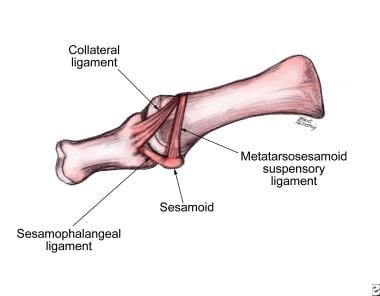 Lateral view of first metatarsophalangeal joint with ligaments of sesamoid complex.
Pathophysiology
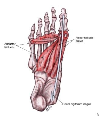
During the gait cycle, the hallux and the digits generally remain parallel to the long axis of the foot, regardless of the degree of forefoot abduction (or pronation) occurring (see the image below). This is because of the pull of the conjoined adductor tendon, EHL tendon, and flexor hallucis longus (FHL) tendon. The tendons gain greater mechanical advantage the further the joint is displaced, with tension created in the medial aspect of the joint and compression laterally.
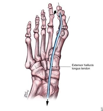 Line of pull of extensor hallucis longus causing metatarsal to deviate medially and hallux to deviate laterally.
Medial tension causes the medial collateral ligaments to pull on the dorsomedial aspect of the first MT head, causing bony proliferation. Lateral tension causes the sesamoid apparatus to fixate in a laterally dislocated position. Remodeling also occurs laterally in addition to medially, as evidenced by the increase of the proximal articular set angle (PASA) or structural remodeling of the cartilage. Therefore, without correction of the biomechanical factors, excessive pronation continues, with propagation of the deformity.
Etiology
HV is known to have numerous etiologies, including biomechanical, traumatic, and metabolic factors. The most common etiology, yet the most difficult to understand, is biomechanical instability. Contributing factors, if present, may include the following:
-
Gastrocnemius or gastrocnemius-soleus equinus
-
Flexible or rigid pes planovalgus
-
Rigid or flexible forefoot varus
-
Dorsiflexed first ray
-
Hypermobility
-
Short first MT
Most often, pronation at the midtarsal and subtalar joints compensates for these factors throughout the gait cycle. However, excessive pronation produces too much midfoot mobility, which decreases stability and prevents resupination and creation of a rigid lever arm; these effects make propulsion difficult, with resultant jamming at the first metatarsophalangeal (MTP) joint.
The hallux and the first MT (also commonly referred to as the first ray) are inherently unstable, secondary to their axial position. Biomechanical stabilizers exist in the form of ligamentous and capsular attachments; dynamic stabilizers include long tendon insertions from the peroneus longus and the instrinsic musculature. [1]
Footwear-based attempts to stabilize this first ray further, however, can keep the hallux in an abducted position if HV is present, causing mechanical stretch and deviation of the medial soft tissue and aggravating symptoms. In addition, tight shoes can cause medial bump pain and nerve entrapment.
During normal propulsion, approximately 65° of dorsiflexion is necessary at the first MTP joint, yet only 20-30° is available from hallux dorsiflexion. Therefore, the first MT must plantarflex at the sesamoid complex to gain the additional 40° of motion needed. Failure to attain the full 65° because of jamming of the joint during pronation subjects the first MTP to intense forces from which hallux valgus can develop.
If the foot is sufficiently hypermobile as a result of excessive pronation, the metatarsal tends to drift medially while the hallux drifts laterally, producing HV. If no hypermobility is present, hallux rigidus develops instead.
Arthritic or metabolic conditions that may cause HV include inflammatory arthropathies such as gouty arthritis, rheumatoid arthritis (see the images below), and psoriatic arthritis, as well as connective tissue disorders such as Ehlers-Danlos syndrome, Marfan syndrome, Down syndrome, and generalized ligamentous laxity.
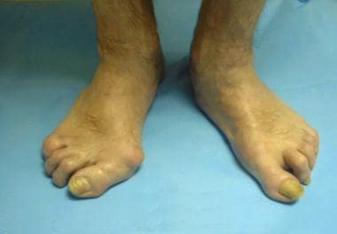 Rheumatoid arthritis. Note greater deformity of right foot (image left) versus left foot (image right).
 Rheumatoid arthritis. Note lateral deviation of hallux, cystic changes of metatarsal head, and hammertoe of lesser digits.
The myriad neuromuscular diseases that may cause HV include multiple sclerosis, Charcot-Marie-Tooth disease, and cerebral palsy.
Traumatic conditions that may cause HV include malunions secondary to first-MT fractures with or without intra-articular damage, soft-tissue injuries of the hallux, and sequelae of dislocations.
Structural deformities that have been associated with HV include malalignment of articular surface or MT shaft, abnormal MT length, metatarsus primus elevatus, external tibial torsion, genu varum or valgum, and femoral retrotorsion.
In a case-control study assessing the role played by the morphology of the first tarsometatarsal (TMT) joint (TMT1) in the etiology of HV, Ji et al compared an HV group (82 feet) with a control group (79 feet). [2] The HV group had a significantly wider middle-facet width (MFW; 9.9 vs 8.7 mm), a lower inferior lateral-facet height (ILFH; 1.7 vs 2.5 mm), a smaller inferior lateral-facet angle (ILFA; 16.3º vs 24.5º), and a larger TMT1 angle (1.9º vs 0.9º). Four types of TMT1 morphology were identified: continuous-flat, separated-flat, continuous-protruded, and separated-protruded. The continuous-flat type was found to be associated with more severe HV and TMT1 instability.
Sovilj et al performed a study (315 feet with HV) aimed at determining whether the anatomic orientation of the first metatarsocuneiform (MTC) joint (oblique, 165 feet; transverse, 145; convex, 5) affected the size of the HV angle (HVA) and the first intermetatarsal angle (IMA) and whether it contributed to the dynamics of the development of HV. [3] An oblique anatomic orientation of the first MTC joint was found to be associated with more severe HV and with faster development of the deformity.
Epidemiology
Although hallux valgus is a common condition that accounts for a significant number of office visits to foot and ankle specialists, its incidence has not been documented accurately. Relatively few studies have been published, and much of the information has consisted of empirical data with low levels of evidence.
The National Health Interview Survey conducted by the National Center for Health Statistics found that hallux valgus affected approximately 1% of adults in the United States. [4] Gould et al found that the incidence increased proportionately with age, from 3% in persons aged 15-30 years to 9% in persons aged 31-60 years to 16% in those older than 60 years. Gould et al also reported a two- to fourfold higher incidence in females than in males. Whether this finding indicates a true sex-based dimorphism or whether it reflects differences in footwear remains to be determined.
The role of genetic predisposition has also been noted, with evidence to suggest familial tendencies.
Prognosis
Hallux valgus is a complex deformity, and various approaches are available. To date, no satisfactory studies have been performed to compare the various procedures and their success rates. If the deformity and etiology are addressed successfully, the benefits of treatment far outweigh the risks. [5, 6]
Schuh et al studied changes of plantar pressure distribution during the stance phase of gait in patients who underwent hallux valgus surgery followed by multimodal rehabilitation. [7] The study included 30 patients who underwent Austin and scarf osteotomy for correction of mild-to-moderate hallux valgus deformity and began a rehabilitation program 4 weeks postoperatively (once weekly for 4-6 weeks). Plantar pressure analysis was performed preoperatively and at 4 weeks, 8 weeks, and 6 months postoperatively. Range of motion (ROM) of the first MTP joint was also measured.
The results of this study included an increased mean American Orthopaedic Foot and Ankle Society (AOFAS) score, from 60.7 points preoperatively to 94.5 points 6 months after surgery, as well as increased ROM of the first MTP joint at 6 months, with a significant increase in isolated dorsiflexion. [7] In the first metatarsal head region, maximum force increased from 117.8 N to 126.4 N, and the force-time integral increased from 37.9 N⋅s to 55.6 N⋅s. In the great-toe region, maximum force increased from 66.1 N to 87.2 N, and the force-time integral increased from 18.7 N⋅s to 24.2 N⋅s.
Ciechanowicz et al assessed ability to resume physical activities in 79 patients who underwent scarf osteotomy for hallux valgus. [8] After the procedure, patients' frequency of physical activity rose by about 21%, and the time they spent on sports (min/wk) rose by about 19%. Their satisfaction with the results of scarf osteotomy averaged 8.2 on a scale of 1 to 10. A majority of the patients (67%) were able to maintain this amount of physical activity postoperatively, and a minority (24%) were able to increase it.
A number of the surgical procedures performed to treat hallux valgus are potentially capable of reducing the increased forefoot width commonly associated with this condition. Panchbhavi et al reported an 8.7-mm reduction in the metatarsal span in 52 patients who underwent distal chevron osteotomy and Akin osteotomy. [9] Conti et al reported mean decreases of 8.9 mm in bony foot width and 6.9 mm in soft-tissue width on radiography in 30 patients (31 feet) who underwent a modified Lapidus procedure combined with a modified McBride and Akin osteotomy. [10]
-
Rheumatoid arthritis. Note greater deformity of right foot (image left) versus left foot (image right).
-
Rheumatoid arthritis. Note lateral deviation of hallux, cystic changes of metatarsal head, and hammertoe of lesser digits.
-
Line of pull of extensor hallucis longus causing metatarsal to deviate medially and hallux to deviate laterally.
-
Nonweightbearing foot. Note medial prominence, contracture of extensor hallucis longus, and callus on second digit.
-
Nonweightbearing foot with range of motion being assessed of first ray, which is currently in neutral (neither plantarflexed or dorsiflexed) position.
-
Lateral view of first metatarsophalangeal joint with ligaments of sesamoid complex.
-
Plantar muscles that contribute to deforming forces.
-
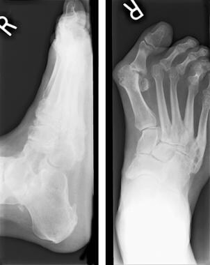
Anteroposterior and lateral radiographs, weightbearing views.
-
Medial bony enlargement is more prominent on this lateral oblique projection than on other views.
-
Template showing angular measurements.
-
Another template showing increased angular relationships.
-
Congruency of first metatarsophalangeal joint.
-
Tibial sesamoid position with bisection of first metatarsal. Positions 1-3 are normal. Positions 4-7 indicate erosion of crista and laterally track-bound, nonreducible hallux valgus.
-
Bunion deformity with minimal joint destruction.
-
Bunion deformity with significant joint destruction.
-
Large intermetatarsal angle and hallux abductovalgus deformity secondary to previous injury. Note increased cortical density of second, third, and fourth metatarsals.
-
Algorithm for choosing surgical correction of hallux abductovalgus. Click image to enlarge.
-
Hallux abductovalgus deformity.
-
Postoperative radiograph obtained after head osteotomy.
-
Preoperative radiograph.
-
Postoperative radiograph shows Keller, or resectional, arthroplasty.
-
Preoperative radiograph shows degenerative joint disease.
-
Postoperative radiograph obtained after resectional arthroplasty and total joint implant placement.
-
Preoperative template for implant placement.
-
Preoperative radiograph shows arthrodesis.
-
Postoperative radiograph show arthrodesis.
-
Preoperative radiograph shows hypermobile first ray.
-
Postoperative radiograph shows arthrodesis of first metatarsocuneiform.
-
Lateral release sequence: (1) release of conjoined adductor hallucis tendon, (2) release of fibular sesamoid ligament, (3) tenotomy of lateral head of flexor hallucis brevis, and (4) excision of fibular sesamoid.
-
Severe hammertoe deformity in second toe overlapping great toe with associated hallux valgus deformity.
-
Preoperative anteroposterior view of hallux valgus.
-
Intraoperative anteroposterior view of correction of hallux valgus via minimally invasive surgical approach.
-
Intraoperative photo showing setup for minimally invasive surgery for hallux valgus.
-
Postoperative anteroposterior view 3 months after hallux valgus surgery.


