Background
Ichthyosis refers to a relatively uncommon group of skin disorders characterized by the presence of excessive amounts of dry surface scales. It is regarded as a disorder of keratinization or cornification, and it is due to abnormal epidermal differentiation or metabolism.
The ichthyosiform dermatoses may be classified according to clinical manifestations, genetic presentation, [1] and histologic findings. Inherited and acquired forms of ichthyosis have been described, and ocular alterations may occur in specific subtypes. Five distinct types of inherited ichthyosis are noted, as follows: ichthyosis vulgaris, lamellar ichthyosis, epidermolytic hyperkeratosis, congenital ichthyosiform erythroderma, and X-linked ichthyosis.
Pathophysiology
Ichthyosis vulgaris is characterized by onset in early childhood, usually between age 3 and 12 months, with fine scales and varying degrees of dryness of the skin. Scaling is most prominent over the trunk, abdomen, buttocks, and legs. The flexural areas, such as the antecubital fossa, are spared. An association may be present between ichthyosis vulgaris and atopic diseases because one third to one half of patients show features of atopic disease and a similar proportion have relatives with atopic disease. A reported 11.5% association is noted between atopic dermatitis and primary hereditary ichthyosis. Ichthyosis vulgaris typically produces no significant ocular findings; however, scaling may be present on the eyelid skin, which could lead to punctate epithelial keratitis and recurrent corneal erosion. Linkage analysis has identified an ichthyosis vulgaris locus on band 1q22.
Two loss-of-function mutations in the coding of the filaggrin (filament aggregating protein) gene have been identified in both ichthyosis vulgaris and atopic dermatitis. Keratohyalin synthesis is affected because of the filaggrin mutation. Filaggrin is an epidermal protein that normally functions as a barrier molecule against environmental allergens, water loss, and infection.
In epidermolytic hyperkeratosis (bullous ichthyosiform erythroderma), a mild generalized erythroderma is present at birth. Bullae formation may occur, which may become infected and give rise to a foul skin odor. Erythroderma fades in infancy while the characteristic grey, waxy scale progresses. They are particularly prominent in the flexural creases. A mutation in the keratin genes (ie, KRT1, KRT10) is the cause of this autosomal dominant disorder.
Lamellar ichthyosis is a rare, autosomal recessive, genetically heterogeneous skin disease caused by mutations involving multiple genetic loci. Type 1 maps to band 14q11.2 and is caused by mutations in the gene for keratinocyte transglutaminase 1, an enzyme responsible for the assembly of the keratinized envelope. [2] Type 2, which is clinically indistinguishable from type 1, maps to band 2q33-q35. In classic lamellar ichthyosis, children with the disease are referred to as collodion babies and are covered at birth by a thickened membrane that subsequently is shed. The scaling of the skin involves the whole body with no sparing of the flexural creases. Approximately one third of children affected with this disorder develop bilateral ectropion of the cicatricial type that appears to result from excessive dryness of the skin and subsequent contracture. Secondary corneal ulceration may occur secondary to long-term exposure.
In X-linked ichthyosis, generalized scaling is present at or shortly after birth. This scaling is most prominent over the extremities, neck, trunk, and buttocks. The flexural creases may be involved but palms, and soles are spared. Irregular stromal corneal opacities that are located anterior to the Descemet membrane are found in 16-50% of male patients, and this finding may be used to distinguish this form of ichthyosis from all other forms. Approximately 25% of female carriers have minor corneal opacities. The corneal opacities are not known to affect visual acuity. An example of findings in X-linked ichthyosis is shown in the image below.
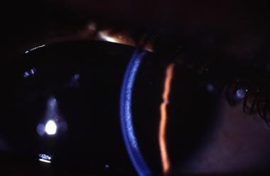 This slit beam illumination photograph of the cornea localizes the corneal opacity to the posterior stroma and the pre-Descemet membrane region. This type of corneal opacity is commonly present in X-linked recessive ichthyosis.
Previous studies have shown a deficiency of steroid sulfatase (STS) in skin fibroblasts and a marked elevation of plasma cholesterol sulfate in patients with X-linked ichthyosis. In most cases, STS deficiency is caused by a partial or complete deletion of the STS gene mapped on band Xp22.3. Deletions of the STS gene have systemic effects, such as corneal opacities, cryptorchidism, and failed progression during labor. Deletions to flank regions of the STS gene have been linked with intellectual disability. These deletions involve portions of the VCXA and VCXB1 genes.
Nonbullous congenital ichthyosiform erythroderma (NBIE) or congenital ichthyosiform erythroderma (CIE) is a milder form of the disease that is autosomal recessive in inheritance. CIE has been found to be caused by mutations in the genes coding for transglutaminase 1, 12R-lipoxygenase, and/or lipoxygenase 3. The lipoxygenase genes play a role in the epidermal permeability layer. As with lamellar ichthyosis, neonates with CIE are referred to as collodion babies, but, as children and adults, they show generalized red skin with thin, white scaling. Other manifestations include persistent ectropion and scarring alopecia.
Multiple congenital ectodermal dysplastic syndromes are associated with scaling and other system defects. The keratitis, ichthyosis, and deafness (KID) syndrome is a congenital disorder of ectoderm that affects not only the epidermis but also other ectodermal tissues, such as the corneal epithelium and the inner ear. [3, 4] KID syndrome may present with the Hutchinson triad (the combination of notched, widely spaced peg teeth, interstitial keratitis, and deafness). KID syndrome has been linked to mutations in the connexin (gap junction protein) 26 gene (GJB2) on band 13q11-q12.
Colobomas of the eye, heart defects, ichthyosiform dermatosis, intellectual disability, and ear defects (CHIME) syndrome comprises a rare neuroectodermal disorder.
Netherton syndrome is an autosomal recessive condition that consists of an ichthyosiform dermatosis with variable erythroderma, hair shaft defects, and atopic features. Netherton syndrome has been linked to a mutation on band 5q32, specifically encoding for LEKTI (lymphoepithelial Kazal-type—related inhibitor), a serine protease inhibitor.
Sjögren-Larsson syndrome is an autosomal recessive condition that comprises ichthyosis, spastic diplegia, pigmentary retinopathy, and intellectual disability.
Congenital hemidysplasia with ichthyosiform erythroderma or nevus and limb defects (CHILD) syndrome is a rare X-linked dominant malformation syndrome characterized by unilaterally distributed ichthyosiform erythroderma or nevi, often sharply delimited at the midline, and ipsilateral limb defects. This syndrome is caused by a loss-of-function mutation of nicotinamide adenine dinucleotide phosphate (NADPH) steroid dehydrogenase-like (NSDHL) protein at band Xq28.
Acquired ichthyosis usually occurs in adults and manifests as small, white, fishlike scales that frequently are concentrated on the extremities but may be seen in a generalized distribution. This form of ichthyosis may be associated with internal neoplasia (eg, Hodgkin lymphoma, leukemia), systemic illness (eg, sarcoidosis, HIV infection, hypothyroidism, chronic hepatitis, malabsorption), bone marrow transplantation, or the intake of certain medications that interfere with sterol synthesis in epidermal cells (eg, nicotinic acid).
Newborns with type 2 Gaucher disease (glucosyl cerebroside lipidosis) may present with ichthyotic skin at birth prior to neurologic manifestations, which could be mistaken for a congenital form of ichthyosis.
Epidemiology
Frequency
United States
Ichthyosis vulgaris is the most common form and is an autosomal dominant trait with an incidence of 1 case per 300 population. Epidermolytic hyperkeratosis is an autosomal dominant disorder with an incidence of 1 case per 300,000 population. Lamellar ichthyosis, a more severe form of dermatosis, is an autosomal recessive trait with an incidence of 1 case per 300,000 population. X-linked recessive ichthyosis has an incidence of 1 case per 6000 males.
International
Studies show that a higher prevalence of X-linked ichthyosis as compared to ichthyosis vulgaris may be noted in Mexico, which is markedly different from the experience in the United States. In the United Kingdom, the incidence of ichthyosis vulgaris was reported to be 1 case per 250 population in a study population of 6501 healthy school children. Similarly, in another large epidemiologic study, the incidence of X-linked recessive ichthyosis was 1 case in 6190 males. In a Danish population study, the predicted incidence of X-linked recessive ichthyosis was 1 case per 2000 males. In a Danish study, Bygum et al concluded that epidermolytic ichthyosis had a prevalence of approximately 1 in 350,000, with a high percentage of de novo mutations (75%). [5] In southern coastal Italy, the frequency of X-linked recessive ichthyosis was estimated at 1.98 in 10,000 males. In China, ichthyosis vulgaris has a prevalence of 2.29%.
Mortality/Morbidity
An increased risk of testicular cancer in men with ichthyosis vulgaris has been suggested. In addition, an increased incidence of testicular maldescent, cryptorchidism, abnormalities of the sperm count or motility leading to infertility, and testicular cancer has been reported in patients with X-linked recessive ichthyosis.
Race
In general, all races may be affected in the inherited and acquired forms of ichthyosis.
Sex
X-linked recessive ichthyosis is much more prevalent in males. It is caused by a deficiency of STS. Because this enzyme plays an important role in androgen metabolism, men with this ailment do not show androgenetic alopecia or develop only mild forms of this common type of hair loss.
Prognosis
The dryness of the eyes can be treated with artificial tears, ointments, bandage contact lenses, punctal occlusion, and possibly surgery, depending on the presence of abnormal lid closure or limbal stem cell deficiency. Any persistent corneal epithelial defect must be treated aggressively to prevent corneal infection.
Patient Education
Patients must realize that this condition is chronic, and they will need long-term therapy. Without long-term therapy, the defective permeability barrier associated with ichthyosis can result in a chronic loss of water and calories, which may impair growth in children.
-
This direct illumination slit lamp photograph discloses a reticular central corneal haze that is seen bilaterally. Visual acuity is 20/20 in both eyes. The patient's chief complaint is photophobia and dry scaly skin.
-
This slit beam illumination photograph of the cornea localizes the corneal opacity to the posterior stroma and the pre-Descemet membrane region. This type of corneal opacity is commonly present in X-linked recessive ichthyosis.


