Overview
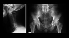
Ankylosing spondylitis is a chronic, progressive inflammatory disorder primarily affecting the axial skeleton. Ankylosing spondylitis is one of the seronegative spondyloarthropathies, a group of multisystem inflammatory diseases that affects the spine, peripheral joints, adjacent soft tissues, and extra-articular structures. Most of these disorders are associated with the human leukocyte antigen B27 (HLA-B27). [1]
In addition to ankylosing spondylitis, the seronegative spondyloarthropathies include the following:
-
Reactive arthritis (also referred to as Reiter syndrome)
-
Inflammatory bowel disease-associated arthritis
-
Psoriatic arthropathy
-
Juvenile spondyloarthropathy
-
Undifferentiated spondyloarthropathies
Several of these disorders share some clinical features, such as sacroiliitis, spondylitis, asymmetric peripheral arthritis, aortitis, clinically evident or occult bowel lesions, and uveitis. Acute anterior uveitis (AAU) occurs in as many as 39% of patients at some time during the course of ankylosing spondylitis, particularly in persons with the HLA-B27 allele, and can precede the onset of spondylitis in half the cases. [2] The uveitis associated with spondyloarthropathies almost always is confined to the anterior segment of the eye, in the form of AAU, but uncommonly can coexist with prominent involment of the vitreous and the posterior segment. [3] Patients with ankylosing spondylitis who have peripheral arthritis also have a higher prevalence of uveitis. [4, 5, 6]
Go to Ankylosing Spondylitis and Undifferentiated Spondyloarthropathy for complete information on this topic.
Complications
Anterior and posterior segment inflammation is greater with uveitis associated with ankylosing spondylitis than it is with idiopathic uveitis, especially if ocular surgery is performed.
Etiology
The exact role of HLA-B27 in the development of uveitis is not precisely known, but several theories for its role in disease exist. The oldest theory is that of molecular mimicry, in which an infectious agent triggers an immune response against autoantigens. The HLA-B27 antigen shares some amino acid homology with proteins from several gram-negative bacteria, including Yersinia enterocolitica, Shigella flexneri, and Chlamydia trachomatis, as well as from gram-negative bacteria found in the GI tract.
Other theories include the arthritogenic peptide theory, in which the immune response is directed against a peptide presented by HLA-B27; the altered self theory, in which a reactive sulfhydryl group on the HLA-B27 molecule interacts with other self peptides, thereby inducing antigenicity; and, finally, the theory that the HLA-B27 allele confers an altered immune response that directly relates to the disease.
Prognosis
HLA-B27 typing is not necessary to establish the diagnosis of ankylosing spondylitis, but it is a useful prognostic indicator in patients with uveitis.
Patients with ankylosing spondylitis and the HLA-B27 antigen tend to develop uveitis at a younger age, have a longer duration of ocular disease, and have more recurrences. Additionally, patients who are HLA-B27 positive appear to have more severe disease in terms of anterior chamber inflammation and vitreitis. More severe and recurrent attacks will tend to cause structural complications such as cataract, anterior and posterior synechiae, elevated intraocular pressure, secondary glaucoma, and CME 15.
The ocular prognosis is dependent on the ability to control the inflammatory response.
Sex and Age Predilections
The male-to-female ratio is estimated to be 2:1. [7]
Ankylosing spondylitis usually begins during the second decade. The associated uveitis typically occurs at an earlier age than does idiopathic uveitis.
Patient History
Take a thorough history.
Patients often complain of acute onset of blurry vision, ocular redness, pain, photophobia, and occasionally tearing. No history of ocular discharge is present.
Most episodes of uveitis are unilateral, but the condition can be bilateral, presenting in an alternating pattern between the eyes.
Attacks are often sudden in onset and have a limited duration. Frequent recurrences with a full remission between episodes are characteristic.
Some patients may present with chronic symptoms of blurring and ocular hyperemia. [2, 8]
Past ocular history
Past ocular history should include the following:
-
Previous episodes, duration, and treatment
-
Previous complications (eg, cataract, glaucoma, cystoid macular edema [CME], epiretinal membrane, band keratopathy)
Physical Examination
Patients suspected of having ankylosing spondylitis should undergo a thorough physical examination, preferably by a rheumatologist.
Perform a complete ophthalmic examination, with special emphasis on the following:
-
Best-corrected distance and near acuity
-
Pupil examination
-
Slit-lamp examination
-
Intraocular pressure - IOP is often low during acute exacerbations, hypotony, and phthisis. IOP may be high in severe attacks, trabeculitis, and secondary glaucoma.
-
Gonioscopy - Peripheral anterior synechiae can form and cause secondary angle closure.
-
Dilated fundus examination - Vitreitis, CME, retinal vasculitis, epiretinal membrane, and papilitis may be seen.
Slit-lamp examination may reveal the following:
-
Conjunctiva and sclera - Conjunctival and limbar hyperemia, watery discharge; no follicles or papillae
-
Cornea - Small to medium white/grey keratic precipitates
-
Anterior chamber - Cells and flare, fibrin, and/or hypopyon not uncommon
-
Iris - Posterior synechiae, peripheral anterior synechiae, pupillary membrane, dilated iris vassels. Rarely spontaneus hyphema
-
Lens - Posterior subcapsular cataract
-
Vitreous - Anterior vitreous cells, vitreitis
-
Optic Nerve: Optic disc swelling
-
Retina: Retinal vasculitis
Differential Diagnosis
Conditions to consider in the differential diagnosis of the ophthalmologic manifestations of ankylosing spondylitis include the following disorders:
-
Infectious anterior uveitis, as ocular manifestations of syphilis or tuberculosis
Ancillary Studies
HLA-B27 typing
A positive result narrows the differential diagnosis to the seronegative spondyloarthropathies. This also may be a useful prognostic indicator for the severity of ocular disease.
Antinuclear antibody
Antinuclear antibody (ANA) results are negative in ankylosing spondylitis but positive in many patients with collagen vascular diseases. Also, the results are positive in children with juvenile idiopathic arthritis (JIA) who are at high risk to develop uveitis.
Rheumatoid factor
Most patients with JIA are rheumatoid factor (RF) negative.
Complete blood count
A mild anemia may be noted in some patients with a seronegative spondyloarthropathy, JIA, or collagen vascular disease.
Syphilis serology
Treponemal tests such as the fluorescent treponemal antibody absorption (FTA-ABS) test should be performed to rule out syphilis as a cause of the uveitis. Nontreponemal tests such as the Venereal Disease Research Laboratory (VDRL) test are useful to monitor the response to treatment and reactivation.
Lyme titer
Lyme titer is useful only in patients with a history of a tick bite and a rash typical for erythema chronicum migrans.
Mycobacterium tuberculosis test
Purified protein derivative (PPD) or interferon gamma release assay (IGRA) should be performed to rule out tuberculosis as a cause of the uveitis.
Angiotensin-converting enzyme
Angiotensin-converting enzyme (ACE) often is elevated in patients with sarcoidosis.
Optical Coherence Tomography
Optical coherence tomography (OCT) may be useful in patients with CME.
Fluorescein angiography
Fluorescein angiography (FA) may be useful in patients with CME and/or retinal vasculitis.
Other tests
Chest Radiograph/Computed tomography: May be usefull to identify findings suggestive of sarcoidosis or pulmonary tuberculosis as a cause of the uveitis.
Medical Therapy
The objectives of treatment for patients with uveitis include the reduction of inflammation (corticosteroids), the relief of symptoms (cycloplegics and anti-inflammatories), and the preservation or restoration of vision.
Corticosteroids
Corticosteroids decrease the recruitment of inflammatory cells and alter cell function. They may be administered topically, via periocular injection, or systemically, and then tapered slowly.
Potent topical steroids are used to treat disease limited to the anterior segment, reducing anterior chamber inflammation. Adverse effects include cataract formation and increased intraocular pressure. Typically, prednisolone acetate 1% is prescribed every hour while awake, with dexamethasone ointment at bedtime, and then slowly taper down over 8 to 10 weeks according to response. Therapeutic levels of corticosteroids cannot be achieved in the vitreous via the topical route; therefore, this method of administration is ineffective for posterior segment disease. An alternative route of delivery may be considered when the anterior uveitis is severe or unresponsive to topical treatment.
Peri and intra ocular corticosteroids may be used to treat severe anterior uveitis, intermediate uveitis, or CME, which is the major cause of inflammatory visual loss in patients with uveitis. Complications include cataract formation and increased intraocular pressure, which may be refractory to all forms of therapy, short of surgical removal of the injected material.
Systemically administered corticosteroids may be considered for vision-threatening uveitis unresponsive to maximal topical and periocular therapy. An internal medicine or rheumatology consultation is advisable in the management of these patients in the absence of an ophthalmologist specialized in uveitis.
If systemic treatment is required, it is necessary to determine whether medical contraindications to systemic corticosteroids exist, particularly in children and in elderly patients. Systemic corticosteroids suppress growth in children. They may exacerbate diabetes mellitus in susceptible individuals. Weight gain and fluid retention are expected effects. Electrolyte imbalance is a common complication. Long-term hazards include osteoporosis, compression of the spine, gastrointestinal hemorrhage, and reduction in immune response to infection, especially tuberculosis.
Cycloplegics
Cycloplegics reduce ciliary spasm and pain and prevent the development of posterior synechiae. Drugs and dosing include the following:
-
Tropicamide 1%, 3 times a day (mild disease)
-
Homatropine 2% or 5%, 2 to 4 times a day (moderate to severe inflammation, hypopyon)
Immunomodulatory therapy
Immunomodulatory therapy often is useful in patients who are unresponsive to short courses of corticosteroids, in patients with chronic uveitis, or in patients who develop adverse effects from corticosteroid therapy.
A number of agents have been used, including methotrexate, azathioprine, cyclosporin A, mycophenolate mofetil, cyclophosphamide, and chlorambucil. Myelosuppression and secondary infection are among the most common adverse effects of these agents.
Tumor necrosis factor-alpha (TNF-alpha) inhibitors may be useful in patients with the seronegative spondyloarthropathies, including ankylosing spondylitis. Available TNF-alpha inhibitors include infliximab and adalimumab. These agents may be useful in reducing the number of flares of anterior uveitis in patients with ankylosing spondylitis. Adalimumab has been shown to reduce the rate of anterior uveitis flares by at least 50% in a large open-label study. [9, 10, 11, 12, 13] New anti TNF-alpha agent golimumab and certolizumab pegol have been used in refractory cases, showing good results in achieving quiescence and improving visual acuity. [14, 15]
An ophthalmologist specialized in uveitis, a rheumatologist, an internist, or an oncologist is an essential member of the team in the management of patients treated with immunomodulatory agents.
Consultations
Rheumatology
Early evaluation for all patients with suspected ankylosing spondylitis is essential to avoid delays in diagnosis and treatment of the systemic disease. Ongoing communication with the rheumatologist is critical for managing patients who require immunomodulatory therapy.
Questions & Answers
Overview
What is ankylosing spondylitis?
What are the possible ophthalmologic complications of ankylosing spondylitis?
What causes the ophthalmologic manifestations of ankylosing spondylitis?
What is the ocular prognosis of ankylosing spondylitis?
Which ocular history findings are characteristic of ankylosing spondylitis?
What are the goals of treatment for ophthalmologic manifestations of ankylosing spondylitis?
Which specialist consultations are beneficial to patients with ankylosing spondylitis?