Background
Neonatal conjunctivitis, also known as ophthalmia neonatorum, presents during the first month of life. It may be aseptic or septic and, if left untreated, may be blinding.
Aseptic neonatal conjunctivitis most often is a chemical conjunctivitis that is induced by silver nitrate solution, which has been used at birth since the late 1800's for prophylaxis of infectious conjunctivitis (a procedure known as Credé's prophylaxis). Chemical conjunctivitis is becoming less common owing to the use of erythromycin ointment or povidone iodide in place of silver nitrate solution for the prophylaxis of infectious conjunctivitis.
Bacterial and viral infections are major causes of septic neonatal conjunctivitis, with Chlamydia being the most common infectious agent and Neisseria being the most visually threatening. Infants may acquire these infective agents as they pass through the birth canal during the birth process.
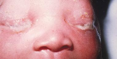 Severe purulent discharge and eyelid edema in a newborn with gonococcal conjunctivitis (confirmed with Gram stain and culture).
See the following for more information:
Anatomy and pathology
The conjunctiva (a thin translucent mucous membrane) can be divided into palpebral, bulbar, and forniceal regions. The conjunctiva contains nonkeratinizing, squamous epithelium and a thin, richly vascularized substantia propria containing lymphatic vessels and cells, such as lymphocytes, plasma cells, mast cells, and macrophages. The conjunctiva also has accessory lacrimal glands and goblet cells.
The pathology of neonatal conjunctivitis is influenced by the anatomy of the conjunctival tissues in the newborn. The inflammation of the conjunctiva may cause blood vessel dilation, potentially dramatic chemosis, and excessive secretion. This infection tends to be more serious in neonates owing to their lack of immunity, lack of lymphoid tissue in the conjunctiva, and absence of tears at birth.
Patient education
Educate parents or care providers to wash their hands frequently to prevent transmission of neonatal conjunctivitis. Educate pregnant women on the importance of regular examinations to detect and treat sexually transmitted infections such as herpes simplex, gonorrhea, and chlamydia in order to decrease the incidence of neonatal conjunctivitis.
For patient education information, see the Eye and Vision Center, as well as Pinkeye.
Etiology
The etiology of neonatal conjunctivitis can be chemical or microbial. Although several noninfectious and infectious agents can inflame the conjunctiva, the most common causes of neonatal conjunctivitis are silver nitrate solution and chlamydial, gonococcal, staphylococcal, and herpetic infections.
Silver nitrate solution
Credé's method of instilling a drop of 2% aqueous solution of silver nitrate into a newborn's eyes was first published in 1881 and significantly advanced the prevention of neonatal conjunctivitis. [1]
Silver nitrate is a surface-active chemical that facilitates agglutination and inactivation of gonococci. Ironically, silver nitrate was later found to be toxic to the conjunctiva, particularly in higher concentrations, potentially causing a sterile neonatal conjunctivitis.
Chlamydial conjunctivitis
Chlamydia trachomatis is an obligate intracellular parasite and has been identified as the most common infectious cause of neonatal conjunctivitis. [2]
The reservoir of the organism is the maternal cervix or urethra. Infants who are born to infected mothers are at high risk (approximately 25%-50%) of developing an infection. [3] Chlamydial pneumonitis may also accompany neonatal conjunctivitis.
Neisserial conjunctivitis
Neisseria gonorrhoeae is a gram-negative diplococcus and is potentially the most dangerous and virulent infectious cause of neonatal conjunctivitis. As with chlamydia, maternal cervical and urethral mucosa provide a reservoir for N gonorrhoeae, which is acquired during birth.
Gonococci can penetrate intact epithelial cells and divide rapidly inside them. Diagnostic Gram or Giemsa stain smears obtained from genitourinary or ocular mucosal scrapings reveal characteristic gram-negative intracellular diplococci.
Gonococcal conjunctivitis must be absolutely excluded in every case of neonatal conjunctivitis to prevent potentially blinding corneal and conjunctival complications.
Other bacteria
The most commonly identified gram-positive organisms include Staphylococcus aureus,Streptococcus pneumoniae,Streptococcus viridans, and Staphylococcus epidermidis. These bacteria make up 30-50% of all cases of infectious neonatal conjunctivitis. [4]
Gram-negative organisms, such as Escherichia coli, Klebsiella pneumoniae, Serratia marcescens, and Proteus, Enterobacter, and Pseudomonas species, also have been implicated. There has been one reported case of Eikenella corrodens neonatal conjunctivitis. [5]
Infants of low birth weight and low gestational age with clinical signs of conjunctivitis in the neonatal intensive care unit (NICU) should be evaluated and treated for a gram-negative etiology. [6]
Herpes simplex
Herpes simplex virus (HSV) is a rare cause of neonatal keratoconjunctivitis, found in less than 1% of cases, [4] and can be associated with a generalized herpes simplex infection.
Most infants with such an infection acquire the disease during the birth process. Caesarean delivery should be strongly considered when active maternal genital herpetic disease is recognized at term since the risk of transmitting HSV to the neonate during vaginal delivery is 25-60%. [7]
Epidemiology
Occurrence in the United States
The incidence of infectious neonatal conjunctivitis ranges from 1-2%, depending on the socioeconomic character of the area.
The epidemiology of neonatal conjunctivitis changed when silver nitrate solution was introduced in the 1800s to prevent gonococcal ophthalmia.
Chlamydia is the most common infectious agent that causes ophthalmia neonatorum in the United States, where 2%-40% of neonatal conjunctivitis cases are caused by Chlamydia. [3]
In contrast, the incidence of gonococcal ophthalmia neonatorum has been reduced dramatically and causes less than 1% of cases of neonatal conjunctivitis. [8]
International occurrence
As in the United States, the incidence of ophthalmia neonatorum in many other countries decreased after silver nitrate solution came into general use.
In Europe, the incidence fell from 10% of births to less than 1%.
The rates of neonatal conjunctivitis vary in different parts of the world. In one hospital in Pakistan, the incidence of neonatal conjunctivitis was reported at 17%. [9]
Race-, sex-, and age-related demographics
No published information is available on race- or sex-related differences in the incidence of neonatal conjunctivitis.
Prognosis
Neonatal conjunctivitis usually responds to appropriate treatment, and the prognosis generally is good.
Antibiotics have significantly altered the prognosis of neonatal conjunctivitis, especially with Neisseria gonorrhoeae infection.
Mortality associated with neonatal conjunctivitis is due to systemic involvement of the infectious agent. No published information is available on mortality.
Complications
If untreated, peripheral corneal ulceration may occur in N gonorrhoeae infection and rapidly progress to corneal perforation.
When unrecognized and not immediately treated, Pseudomonas infection may lead to endophthalmitis and subsequent death.
Pneumonia has been reported in 10-20% of infants with chlamydial conjunctivitis. [10]
HSV keratoconjunctivitis can cause corneal scarring and ulceration. Additionally, disseminated HSV infection often includes central nervous system involvement. [7]
-
Severe purulent discharge and eyelid edema in a newborn with gonococcal conjunctivitis (confirmed with Gram stain and culture).
-
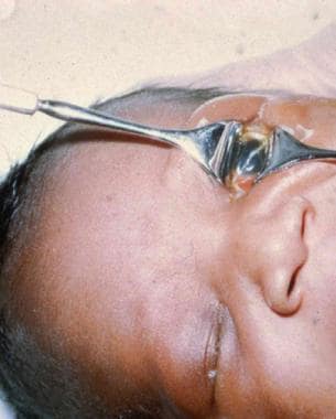
Cloudy cornea without ulcer in neonatal gonococcal conjunctivitis.

