Practice Essentials
Urethral prolapse is a circumferential protrusion of the distal urethra through the external urethral meatus. It is a rarely diagnosed condition that occurs most commonly in prepubertal girls and postmenopausal women. Even less common is strangulated urethral prolapse.
Vaginal bleeding is the most common presenting symptom of urethral prolapse. Upon examination, round, often doughnut-shaped protrusion mucosa is observed obscuring urethral opening.
Because urethral prolapse is rare, the rate of misdiagnosis is high. The differential diagnoses of a urethral mass are broad, ranging from a simple urethral caruncle to rhabdomyosarcoma. Increased physician awareness and early recognition of urethral prolapse avoids unnecessary examinations and patient anxiety.
Management of urethral prolapse ranges from medical therapy that consists of topical estrogen use to conservative surgical excision when medical therapies fail. [1]
History of the Procedure
Solingen first described urethral prolapse in 1732. Most authors believe urethral prolapse is restricted to the distal urethra.
Various treatments have been used for this type of lesion. In the past, treatment was primarily surgical. Currently, conservative therapy with topical agents is the preferred initial therapy for uncomplicated urethral prolapse.
Failure of medical therapy for strangulated urethral prolapse warrants surgical intervention. Many different surgical procedures, ranging from simple manual reduction to complete surgical excision, have been described.
Surgical excision has a high cure rate and is the most definitive therapy. Procedures such as cautery, cryotherapy, and ligation of the prolapsed urethra over a Foley catheter are no longer routinely practiced.
Problem
Urethral prolapse is defined as the complete eversion of the terminal urethra from the external meatus (see image below). Although infrequently reported in the literature, it may in fact be a common disorder among postmenopausal women and prepubertal girls. Urethral prolapse must be distinguished from urethral caruncle, in which only a non-circumferential portion of the urethral mucosa intially protrudes.
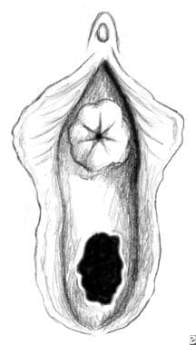 Urethral prolapse. Note the complete circular eversion of the distal urethral mucosa.
Epidemiology
Frequency
Epidemiologic features are as follows:
-
Urethral prolapse is relatively uncommon and has a bimodal age distribution.
-
It children, it occurs in prepubertal girls, with an average age at presentation of 4 years.
-
In adults, urethral prolapse most commonly occurs in women after menopause.
-
The English literature reports 270 cases involving children and 46 cases involving adults.
-
One report of urethral prolapse in identical twins suggests that heredity may play a role. [2]
-
Strangulated urethral prolapse is more common in adults than in children. [3]
Etiology
The exact cause of urethral prolapse remains unknown; however, several theories have been proposed. These theories may be divided into congenital or acquired factors.
Congenital factors include weak pelvic floor structures leading to inadequate genitourinary support and urethral hypermobility. Proposed theories include intrinsic abnormalities of the urethra (eg, an abnormally patulous urethra, a wide urethra, redundant mucosa). Other hypotheses include neuromuscular disorders, urethral malposition, submucosal weakness, or deficient elastic tissue.
Acquired factors include trauma during birth, such as prolonged vaginal delivery or obstetric lacerations. Other possibilities include poor tissue quality in debilitated and malnourished individuals. Periurethral bulking agent injection is an unusual cause of urethral prolapse. [4] A popular theory involves a weakened attachment between the inner longitudinal and outer circular-oblique smooth muscle layers of the urethra. Separation of the 2 muscle layers, coincident with episodic increases in intra-abdominal pressure, may predispose to urethral prolapse.
Risk factors for urethral prolapse in children include increased intra-abdominal pressure as a result of chronic coughing or constipation. One retrospective multicenter review of symptomatic urethral prolapse in children found no predisposing factors, but noted that most patients had a mean weight, height, and body mass index greater than the 50th percentile. [5]
In elderly women, poor nutrition and hygiene have been reported as additional possible risk factors. Loss of estrogen at menopause has also been cited as a risk factor. Urethral prolapse in elderly women has become much less common since the introduction of estrogen replacement therapy.
Pathophysiology
Urethral prolapse primarily involves the distal female urethra. The urethra is composed of inner longitudinal and outer circular-oblique smooth muscle layers. Usually, a natural cleavage plane is present between the inner and outer muscle layers. In a healthy female urethra, this cleavage plane firmly adheres to the opposing muscle layers.
A prolapsed urethra may result from these 2 muscle layers separating after a sudden episodic increase in intra-abdominal pressure. Disruption of these muscle layers results in complete and circular eversion of the urethral mucosa through the external meatus and leads to urethral prolapse. Swelling and congestion of the prolapsed mucosa create a purse-string effect around the distal urethra, impeding venous return and exacerbating vascular congestion. If left untreated, urethral prolapse may progress to strangulation and eventual necrosis of the protruding tissues.
The fundamental anatomical defect of urethral prolapse is the separation of the longitudinal and circular-oblique smooth muscle layers. Surgical apposition of these smooth muscle layers is curative.
Presentation
The two types of urethral prolapse include prepubertal and postmenopausal. Clinical presentation is different for both groups.
Prepubertal urethral prolapse is predominantly asymptomatic. Often, urethral prolapse is an incidental finding during routine examination. The most common presentation is vaginal bleeding associated with a periurethral mass. [6] Symptomatic children present with bloody spotting on their underwear or diapers. Hematuria is uncommon.
Until urethral prolapse is definitively diagnosed, the presence of blood in the genital area in girls should raise suspicion of sexual abuse and warrant ruling out this possibility. Care should be taken before reporting such cases, however, as the prevalence of injuries following sexual abuse is low and the differential diagnosis of prepubertal vaginal bleeding is broad, including foreign bodies and vulvovaginitis as well as rare conditions such as urethral prolapse and failed fusion of the perineum. [7]
Voiding disturbances are typically rare in the pediatric population, but when they are present, patients may report dysuria, urinary frequency, or introital pain. Again, hematuria is uncommon. Children may report genital pain if the prolapsed mucosa becomes very large or if thrombosis and necrosis have developed. Furthermore, although urethral prolapse is reportedly a nonobstructive lesion, acute urinary retention secondary to urethral prolapse has been reported. [8]
In contrast, patients with postmenopausal urethral prolapse are often symptomatic. Vaginal bleeding associated with voiding symptoms is fairly common. Affected women may report dysuria, urinary frequency or urgency, and nocturia. Either microscopic or gross hematuria may be present. If the urethral prolapse is large, the mucosal mass may become strangulated, which results in venous obstruction, thrombosis, and necrosis of the prolapsed tissue. [9] Patients with strangulated urethral prolapse may present with suprapubic pain, dysuria, hematuria, and urethral bleeding. Attendant urinary tract infection is also possible.
Upon physical examination, urethral prolapse appears as a doughnut-shaped mass protruding from the anterior vaginal wall . In children, a pinkish orange congested mass may be observed at the center of the urethral meatus. The mass may be painful and tender to palpation. The mucosa is ulcerated in most cases and usually bleeds upon contact. In adults, urethral prolapse appears as erythematous inflamed mucosa protruding from the urethral meatus (see the image below). The congested mucosa may appear bright red or dark and cyanotic. Depending on the evolution of the process, the prolapsed tissue may appear infected, ulcerated, or necrotic.
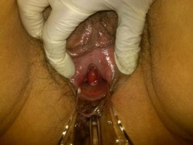 On pelvic examination, urethral prolapse appears as a doughnut-shaped mass protruding from the anterior vaginal wall.
Urethral prolapse is diagnosed by verifying that a central opening is present within the prolapsed tissue and that this opening is the urethral meatus. In children, observation during voiding or catheterization of the central opening is diagnostic. In adults, urethral catheterization or cystourethroscopy helps verify the presence of the urethral meatus. The absence of a urethral meatus at the center of the prolapsed mucosa precludes the diagnosis of urethral prolapse.
The initial diagnosis of urethral prolapse made by a referring pediatrician or emergency department physician warrants scrutiny because of the rarity of this condition. When evaluating a urethral mass, the differential diagnoses should include urethral or vaginal malignancy, urethral caruncle, ectopic ureterocele, [10] condyloma, and rhabdomyosarcoma. Routine evaluation with urograms and voiding cystourethrograms is unnecessary except in patients who may have evidence of malignancy, prolapsed ectopic ureterocele, or abnormalities in the ureterovesical junction.
Urethral masses that were actually sarcoma botryoides or endodermal sinus tumors have been misdiagnosed as urethral prolapse. Urethral leiomyomas and malakoplakia have also been misdiagnosed as urethral prolapse. [11]
If the diagnosis is not completely certain after a detailed history and careful physical examination, the patient should be examined under general anesthesia to rule out more serious lesions. Surgical excision and pathologic examination confirm the diagnosis.
Although urethral prolapse does not have pathognomonic features, histological examination often reveals ulcerated polypoid tissue composed of fibrovascular stroma with dilated veins and a few organized thrombi. Mucosal edema, vascular thrombosis, and inflammatory cells may also be present.
Strangulated urethral prolapse appears to be more common in the adult population than in the pediatric population. Patients with strangulated urethral prolapse may report suprapubic tenderness and severe pain during urination. Urethral prolapse is often associated with urinary tract infection. The diagnosis of strangulated urethral prolapse is predicated upon the discovery of an extremely painful, cyanotic, circular mass surrounding the external meatus, and this condition requires surgical excision.
For more information, see Urethral Anomalies and Urethral Prolapse in Children
Indications
Surgical excision is justified in young patients with symptomatic urethral prolapse or with recurrent urethral prolapse. Additionally, management of predisposing factors, such as constipation or dysfunctional voiding (causing straining) is warranted.
Optimal treatment for postmenopausal women presenting with symptoms of urethral prolapse or strangulated urethral prolapse ranges from long-term topical vaginal estrogen therapy to simple excision followed by a short period of urethral catheterization. Long-term treatment following simple surgical excision would then consist of topical vaginal estrogen cream.
Relevant Anatomy
The adult female urethra is approximately 4 cm long and extends from the bladder neck to the external meatus. The mucosa of the female urethra is lined by transitional cell epithelium that gradually changes to nonkeratinizing squamous epithelium from the bladder neck to the external urethral meatus. Small periurethral secretory glands line the wall of the urethra to provide lubrication to the urethral mucosa. These periurethral glands converge at the distal urethra as Skene glands and empty through 2 small ducts on either side of the external meatus.
The submucosa of the female urethra is composed of a rich vascular network of spongy tissue. It nourishes the urethral epithelium and the underlying mucous glands. Both the mucosa and the submucosa are responsible for providing a part of the female continence mechanism, the mucosal seal.
The mucosal epithelium and the submucosal vascular plexus are highly responsive to estrogen. Loss of estrogen at menopause may result in atrophy and loss of the mucosal seal, elmininating an element that protects against urinary incontinence.
The female urethra contains 2 layers of smooth muscle: the inner longitudinal layer and the outer circular-oblique layer. The inner longitudinal smooth muscle layer is thicker and continues from the bladder neck to the external meatus. The outer circular-oblique smooth muscle layer encases the longitudinal fibers throughout the length of the urethra. Usually, these 2 layers adhere to each other by means of strong connective tissue. Weakening or separation of these 2 layers leads to complete urethral prolapse.
Little sympathetic innervation is found in the female urethra, but parasympathetic cholinergic fibers are found throughout the smooth muscle fibers. Activation of the parasympathetic fibers causes the inner longitudinal smooth muscle of the urethra to contract synchronously with the detrusor. Contraction of the longitudinal fibers shortens and widens the urethra to allow normal urination.
Contraindications
Patients in whom topical estrogen vaginal cream use is contraindicated may include women who have survived breast cancer. Consulting with the patient's oncologist and urologist is necessary in such cases. Those in whom surgery is contraindicated include women and children with minimal symptoms who are experiencing urethral prolapse for the first time and those who cannot tolerate a local, regional, or general anesthesia.
Prognosis
The prognosis for patients with urethral prolapse is excellent, when it is treated appropriately. In children, elimination of risk factors, such as constipation and dysfunctional voiding (causing abdominal straining), can help eliminate recurrence. In postmenopausal women, mild cases often respond well to topical hormone therapy alone. Patients with more substantial or emergent prolapse warranting surgical excision generally recover well with post-procedural catheterization for a short period; subsequently, these patients can remain recurrence free with topical hormone therapy and resoration of the robustness that estrogen maintains in the urogenital tissues.
Delayed or incorrect diagnosis can lead to the more severe presentation of strangulated urethral prolapse. Other reported complications include [12] :
-
Severe pain and bleeding
-
Infection
-
Urinary retention
-
Urethral prolapse. Note the complete circular eversion of the distal urethral mucosa.
-
Urethral prolapse. Intraoperatively, the prolapsed mucosa is excised in quadrants, and the 2 layers of smooth muscle are apposed together.
-
Urethral prolapse. Postoperative depiction of a normal-appearing urethra after surgical excision.
-
On pelvic examination, urethral prolapse appears as a doughnut-shaped mass protruding from the anterior vaginal wall.