Background
A diverse spectrum of diseases affects the biliary system, often presenting with similar clinical signs and symptoms. These conditions include gallstones, acute calculus cholecystitis, acute acalculus cholecystitis, Mirizzi syndrome, chronic cholecystitis, cholangitis (recurrent pyogenic, primary sclerosing, primary biliary, autoimmune), biliary tract malignancies, biliary tract cysts, and others.
See the images below.
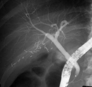
 Biliary disease. In a patient with persistent elevation of liver-associated enzymes, the contrast medium entering the biliary ductal system preferentially enters the cystic duct.
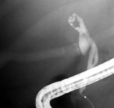
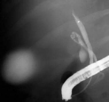 Biliary disease. Even when the catheter is advanced to the proximal common hepatic duct, contrast dye preferentially fills the cystic duct and gallbladder rather than allowing visualization of the intrahepatic ductal system (same patient as in previous image).
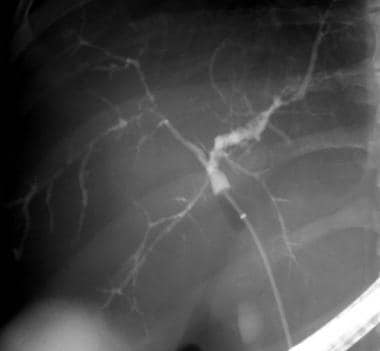 Biliary disease. In this image, the common bile duct is occluded with a balloon-tipped catheter. Contrast material fills the intrahepatic ductal system to reveal diffuse intrahepatic sclerosing cholangitis.
For patient education resources, see Digestive Disorders Center and Cholesterol Center, as well as Gallstones, Primary Biliary Cirrhosis (PBC), Cirrhosis (Liver, Symptoms, Stages, and Diet), and Primary Sclerosing Cholangitis.
Pathophysiology
Bile is produced by the liver and is channeled by the biliary ductal system into the intestinal tract for the emulsification and absorption of fats. Biliary disease is caused by abnormalities in bile composition, biliary anatomy, or function. The liver determines the chemical composition of bile, and this may be modified later by the gallbladder and the biliary epithelium. Cholesterol, ordinarily insoluble in water, comes into solution by forming vesicles with phospholipids (principally lecithin) or mixed micelles with bile salts and phospholipids.
When the ratio of cholesterol, phospholipids, and bile salts is outside an optimum range, cholesterol monohydrate crystals may come out of solution from multilamellar vesicles. Cholesterol supersaturation of bile appears to be a prerequisite for gallstone formation, which involves a variety of factors that affect the activity of low-density lipoprotein (LDL) uptake, hepatic 3-methylglutaryl coenzyme A reductase (HMG CoA), acyl cholesterol-lecithin acyltransferase, and 7-alpha hydroxylase.
By itself, cholesterol supersaturation is inadequate for explaining gallstone pathogenesis. Nucleation, the initial step in gallstone formation, is the transition of cholesterol from a soluble state into a solid crystalline form. Within the gallbladder bile, biologic molecules influence the process in a positive or negative fashion.
For example, mucus may function to promote nucleation, whereas bile-specific glycoproteins may function to inhibit nucleation. Mucin hypersecretion by the gallbladder mucosa creates a viscoelastic gel that fosters nucleation. Arachidonyl lecithin, which is absorbed from the alimentary tract and secreted into the bile, stimulates prostanoid synthesis by gallbladder mucosa and promotes mucus hypersecretion, while inhibitors of prostaglandin inhibit mucus secretion.
Finally, gallbladder hypomotility and bile stasis appear to promote gallstone formation and growth, which may be important in diabetes, pregnancy, oral contraceptive use in women, and prolonged fasting in critically ill patients on total parenteral nutrition.
More recent research suggests that elevated levels of four circulating interleukins (IL) (IL-6, IL-10, IL-12 [p70], IL-13) are associated with an increased risk of gallstones [1] and/or there may be a genetic predisposition that affects the supersaturation of bile with insoluble compounds (eg, cholesterol) thereby also raising the risk of gallstone disease. [2]
Etiology
Gallstones
In about 80% of patients, gallstones are clinically silent. Approximately 20% of patients develop symptoms over 15-20 years, that is, about 1% per year, and almost all become symptomatic before complications develop. Biliary-type pain, the typical clinical presentation, is due to the obstruction of the bile duct lumen. The predictive value of other complaints (eg, intolerance to fatty food, indigestion) is too low to be clinically helpful. The incidence of gallbladder cancer developing in the setting of cholelithiasis is low, about 0.1% per year. Two main types of gallstones exist.
Cholesterol stones (85%)
These are divided into two subtypes—pure (90%-100% cholesterol) or mixed (50%-90% cholesterol).
Pure stones often are solitary, whitish, and larger than 2.5 cm in diameter. Mixed stones usually are smaller, multiple in number, and occur in various shapes and colors. They tend to be arranged in laminated layers of an alternating thicker whitish cholesterol and a thinner dark pigment in a concentric pattern around a pigmented center (similar to the rings visible on the cross section of a tree). These stones tend to occur in residents of Western countries, and they usually are found in the gallbladder.
The risk factors associated with the development of cholesterol gallstones include obesity, a high-calorie diet, clofibrate therapy, gastrointestinal disorders involving major malabsorption of bile acids, cystic fibrosis with pancreatic insufficiency, and female sex and the use of oral contraceptives and other estrogenic medications. Coffee and ascorbic acid have been shown to reduce the risk of symptomatic cholesterol gallstones.
Pigment stones (15%)
Pigment stones occur in 2 subtypes—brown and black.
Brown stones are made up of calcium bilirubinate and calcium-soaps. Bacteria are involved in their formation via secretion of beta glucuronidase and phospholipase. The bacterial glycocalyx aggregates with the bile pigment and precipitates out of solution. These stones are more common in Asia and tend to form within the bile ducts. They frequently are associated with periampullary duodenal diverticula.
Black stones typically form in the gallbladder and result when excess bilirubin enters the bile and polymerizes into calcium bilirubinate. These stones are more common in patients with chronic hemolysis, alcoholic cirrhosis, and advanced age.
Acute calculus cholecystitis
Acute calculus cholecystitis is an inflammation of the gallbladder that develops in the setting of an obstructed cystic or bile duct. It usually develops after 5 hours of biliary-type pain. The initial inflammation is caused by chemical irritation, and bacterial infection probably is a secondary event. A change in the perception of pain, classically a migration to the right upper quadrant, suggests transmural inflammation of the gallbladder, with involvement of the parietal peritoneum. Nausea and vomiting are common associated symptoms, and most patients are afebrile early in the course of the disease.
Mirizzi syndrome
Mirizzi syndrome refers to common hepatic duct obstruction caused by an extrinsic compression from an impacted stone in the cystic duct. [3] It has been estimated to occur in 0.7-1.4% of all cholecystectomies. It is often not recognized preoperatively, which can lead to significant morbidity and biliary injury, particularly with laparoscopic surgery.
Acute acalculous cholecystitis
Acute acalculous cholecystitis is the presence of an inflamed gallbladder in the absence of an obstructed cystic or common bile duct. It typically occurs in the setting of a critically ill patient (eg, severe burns, multiple traumas, lengthy postoperative care, prolonged intensive care) and accounts for 5% of cholecystectomies. Because abdominal pain, fever, and leukocytosis are relatively common in these patients and the signs and symptoms are not specific for acalculous cholecystitis, the physician must have a high index of suspicion to make the diagnosis. The etiology is thought to have an ischemic basis, and a gangrenous gallbladder may result. This condition has an increased rate of complications and mortality. An uncommon subtype known as acute emphysematous cholecystitis generally is caused by infection with clostridial organisms and occlusion of the cystic artery associated with atherosclerotic vascular disease and, often, diabetes.
Chronic cholecystitis
Chronic cholecystitis is a common disorder that frequently is associated with gallstones. The clinical features are nonspecific, and cholescintigraphy initially may suggest the diagnosis. The pathogenesis is poorly understood but may be due to abnormal bile composition leading to chemical injury of the gallbladder mucosa. Histologic evidence of a mononuclear infiltrate, fibrosis, and epithelial metaplasia confirms the diagnosis. A subset of patients develops dystrophic calcifications within the fibrosis, leading to a porcelain gallbladder, which is a risk factor for gallbladder carcinoma.
Cholangitis
Cholangitis is an infection of the biliary system, complicating benign and malignant obstruction of the biliary tract. The clinical presentation is quite variable depending on the nature of the illness, patient age, and condition of the patient. Charcot triad (ie, fever, right upper quadrant pain, jaundice) occurs in only 20%-70% of cases. Hypotension and mental status changes also may accompany severe infection, a pentad described by Reynolds in 1959. [4] The organisms typically identified are enteric in origin, notably Escherichia coli, Streptococcus faecalis, Clostridium species, Klebsiella species, Enterobacter species, Pseudomonas species, and Proteus species. They probably enter the biliary system via portal bacteremia. No correlation exists between the severity of the clinical manifestations and the presence or absence of pus in the biliary system; however, suppurative cholangitis is associated with a higher mortality rate.
Recurrent pyogenic cholangitis
Recurrent pyogenic cholangitis, also known as "oriental cholangiohepatitis," is prevalent in several parts of Asia and the Pacific Rim countries. It is limited to Asian immigrants in America, occurs in the second to fourth decades of life, and is associated with a lower socioeconomic class. It is initiated by parasitic infestation of the biliary ducts by Opisthorchis sinensis (formerly Clonorchis sinensis), in which the adult fluke may impair bile flow. In the setting of bile stasis and secondary bacterial infection, pigment stones form around ova and sets the stage for the intermittent obstruction leading to recurrent pyogenic cholangitis. Pathologic changes principally affect the intrahepatic bile ducts (curiously, more often the left duct).
Primary sclerosing cholangitis
PSC is a chronic cholestatic biliary disease characterized by nonsuppurative inflammation and fibrosis of the biliary ductal system. The cause is unknown but is associated with autoimmune inflammatory diseases, such as chronic ulcerative colitis and Crohn colitis (less commonly), and rare conditions, such as Riedel thyroiditis and retroperitoneal fibrosis. Most patients present with fatigue and pruritus and, occasionally, jaundice. The natural history is variable but involves progressive destruction of the bile ducts, leading to cirrhosis and liver failure. The clinical features of cholangitis (ie, fever, right upper quadrant pain, jaundice) are uncommon unless the biliary system has been instrumented.
Primary biliary cholangitis
Primary biliary cholangitis (PBC), formerly known as primary biliary cirrhosis, is a progressive cholestatic biliary disease that presents with fatigue and itching or asymptomatic elevation of the alkaline phosphatase. The name change reflects the fact that cirrhosis occurs only in the late stage and therefore does not correctly identify patients with early-stage disease. [5] Jaundice develops with progressive destruction of bile ductules that eventually leads to liver cirrhosis and hepatic failure. This autoimmune illness has a familial predisposition, in which even unaffected family members may have immunologic abnormalities, especially an increased serum immunoglobulin M (IgM) and an association with human leucocyte antigen (HLA)-DR8. [6, 7]
Although numerous autoantibodies have been identified, antimitochondrial antibodies (AMA) are present in 95% of patients. AMA is a family of antibodies; those directed against the inner mitochondrial membrane antigen M2 in the 2-oxo-acid dehydrogenase complex are most specific for PBC. [8] Circulating immune complexes also have been identified but are unlikely to play a pathogenic role. Circulating T lymphocyte levels initially are within the reference range and decline as the disease progresses. The histologic appearance of the bile duct destruction resembles hepatic allograft rejection and graft-versus-host disease of the liver and appears to be mediated by cytotoxic T lymphocytes.
Autoimmune cholangitis
Autoimmune cholangitis represents a rare, distinct disease entity. While it shares some features with PBC, the results of tests for AMA are negative, the levels of gamma globulin and IgM are lower, and the results of tests for fluorescent antinuclear antibody (FANA) and anti–smooth muscle antibody (ASMA) are positive more commonly.
Neoplasms of the biliary tract
Carcinoma of the biliary system manifests with clinical symptoms of weight loss (77%), nausea (60%), anorexia (56%), abdominal pain (56%), fatigue (63%), pruritus (51%), fever (21%), malaise (19%), diarrhea (19%), constipation (16%), and abdominal fullness (16%). Symptomatic patients usually have advanced disease, with spread to hilar lymph nodes before obstructive jaundice occurs. It is associated with a poor prognosis.
Gallbladder cancer
This uncommon malignancy affects 2.5 individuals per 100,000 population. It represents 54% of biliary tract cancers, and more than 6500 patients die from this disease in the United States each year. Cancer that develops in the infundibulum (neck of the gallbladder) can produce hydrops of the gallbladder that is clinically indistinguishable from an obstructing stone.
Cholangiocarcinoma
Cholangiocarcinoma is an adenocarcinoma of the bile ducts. [9] It may occur without associated risk factors, but it is associated more commonly with chronic cholestatic liver disease such as PSC, choledochal cysts, oriental cholangiohepatitis, and work-related handling of asbestos. Cholangiocarcinoma accounts for 25% of biliary tract cancers. Patients usually present with jaundice, a vague upper or right upper quadrant abdominal pain associated with anorexia, weight loss, and pruritus.
Ampullary cancer
Ampullary cancer accounts for 8% of biliary tract cancers. It most commonly presents with painless jaundice or acute pancreatitis.
Biliary tract cysts
Cystic dilatation of the biliary tree is an uncommon abnormality. About half of the patients present with some combination of jaundice, abdominal pain, and an abdominal mass. The presence of these cysts is often associated with an anomalous union of the pancreatic and biliary ductal system. This suggests that pancreatic juice enters the bile duct, causes a proteolytic and inflammatory injury to the duct wall, and leads to biliary cyst formation. The most commonly used classification scheme was proposed by Todani, which defines 5 cyst types, with groups I and IV having subtypes.
Type I involves a cystic dilatation of the extrahepatic biliary system. In subtype 1a (most common), the entire extrahepatic duct is diffusely involved. In subtype 1b (rare), a localized portion of the common bile duct is segmentally cystic. In subtype 1c (uncommon), the common bile duct is diffusely dilated.
Type II (rare) is a diverticulum of the extrahepatic bile duct.
Type III (uncommon) is a cystic dilatation of the intraduodenal portion of the common bile duct (sometimes referred to as a choledochocele).
Type IV has multiple cysts. Subtype IVa (uncommon) involves both the intrahepatic and extrahepatic biliary system, while subtype IVb (rare) has multiple cysts confined to the extrahepatic system.
Type V (rare) is characterized by single or multiple cysts involving the intrahepatic bile ducts (usually referred to as Caroli disease). Clinical symptoms usually are the result of associated complications such as cholangitis, choledocholithiasis, pancreatitis, hepatic abscess, cirrhosis, and biliary malignancy.
Epidemiology
United States statistics
Gallstone disease is one of the most common and costly of all digestive diseases. The third National Health and Nutrition Examination Survey estimated that, in the United States, 6.3 million men and 14.2 million women aged 20-74 years have gallbladder disease.
The incidence of gallstones is 1 million new cases per year. The prevalence is 20 million cases among Americans.
Approximately 2-7 cases per 100,000 population of primary sclerosing cholangitis (PSC) exist. About 5% of patients with chronic ulcerative colitis develop PSC.
The incidence of gallbladder cancer is 2.5 cases per 100,000 population.
International statistics
The incidence of primary biliary cholangitis (PBC) is 5.8-15 cases per 1 million population. The incidence of PBC appears to be increasing, but the cause of the increase is unclear. However, the increase is possibly due to better detection and increased awareness rather than a true change in disease incidence.
According to an international multicenter study comprising 4805 patients with primary biliary cholangitis (primary biliary cirrhosis) over 44 years (1970-2014), there was an incremental increase in mean age at diagnosis: 2-3 years per decade from 46.9 ± 10.1 years in the 1970s to 57.0 ± 12.1 years from 2010 onward (P<0.001).<ref>10 </ref>In addition, although there were no significant changes in female predominance (female-male ratio of 9:1) and antimitochondrial antibody positivity (90%), increases in the incidence of mild biochemical disease and mild histologic stage at diagnosis were observed, as well as lower rates of decompensation and higher 10-year transplant-free survival with each decade forward. [10]
Race-related demographics
Mexican Americans and several American Indian tribes, particularly the Pima Indians in the Southwest, have very high prevalence rates of cholesterol gallstones. Decreased bile acid secretion is believed to be the common denominator in these ethnic groups.
Gallbladder cancer is the most common gastrointestinal malignancy in both Southwestern Native Americans and Mexican Americans. A prominent geographic variability exists in the incidence of gallbladder cancer that correlates with the prevalence of cholelithiasis. High rates of gallbladder cancer are also seen in South American countries, particularly Chile and Bolivia. These populations all share a high prevalence of gallstones and/or Salmonella infection, both recognized risk factors for gallbladder cancer.
Sex-related demographics
The prevalence of cholesterol gallstones is higher among females than males (lifetime risk of 35% vs 20%, respectively). This likely is due to endogenous sex hormones, which enhance cholesterol secretion and increase bile cholesterol saturation. Progesterone also may contribute by relaxing gallbladder smooth muscle and impairing gallbladder emptying. Note the following:
-
PSC: Males (primarily young to middle-aged [11] ) are affected twice as frequently as females.
-
PBC: Females are affected nine times as often as males.
Age-related demographics
Increased age is associated with lithogenic bile and an increased rate of gallstones. Note the following:
-
PSC: Mean age of onset is 40 years.
-
PBC: Among the autoimmune diseases, PBC is unique in that it never occurs in childhood and is rarely found before age 30 years. The onset is usually between the ages of 30-65 years, but the disease has been reported in women as young as 22 years and as old as 93 years.
Prognosis
In primary sclerosing cholangitis (PSC), several factors suggest a high risk of death. These include advancing age, serum bilirubin, serum albumin, presence or absence of inflammatory bowel disease, and histologic stage on liver biopsy.
Morbidity/mortality
Gallstones are a rare cause of mortality, accounting for 5000 of the 2.2 million deaths annually in the United States.
Primary biliary cholangitis (PBC) accounts for 0.6%-2% of deaths from cirrhosis worldwide. The median time of patient survival was 9.3 years from diagnosis. Independent predictors of survival include age and serum levels of alkaline phosphatase, albumin, and bilirubin. Liver failure develops in 26% of patients by 10 years after diagnosis. Neither the presence of antimitochondrial antibodies nor their titer affects disease progression or survival.
PSC is a leading reason for liver transplantation. Median survival without liver transplantation after diagnosis is approximately 12 years. Variables that appear to predict prognosis in PSC include age, histologic stage, hepatomegaly, splenomegaly, and serum alkaline phosphatase and serum bilirubin levels.
Complications
The complications common to all of the chronic cholestatic liver diseases, such as PSC and PBC, include fatigue, pruritus, steatorrhea, fat-soluble vitamin deficiencies (A, D, E, and K), metabolic bone disease, hypercholesterolemia, xanthomas, hypothyroidism, and anemia. There is a reported association of PBC with Sjögren syndrome, Raynaud phenomenon, and sicca symptoms.
Approximately 20% of patients with PSC develop a dominant stricture in the intrahepatic or extrahepatic biliary tree. Medical therapy to treat biliary strictures has been ineffective. Nonsurgical modalities to relieve biliary obstruction, such as endoscopically- or radiologically–guided balloon dilation of strictures or placement of prosthetic stents across strictures, should be attempted initially.
Choledocholithiasis and cholelithiasis due to cholesterol and/or pigment stones may be present in up to one third of patients with PSC. Bacterial cholangitis can occur in patients with PSC.
Cholangiocarcinoma eventually develops in about 20% of patients with PSC, principally late in the course of long-standing ulcerative colitis and the cirrhotic stage of biliary disease. About half of patients with PSC are diagnosed with cholangiocarcinoma within 2 years of the initial diagnosis, with an associated poor prognosis owing to advanced disease at the time of diagnosis. [12] This complication is difficult to detect, as evidenced by the finding of cholangiocarcinoma in 10% of patients undergoing liver transplantation for PSC. [13]
The incidence of hepatocellular carcinoma is increased in patients with PBC who have had stage IV disease for many years. [14]
Patients with both PSC and ulcerative colitis have an increased risk of colon cancer and progression of neoplastic transformation.
-
A normal postcholecystectomy cholangiogram.
-
Biliary disease. In a patient with persistent elevation of liver-associated enzymes, the contrast medium entering the biliary ductal system preferentially enters the cystic duct.
-
Biliary disease. Even when the catheter is advanced to the proximal common hepatic duct, contrast dye preferentially fills the cystic duct and gallbladder rather than allowing visualization of the intrahepatic ductal system (same patient as in previous image).
-
Biliary disease. In this image, the common bile duct is occluded with a balloon-tipped catheter. Contrast material fills the intrahepatic ductal system to reveal diffuse intrahepatic sclerosing cholangitis.
-
Biliary disease. Common bile duct stones are among the most frequent problems occurring in the biliary system. In this cholangiogram, the stones line up like peas in a pod.
-
Biliary disease. After a biliary sphincterotomy, a balloon-tipped catheter is used to remove the stones one by one.
-
Biliary disease. This clearing cholangiogram shows a common bile duct free of filling defects and good flow into the duodenum. The stones are visible as filling defects in the duodenal bulb.
-
Biliary disease. A patient with pancreatic cancer developed jaundice during his treatment. The cholangiogram shows a stricture in the distal common bile duct.
-
Biliary disease. A patient with pancreatic cancer developed jaundice during his treatment (same patient as in previous image). To palliate the jaundice, the biliary stricture is dilated and stented with a 10F plastic stent. Note the contrast dye flowing down the stent.
-
Biliary disease. This computed tomography scan of the abdomen shows a large tumor mass in the head of the pancreas. The brightly colored object within the mass is the biliary stent placed by endoscopic retrograde cholangiopancreatography.
-
Biliary disease. This abdominal computed tomography scan shows mild intrahepatic biliary ductal dilatation.
-
Biliary disease. Abdominal computed tomography scanning in a patient with jaundice revealed polycystic liver disease.
-
Biliary disease. Findings on this endoscopic retrograde cholangiopancreatogram exclude extrahepatic biliary obstruction but demonstrate that the intrahepatic biliary ductal system is splayed by multiple hepatic cysts.
-
Biliary disease. This cholangiogram shows a choledochal cyst. Fusiform dilatation of the entire extrahepatic bile duct is present.
-
A 92-year-old woman had recurrent abdominal pain and jaundice. A right upper quadrant ultrasonogram showed a dilated biliary duct with no stones. She had a previous Roux-en-Y surgery that made endoscopic retrograde cholangiopancreatography impossible. Critical aortic stenosis increased the risk of most interventions. This percutaneous cholangiogram, performed under conscious sedation in the operating room, revealed a large stone missed by the ultrasonogram. It was removed successfully with percutaneous choledochoscopy and electrohydraulic lithotripsy.
-
Biliary disease. This cholangiogram shows a stone too large to deliver through a standard biliary sphincterotomy.
-
Biliary disease. Here, a mechanical lithotripter is used to grab a stone too large to deliver through a standard biliary sphincterotomy and crush it into small pieces (same patient as in previous image). The smaller pieces then are removed with a balloon-tipped catheter.
-
Biliary disease. A patient had malignant strictures of the biliary system palliated with metal mesh stents. Unfortunately, the tumor grew through the metal mesh to reobstruct the biliary system.
-
Biliary disease. A patient had malignant strictures of the biliary system that were palliated with metal mesh stents, but the tumor grew through the metal mesh to reobstruct the biliary system (same patient as in previous image). In this image, after a wire was passed through the lumen, a balloon-dilating catheter was passed into the metal mesh stents and inflated to enlarge the lumen.
-
Biliary disease. A patient had malignant strictures of the biliary system that were palliated with metal mesh stents, but the tumor grew through the metal mesh to reobstruct the biliary system (same patient as in previous image). After a wire was passed through the lumen, a balloon-dilating catheter was passed into the metal mesh stents and inflated to enlarge the lumen. In this image, two plastic stents were passed into the intrahepatic ductal system to again palliate the obstruction.


