Practice Essentials
Splenomegaly is defined as enlargement of the spleen, measured by size or weight. [1] In the past, splenomegaly was a clinical finding, but in recent years, imaging studies have also helped to assess for or confirm mild splenomegaly.
The spleen is a functionally diverse organ with active roles in immunosurveillance and hematopoiesis. It lies within the left upper quadrant of the peritoneal cavity and abuts ribs 9-12, the stomach, the left kidney, the splenic flexure of the colon, and the tail of the pancreas. A normal spleen weighs 150 g and is approximately 11 cm in craniocaudal length. [2]
The normal spleen is usually not palpable, although it can sometimes be palpated in adolescents and individuals with a slender build. If the spleen is not palpable, the clinician can assess for splenomegaly by percussing the area known as Traube's semilunar space, which is defined by the left sternal border, the costal margin, and the lower border of the 9th rib; dullness to percussion there (as opposed to tympany, which is normally present and represents the air-filled stomach) is consistent with mild to moderate splenomegaly. More pronounced splenomegaly can be palpated below the level of the costal margin and can even extend down to the pelvic brim. Of note, an enlarged or palpable spleen is not necessarily of clinical significance. For example, certain individuals with broadly splayed costal margins have readily palpable, but small, spleens. (See Presentation.)
A spleen weight of 400-500 g indicates splenomegaly, while a weight of more than 1000 g is labelled as massive splenomegaly. Poulin et al defined splenomegaly as moderate if the largest dimension is 11-20 cm, and severe if the largest dimension is greater than 20 cm. [3]
Importantly, while lymph nodes can be thought of as the draining secondary lymphoid organs of the respective anatomic compartments via afferent lymphatics (ie the lung drains into the mediastinum), the spleen can be thought of as the primary draining secondary lymphoid organ of the systemic circulatory system with the afferent inflow occurring via the splenic artery instead of afferent lymphatics. [4]
In many instances, the spleen enlarges as it performs its normal functions. The most important normal functions of the spleen are as follows: [5]
-
Filtering both abnormal and senescent red blood cells (RBCs), as well as particulates and microorganisms
-
Immune function – Providing an interface between adapative and innate immunity, with synthesis of immunoglobulin M (IgM), properdin (an essential component of the alternate pathway of complement activation), and tuftsin (an immunostimulatory tetrapeptide)
-
Erythropoiesis, particularly early in fetal life and as an adaptive response to bone marrow failure
-
Providing a resevoir of blood cells, including RBCs and platelets that can be utilized under stress
-
Iron metabolism
As the spleen enlarges, cytopenias often result due to sequestration of cells; this condition is called hypersplenism. Importantly, the resultant thrombocytopenia in a patient with hypersplenism does not result in a proportional defect in hemostasis, as the total body platelet count largely remains unchanged but rather is redistributed.
Increasingly, most cases of splenomegaly are recognized as being secondary to some other condition. Successful medical treatment of the primary disorder in such cases can lead to regression of the hypersplenism without the need for surgery. Splenectomy has a decreasing number of indications, but can still be used to help control or stage the underlying disease in cases of splenomegaly.
For discussion of splenomegaly in children, see Pediatric Splenomegaly. For discussion of hyperreactive malarial syndrome, see Tropical Splenomegaly Syndrome. For patient education information, see Enlarged Spleen: Causes, Symptoms, and Treatments.
Pathophysiology
Eichner et al proposed an early taxonomy for the pathophyisiology of splenomegaly as follows [6] :
-
Immune response work hypertrophy
-
Red blood cell (RBC) work hypertrophy
-
Congestive splenomegaly
-
Infiltrative splenomegaly
-
Neoplastic splenomegaly
-
Miscellaneous
Immune response work hypertrophy
Acute enlargement of the spleen due to various infections or inflammatory processes results from an increase in the defense activities of the organ. The demand for increased antigen clearance from the blood may lead to increased numbers of reticuloendothelial cells in the spleen and stimulate accelerated antibody production, with resultant lymphoid hyperplasia. Examples include splenomegaly from subacute bacterial endocarditis, lupus, and Felty syndrome, and from viral infections such as Epstein-Barr virus–induced mononucleosis.
Red blood cell work hypertrophy
A high rate of removal of abnormal blood cells from the circulation (either cells with intrinsic defects or cells coated with antibody) is the usual source of RBC work hypertrophy. This occurs in thalassemia major, hereditary spherocytosis, and pyruvate kinase deficiency. Thalassemia major has the additional mechanism of leading to splenomegaly due to extramedullary hematopoiesis as a result of intramedullary hemolysis.
Congestive splenomegaly
Cirrhosis with portal hypertension, splenic vein occlusion (thrombosis), or chronic heart failure (CHF) with increased venous pressure causes congestive splenomegaly. In patients receiving oxaliplatin-based chemotherapy, splenomegaly may result from hepatic sinusoidal obstructive syndrome caused by the chemotherapy; use of bevacizumab may reduce the splenomegaly in these cases. [7]
Infiltrative splenomegaly
Infiltrative splenomegaly is the result of engorgement of macrophages with indigestible materials. It may be seen in conditions such as sarcoidosis, Gaucher disease, and amyloidosis.
Neoplastic splenomegaly
Hematologic neoplasms make up the bulk of cancer-related causes of splenomegaly. This category includes both lymphoproliferative (eg, lymphomas, leukemias) as well as myeloproliferative (eg, chronic myeloid leukemia, primary myeloid fibrosis, essential thrombocythemia, polycythemia vera) neoplasms. Rarely, sarcoma can occur in the spleen, or primary solid tumors can metastasize to the spleen.
Miscellaneous
Additional causes of splenomegaly include trauma, splenic cysts, and hemangiomas.
Splenic filtering of blood-borne pathogens, especially encapsulated organisms, may lead to abscess formation. Because many splenic abscesses may be indolent in presentation, splenic size may increase as the abscess enlarges. This is a relatively uncommon, but important, process to recognize and treat.
Acute splenic sequestration crisis (ASSC) is a major cause of morbididty and mortality in children with sickle cell disease and other hereditary hemolytic anemias. ASSC is characterized by sudden enlargement of the spleen due to trapping of a significant proportion of the blood volume, rapid drop in the hematocrit with hypovolemia, and thrombocytopenia. ASSC is rare in adults with sickle cel disease or beta thalassemia, despite the frequent presence of spleneomegaly in this population. Infection and high-altitude exposure are known precipitating factors for ASSC. [8]
Etiology
Many of the mechanisms leading to splenomegaly are exaggerated forms of normal splenic function. Although a wide variety of diseases are associated with enlargement of the spleen, the following six etiologies of splenomegaly are considered primary:
-
Immune response work hypertrophy - Such as in subacute bacterial endocarditis or infectious mononucleosis
-
RBC destruction work hypertrophy - Such as in hereditary spherocytosis or thalassemia major
-
Congestive - Such as in splenic vein thrombosis, portal hypertension, or Banti disease
-
Infiltrative - Such as in sarcoidosis and some neoplasms
-
Neoplastic - Lymphoproliferative disorders such as chronic lymphocytic leukemia, hairy cell leukemia, and lymphomas, as well as myeloproliferative disorders including chronic myeloid leukemia, primary myeloid fibrosis, essential thrombocythemia, and polycythemia vera
-
Miscellaneous - Such as trauma, cysts, hemangiomas, abscesses (see the images below), certain drugs (ie RhoGAM)
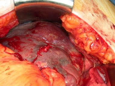 This patient has a splenic abscess due to pneumococcal bacteremia. Note that the massively enlarged spleen is readily visible, with minimal retraction in the left upper quadrant.
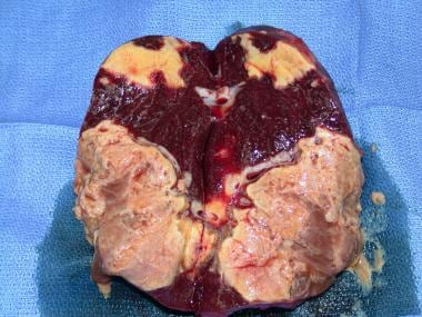 Resected specimen from the patient in the previous image. Note the discrete abscesses adjacent to normal parenchyma.
Epidemiology
In the United States, one large series reported a palpable spleen in 2% of patients and another in 5.6% of patients. [9] Tropical splenomegaly syndrome occurs most often in persons indigenous to the malarial belt of tropical Africa and in visitors to that region.
Race-, Sex-, and Age-related demographics
No race predilection is recognized for splenomegaly. However, note that blacks may have hemoglobin SC disease, a disorder related to sickle cell disease. Unlike sickle cell disease, which results in a small, autoinfarcted spleen, patients with hemoglobin SC disease may have splenomegaly that accompanies their pigment gallstones.
Tropical splenomegaly syndrome (or hyperactive malarial syndrome) has a female-to-male incidence ratio of 2:1. Otherwise, no sex predilection is documented for splenomegaly.
The capsules of older spleens are much thinner than their younger counterparts. The combination of capsular thinning with increased spleen weight and size makes splenic injury more common in elderly persons. These factors account for the increased likelihood of splenectomy for trauma in this subgroup.
Prognosis
The prognosis for patients with splenomegaly is usually excellent and not substantially different from age-matched controls, but it is impacted by the underlying disease state rather than the presence of splenomegaly or the postsplenectomy state.
Morbidity and mortality
Morbidity and mortality in cases of splenomegaly principally stem from associated disease states or surgical procedures, rather than from the splenomegaly itself. The rates for morbidity and mortality vary widely and relate to the presence or absence of comorbidities, hemorrhage, and organ failure.
Patients with enlarged spleens are more likely to have splenic rupture from blunt abdominal or low thoracic trauma. These patients are more likely to be exposed to emergent operative splenectomy and its attendant risks.
Patient Education
Patients with splenomegaly need education with regard to decreasing their risk of splenic trauma and rupture. These patients must be cautioned about contact sports and other activities that may acutely increase intra-abdominal pressure or place excessive forces on the left upper quadrant, left flank, or lateral back. This decreases the likelihood of splenic rupture in a patient with an abnormal splenic mass and capsule. The routine use of seat belts is essential while driving or riding in a motor vehicle.
For patient education information, see Enlarged Spleen.
Patients whose splenomegaly has led to splenectomy require education regarding the signs and symptoms of postsplenectomy sepsis. Education represents a mandatory strategy in the prevention of overwhelming postsplenectomy infection. Asplenic patients should be encouraged to wear a medical alert bracelet and carry a wallet card explaining their lack of a spleen. These patients should also be aware of the need to notify their physician in the event of an acute febrile illness, especially if it is associated with rigors or systemic symptoms. Prompt antibiotic therapy may be lifesaving.
-
This patient has a splenic abscess due to pneumococcal bacteremia. Note that the massively enlarged spleen is readily visible, with minimal retraction in the left upper quadrant.
-
Resected specimen from the patient in the previous image. Note the discrete abscesses adjacent to normal parenchyma.
-
The margins of this massive spleen were palpated easily preoperatively. Medially, the 3.18 kg (7 lb) spleen crosses the midline. Inferiorly, it extends into the pelvis.
-
Massive splenomegaly does not preclude splenectomy through a minimally invasive approach. This photograph depicts a fragmented 3.2 kg (7.05 lb) spleen after removal via a hand-assisted laparoscopic technique.
-
A portion of a massive spleen is extracted via hand-assisted laparoscopy.
-
Intraoperative photograph of a laparoscopic splenectomy being taken down using the hanging-pedicle technique. The tip of the spleen is visualized in the background, whereas the stapler is detailed in the foreground across a segment of the pedicle.
-
A massive spleen that was removed from an elderly woman with lymphoma.