Background
Rhinosporidiosis is a chronic granulomatous infection of the mucous membranes that usually manifests as vascular friable polyps that arise from the nasal mucosa or external structures of the eye.
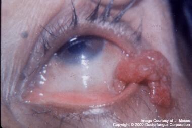 Granulomatous mass involving structures of the eye. Image used with permission from doctorfungus.org.
Initially described by Seeber in 1900 in an individual from Argentina, [1] rhinosporidiosis is endemic in India, Sri Lanka, South America, and Africa. [2, 3, 4, 5, 6, 7, 8, 9, 10, 11, 12, 13, 14] Cases from the United States and Southeast Asia, as well as scattered occurrences throughout the world, have also been reported. Most cases of rhinosporidiosis occur in persons from or residing on the Indian subcontinent or Sri Lanka. In addition to humans, rhinosporidiosis has been noted in cats, cattle, dogs, ducks, goats, horses, mules, parrots, and swan. [15]
The etiologic agent, Rhinosporidium seeberi, has never been successfully propagated in vitro. Initially thought to be a parasite, for more than 50 years R seeberi had been considered to be a water mold. [16] Molecular biological techniques have more recently demonstrated this organism to be an aquatic protistan parasite, and it has been placed into a new class, the Mesomycetozoea, along with organisms that cause similar infections in amphibians and fish. [17, 18, 19] This reclassification is not without controversy, as other researchers have presented data that R seeberi is a cyanobacterium, further demonstrating the difficulties that arise when working with pathogens that cannot be maintained in the laboratory setting. [20, 21] Other molecular work has demonstrated evidence that R seeberi may have host-specific strains (eg, human vs dog vs swan). [22]
Pathophysiology
Rhinosporidiosis is an infection that typically is limited to the mucosal epithelium. Infection usually results from a local traumatic inoculation with the organism. The disease progresses with the local replication of R seeberi and associated hyperplastic growth of host tissue and a localized immune response.
Infection of the nose and nasopharynx is observed in 70% of persons with rhinosporidiosis; infection of the palpebral conjunctivae or associated structures (including the lacrimal apparatus) is observed in 15%. [23]
Other structures of the mouth, upper airway, and eye may be sites of disease. Disease of the skin, ear, larynx, trachea, bronchi, genitals, and rectum has also been described. [24] Genital rhinosporidiosis has been described in the vagina, penile urethra or meatus, and scrotum. [25] Dissemination with cutaneous and multisite disease also is reported, but this is much less common. Isolated cases of dissemination involving deep organs rarely have been reported. [26, 27]
Epidemiology
Frequency
United States
Rhinosporidiosis cases in the United States are rare, but are more common in Texas and the Southeast. From 1896 through 2019, less than 50 cases have been reported from the United States. [28]
International
Rhinosporidiosis usually affects persons in or from southern India and Sri Lanka. Cases have been reported worldwide, with an increased incidence in South America and Africa.
Mortality/Morbidity
Rhinosporidiosis can cause prolonged painless disease with limited morbidity. Disease of up to 30 years' duration has been reported. Secondary bacterial infection can cause morbidity. Death has been reported in only the few rare reports of disseminated rhinosporidiosis.
Race
Rhinosporidiosis has no known racial predilection.
Sex
Men are affected more commonly than women, with a male-to-female ratio of 4:1.
Age
Rhinosporidiosis most commonly occurs in children and in individuals aged 15-40 years.
Risk Factors
Rhinosporidiosis has been associated with rural residence, occupation in farming or agriculture, and bathing in ponds or rivers. [29, 30, 31]
Prognosis
The prognosis of rhinosporidiosis is excellent, except with dissemination, as described above (see Mortality/Morbidity).
Patient Education
Patients with rhinosporidiosis should be advised that recurrence is possible. They should be instructed to return for further evaluation if symptoms recur or new symptoms arise.
Cases of dissemination or more extensive disease have been associated with a prior history of both treated and untreated nasal disease. Accordingly, ensure that patients are instructed to (1) be vigilant for a recurrence of symptoms or new lesions and (2) promptly consult with their physician if these are noted.
-
Granulomatous mass involving structures of the eye. Image used with permission from doctorfungus.org.
-
Sporangia of Rhinosporidium seeberi within nasal polyp (periodic acid-Schiff [PAS] stain). Image used with permission from doctorfungus.org.
-
Sporangia of Rhinosporidium seeberi in polyp (Gomori methenamine silver [GMS]) stain. Image used with permission from doctorfungus.org.


