Practice Essentials
Chronic pancreatitis is commonly defined as a continuing, chronic, inflammatory process of the pancreas, characterized by irreversible morphologic changes.
Signs and symptoms
For most patients with chronic pancreatitis, abdominal pain is the presenting symptom. The patient experiences intermittent attacks of severe pain, often in the mid-abdomen or left upper abdomen and occasionally radiating in a bandlike fashion or localized to the midback. The pain may occur either after meals or independently of meals, but it is not fleeting or transient and tends to last at least several hours.
Other symptoms associated with chronic pancreatitis include diarrhea and weight loss.
See Presentation for more detail.
Diagnosis
Diagnosis is based on tests of pancreatic structure and function.
Imaging tests
Imaging studies such as abdominal radiography and CT scanning can show inflammation or calcium deposits in the pancreas or changes in the pancreatic ducts. Pancreatic calcifications, often considered pathognomonic of chronic pancreatitis, are observed in approximately 30% of cases.
Endoscopic retrograde cholangiopancreatography
The endoscopic retrograde cholangiopancreatography (ERCP) test provides the most accurate visualization of the pancreatic ductal system and has been regarded as the criterion standard for diagnosing chronic pancreatitis. It combines the use of endoscopy and fluoroscopy to visualize and treat problems of the biliary and pancreatic ducts. See the image below.
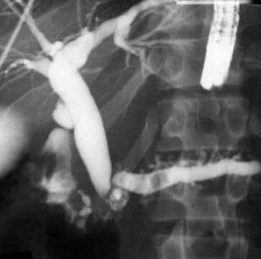 This endoscopic retrograde cholangiopancreatography (ERCP) shows advanced chronic pancreatitis. The pancreatogram has blunting of the lateral branches, dilation of the main pancreatic duct, and filling defects consistent with pancreatolithiasis. The cholangiogram also shows a stenosis of the distal bile duct and a dilated biliary tree.
Magnetic resonance cholangiopancreatography
MRCP provides information on the pancreatic parenchyma and adjacent abdominal viscera, and it uses heavily T2-weighted images to visualize the biliary and pancreatic ductal systems. This procedure is relatively safe, reasonably accurate, noninvasive, fast, and very useful in planning surgical or endoscopic intervention.
Endoscopic ultrasonography
The most predictive endosonographic feature of chronic pancreatitis is the presence of stones. Other suggestive features include the following:
-
Visible side branches
-
Cysts
-
Lobularity
-
An irregular main pancreatic duct
-
Hyperechoic foci and strands
-
Dilation of the main pancreatic duct
-
Hyperechoic margins of the main pancreatic duct
See Workup for more detail.
Management
Treatment is typically directed at the underlying cause of the pancreatitis and to relieve pain and malabsorption.
Pain relief
Pancreatic enzyme supplementation may be helpful in reducing pain. The hypothesis is that stimulation of the pancreas by food causes pain. Cholecystokinin (CCK) is one of the possible mediators of this response. When exogenous pancreatic enzymes are taken with a meal, CCK-releasing factors are degraded and CCK release in response to a meal is reduced. This decreases pancreatic stimulation and pain.
If conventional medical therapy is unsuccessful and the patient has severe, intractable pain, celiac ganglion blockade can be considered. [1] This approach tries to alleviate pain by modifying the afferent sensory nerves in the celiac plexus, using agents that anesthetize, reduce inflammation, or destroy the nerve fibers.
Endoscopic therapy aimed at decompressing an obstructed pancreatic duct can be associated with pain relief in some patients. The rationale for this approach is based on the hypothesis that ductal hypertension due to strictures of the main pancreatic duct leads to pain. [2, 3]
Surgery
The choice of operation depends on the clinical problem and the preoperative assessment of the abnormality. In general, the approach aims either to improve pancreatic duct drainage or to resect the diseased organ. Data suggest that surgical drainage of the pancreatic duct is more effective than endoscopic drainage in patients with obstruction of the pancreatic duct due to chronic pancreatitis.
In patients with a dilated pancreatic duct, a Roux-en-Y side-to-side pancreaticojejunostomy is indicated. If the disease is limited to the head of the pancreas, a Whipple operation (pancreaticoduodenectomy) can produce good results.
See Treatment and Medication for more detail.
Background
Chronic pancreatitis is commonly defined as a continuing, chronic, inflammatory process of the pancreas, characterized by irreversible morphologic changes. [4] This chronic inflammation can lead to chronic abdominal pain and/or impairment of the endocrine and exocrine functions of the pancreas. (See Pathophysiology and Etiology.)
Chronic pancreatitis usually is envisioned as an atrophic fibrotic gland with dilated ducts and calcifications. However, the findings on conventional diagnostic studies may be normal in the early stages of chronic pancreatitis, as the inflammatory changes can be seen only by histologic examination (see the images below). (See Workup.)
This endoscopic retrograde cholangiopancreatography (ERCP) shows advanced chronic pancreatitis. The pancreatogram has blunting of the lateral branches, dilation of the main pancreatic duct, and filling defects consistent with pancreatolithiasis. The cholangiogram also shows a stenosis of the distal bile duct and a dilated biliary tree.
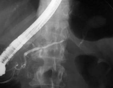 This patient has recurrent abdominal pain. She used alcohol heavily in the past and was involved in a motor vehicle accident. The pancreatogram shows subtle blunting of the side branches consistent with chronic pancreatitis. A stricture also is present in the body of the pancreas where it drapes over the spine, probably resulting from the trauma she sustained in the motor vehicle accident. Air in the stomach makes it difficult to observe that contrast is filling a pseudocyst on the other side of the stricture. These findings are not amenable to endoscopic intervention, and the patient was sent for a distal pancreatectomy.
By definition, chronic pancreatitis is a completely different process from acute pancreatitis. [5] In acute pancreatitis, the patient presents with acute and severe abdominal pain, nausea, and vomiting. The pancreas is acutely inflamed (neutrophils and edema), and the serum levels of the pancreatic enzymes (amylase and lipase) are elevated. Full recovery is observed in most patients with acute pancreatitis, whereas in chronic pancreatitis, the primary process is a chronic, irreversible inflammation (monocyte and lymphocyte) that leads to fibrosis with calcification. (See Pathophysiology, Etiology, Presentation, and Workup.)
The patient with chronic pancreatitis clinically presents with chronic abdominal pain and normal or mildly elevated pancreatic enzyme levels. When the pancreas loses its endocrine and exocrine function, the patient presents with diabetes mellitus and steatorrhea. (See Presentation and Workup.)
Patient education
For patient education information, see the Digestive Disorders Center and the Cancer Center, as well as Pancreatitis and Pancreatic Cancer.
Pathophysiology
Whatever the etiology of chronic pancreatitis, [6] pancreatic fibrogenesis appears to be a typical response to injury. This involves a complex interplay of growth factors, cytokines, and chemokines, leading to the deposition of extracellular matrix and fibroblast proliferation. In pancreatic injury, the local expression and release of transforming growth factor beta (TGF-beta) stimulates the growth of cells of mesenchymal origin and enhances the synthesis of extracellular matrix proteins, such as collagens, fibronectin, and proteoglycans.
Evidence indicates the involvement of distinct chemokines in the initiation and perpetuation of chronic pancreatitis.
Etiology
The cause of chronic pancreatitis usually is metabolic in nature. The proposed pathologic mechanisms of chronic pancreatitis are as follows:
-
Intraductal plugging and obstruction - Eg, ethanol (ETOH) abuse, stones, tumors
-
Direct toxins and toxic metabolites - These act on the pancreatic acinar cells to stimulate the release of cytokines, which stimulate the stellate cells to produce collagen and to lay down fibrous tissue; cytokines also act to stimulate inflammation by neutrophils, macrophages, and lymphocytes (eg, ETOH, tropical sprue)
-
Oxidative stress - Eg, idiopathic pancreatitis
-
Necrosis-fibrosis - Recurrent acute pancreatitis that heals with fibrosis
-
Ischemia - From obstruction and fibrosis; important in exacerbating or perpetuating disease rather than in initiating disease
-
Autoimmune disorders - Chronic pancreatitis has been found in association with other autoimmune diseases, such as Sjögren syndrome, primary biliary cirrhosis, and renal tubular acidosis.
-
Secondary forms of autoimmune chronic pancreatitis are associated with primary biliary cirrhosis, primary sclerosing cholangitis, and Sjögren syndrome.
-
Although alcohol greatly influences the understanding of its pathophysiology because it is the most common etiology (60-70%), approximately 20-30% of cases are idiopathic and 10% of cases are due to rare diseases.
Autoimmune pancreatitis
Autoimmune pancreatitis is a more recently described entity. Clinical characteristics include symptomatic or asymptomatic, diffuse enlargement of the pancreas, diffuse and irregular narrowing of the main pancreatic duct, increased circulating levels of gamma globulin, the presence of autoantibodies, and a possible association with other autoimmune diseases. Fibrosis with lymphocytic infiltration is seen on pathology. The disorder is associated with elevated immunoglobulin G4 (IgG4) concentrations.
In a study of 51 patients with autoimmune pancreatitis, Kawa et al suggested that a strong link exists between pancreatic stone formation and the recurrence of autoimmune pancreatitis and that following several recurrences, this disease may develop into chronic pancreatitis. In the study, the authors found that during a long-term follow-up period, 21 patients suffered a recurrence of the condition and 9 of the 51 patients developed pancreatic stones. [7]
The investigators also found that the stones developed more frequently in the recurrence group (7 [33%] of 21 patients) than in the other patients (2 [7%] of 30 patients). In addition, within a group of 175 patients with chronic hepatitis, 13 patients were found to have high serum concentrations of IgG4.
Alcoholic chronic pancreatitis
Excessive alcohol consumption is the most common cause of chronic pancreatitis, accounting for about 60% of all cases.
In the affected gland, alcohol appears to increase protein secretion from the acinar cells while decreasing fluid and bicarbonate production from the ductal epithelial cells. The resulting viscous fluid results in proteinaceous debris becoming inspissated within the lumen, causing ductular obstruction, upstream acinar atrophy, and fibrosis. GP2, which is secreted from the acinar cell and is homologous to a protein involved in renal tubular casts, is an integral component of these ductal plugs.
Lithostathine (formerly called pancreatic stone protein), which also is produced by the acinar cells, accounts for about 5% of secretory protein and inhibits the growth of calcium carbonate crystals. Abnormal lithostathine S1, whether inherited or acquired through trypsin digestion, appears to play a role in stone formation; it is insoluble at the neutral pH of pancreatic juice and is the major constituent of pancreatic stones.
A competing theory suggests that the persistent demands of metabolizing alcohol (and probably other xenobiotics, such as drugs, tobacco smoke, environmental toxins, and pollution) cause oxidative stress within the pancreas and may lead to cellular injury and organ damage, especially in the setting of malnutrition. Oxidative and nonoxidative pathways metabolize ethanol. Alcohol dehydrogenase oxidatively metabolizes ethanol first to acetaldehyde and then to acetate. When the alcohol concentration increases, cytochrome P-450 2E1 is induced to meet the metabolic demands.
Although these reactions occur principally in the liver, further increases in ethanol concentration induce pancreatic cytochrome P-450 2E1, and the level of acetate within the pancreas begins to approach that observed in the liver. Reactive oxygen species produced by this reaction may overwhelm cellular defenses and damage important cellular processes.
Although nonoxidative metabolism of ethanol is a minor pathway, the fatty acid ethyl esters produced by this reaction may cause cellular injury and are synthesized in the pancreas to a greater extent than in other organ systems.
Because fewer than 5-10% of people with alcoholism develop chronic pancreatitis, other factor(s) must place these individuals at risk. Researchers have studied genetic polymorphisms of ethanol-oxidizing enzymes, but to date, none have correlated with a susceptibility to alcohol-induced pancreatitis.
A mutation in the gene encoding the serine protease inhibitor, Kazal type 1, has been identified in patients with chronic pancreatitis. The N34S mutation was detected in 5.8% of 274 patients with alcoholic chronic pancreatitis, compared with 1.0% of people with alcoholism without pancreatitis. Although all patients were heterozygous for the mutation, it provides evidence for abnormalities in the pancreatic protease/protease inhibitor system playing a role in the pathogenesis of alcoholic chronic pancreatitis.
Hereditary pancreatitis
Several inherited disorders also are considered metabolic in origin. [8] Hereditary pancreatitis is an autosomal dominant disorder with an 80% penetrance, accounting for about 1% of cases. Research of families with hereditary pancreatitis has led to the identification of several mutations in the cationic trypsinogen gene on chromosome 7. These mutations apparently render the activated enzyme resistant to the second-line proteolytic control mechanisms. Mutations were found in the pancreatic secretory serine protease inhibitor Kazal type 1 (SPINK1) gene in 18 of 96 patients with idiopathic or hereditary chronic pancreatitis.
Cystic fibrosis in pancreatitis
Cystic fibrosis, one of the most common genetic abnormalities, is an autosomal recessive disorder accounting for a small percent of patients with chronic pancreatitis. The cystic fibrosis transmembrane regulator (CFTR) gene transcribes a protein important in regulating chloride transport across cellular membranes.
Several hundred mutations of the CFTR gene have been identified, and the clinical manifestation of any given mutation depends on how severely it affects the protein's ability to regulate chloride transport. Different mutations in CFTR are associated with different functional statuses of the exocrine pancreas.
Specific CFTR genotypes are significantly associated with pancreatitis. Patients with genotypes associated with mild phenotypic effects have a greater risk of developing pancreatitis than do patients with genotypes associated with moderate-severe phenotypes. [9]
Idiopathic chronic pancreatitis
This form of chronic pancreatitis accounts for approximately 30% of cases. It has been arbitrarily divided into early onset and late-onset forms. While the cause of idiopathic chronic pancreatitis is not yet known, some evidence points to atypical genetic mutations in CFTR, cationic trypsinogen, and other proteins.
Congenital abnormalities in chronic pancreatitis
Congenital abnormalities, such as pancreas divisum and annular pancreas, are uncommon (even rare) causes of chronic pancreatitis and usually require an additional factor to induce chronic pancreatitis. For example, while pancreas divisum usually does not cause chronic pancreatitis, patients with divisum and minor papilla stenosis are at risk. In these patients, clear evidence of disease exists in the dorsal pancreas, whereas the ventral pancreas is normal histologically.
Acquired obstructive chronic pancreatitis
Acquired obstructive forms typically result from blunt abdominal trauma or accidents involving motor vehicles, bicycles, horses, or, on occasion, severe falls. In these cases, the pancreas is whiplashed against the spine, causing trauma to the ductal system, resulting in a stricture close to the surgical genu. In rare instances, chronic inflammatory conditions affecting the duodenum, or primarily the duodenal papilla, can induce fibrosis and papillary stenosis in a subset of patients, leading to chronic pancreatitis.
Additional causes
Other causes of chronic pancreatitis include the following:
-
Hyperlipidemia (usually type I and type V) - However, hyperlipidemia usually presents with repeated attacks of acute pancreatitis
-
Hypercalcemia due to hyperparathyroidism - Now is a rare cause of chronic pancreatitis, probably because automation of serum chemistries reveals hypercalcemia before it results in pancreatitis
-
Nutritional, or tropical, chronic pancreatitis - Rare in the United States, but an important cause of disease in other parts of the world
-
Medications - An infrequent, or possibly underrecognized, cause of chronic pancreatitis
-
Obstruction of the flow of pancreatic juice can cause chronic pancreatitis. Obstructive forms account for less than 10% of cases and may be congenital or acquired.
Epidemiology
Based on the estimates from hospital discharge data in the United States, approximately 87,000 cases of pancreatitis occur annually.
Comparing the hospital admissions data from several cities around the globe, the overall frequency is similar. Expressed as number of cases per 1000 hospital admissions, the value for Marseille is 3.1; for Cape Town, 4.4; for Sao Paulo, 4.9; and for Mexico City, 4.4. When the data from several centers were compared over time, the incidence of chronic pancreatitis from 1945-1985 appeared to be increasing.
Race-, sex-, and age-related demographics
Hospitalization rates for blacks are 3 times higher than for whites in the United States. In population studies, males are affected more commonly than females (6.7 vs 3.2 per 100,000 population).
Differences in the hospitalization rates of patients with chronic pancreatitis exist with respect to sex. Rates in males peak between ages 45 and 54 years and then decline; female rates reach a plateau, which remains stable after age 35 years.
Sex differences with respect to etiology also exist. Alcohol-induced illness is more prevalent in males, idiopathic and hyperlipidemic-induced pancreatitis is more prevalent in females, and equal sex ratios are observed in chronic pancreatitis associated with hereditary pancreatitis.
In aggregate, the mean age at diagnosis is 46 years, plus or minus 13 years. In idiopathic chronic pancreatitis, a bimodal age distribution has been reported, designated as the early onset form (median age 19.2 y) and the late-onset form (median age 56.2 y).
Prognosis
The prognostic factors associated with chronic pancreatitis are age at diagnosis, smoking, continued use of alcohol, and the presence of liver cirrhosis.
The overall survival rate is 70% at 10 years and 45% at 20 years. In an international study, 559 deaths occurred among patients with chronic pancreatitis, compared with an expected number of 157, which creates a standard mortality ratio of 3.6. Taking the opposite view, the 10-year mortality rate is 30%, and the 20-year mortality rate is 55%. The risk of developing pancreatic cancer is approximately 4% at 20 years.
The most common complications of chronic pancreatitis are pseudocyst formation and mechanical obstruction of the duodenum and common bile duct. Less frequent complications include pancreatic ascites or pleural effusion, splenic vein thrombosis with portal hypertension, and pseudoaneurysm formation of the splenic artery.
Pseudocyst
A pseudocyst is a collection of pancreatic juice enclosed by a wall of fibrous or granulation tissue. It arises as a consequence of acute pancreatitis, pancreatic trauma, or chronic pancreatitis. The clinical challenge is to diagnose a cystic pancreatic structure correctly as a pseudocyst. As many as 5% of cysts are retention cysts, another 5% of these cysts are either congenital in origin or acquired (as in von Hippel-Lindau syndrome), and 10% are neoplastic in origin (mucinous vs serous cyst).
Pseudocysts develop in approximately 10% of patients with chronic pancreatitis. They develop as a result of ductal disruptions rather than from peripancreatic fluid accumulations that lead to pseudocyst formation in the setting of acute pancreatitis. Pseudocysts may be single or multiple and can be small or large, and they can be located either within or outside of the pancreas. Most pseudocysts communicate with the pancreatic ductal system and contain high concentrations of digestive enzymes.
The walls of pseudocysts are formed by adjacent structures, such as the stomach, transverse mesocolon, gastrocolic omentum, and pancreas. The lining of the pancreatic pseudocysts consists of fibrous and granulation tissue; the lack of an epithelial lining distinguishes pseudocysts from true cystic lesions of the pancreas. Most pseudocysts are asymptomatic. They can, however, produce a wide range of clinical problems, depending upon the location and extent of the fluid collection.
Expansion of the pseudocyst can produce abdominal pain, duodenal or biliary obstruction, vascular occlusion, or fistula formation into adjacent viscera, the pleural space, or pericardium. Spontaneous infection with abscess formation can occur. (See the images below.)
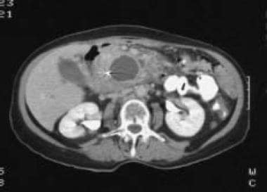 Chronic pancreatitis. Abdominal CT scan showing a pancreatic pseudocyst causing distortion of the ductal system.
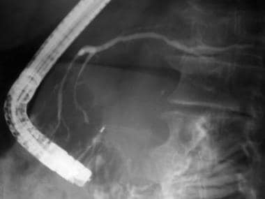 Chronic pancreatitis. Pancreatogram in a patient with a pancreatic pseudocyst. Note how the pancreatic ducts are extrinsically distorted by a mass lesion.
Digestion of an adjacent vessel can result in a pseudoaneurysm, which can produce a sudden expansion of the cyst or gastrointestinal bleeding due to bleeding into the pancreatic duct (hemosuccus pancreaticus).
Pancreatic ascites and pleural effusion can result from disruption of the pancreatic duct, leading to fistula formation to the abdomen or chest, or rupture of a pseudocyst with tracking of pancreatic juice into the peritoneal cavity or pleural space.
The indications for drainage of pseudocysts include rapid enlargement, compression of surrounding structures, pain, or signs of infection. Endoscopic retrograde pancreatograms may be helpful prior to drainage to rule out a stricture of the pancreatic duct, which can lead to persistent drainage from the pseudocyst.
Bile obstruction and duodenal obstruction
Symptomatic obstruction of the bile duct and/or duodenum develops in 5-10% of patients with chronic pancreatitis. Postprandial pain and early satiety are characteristic of duodenal obstruction, while pain and abnormal liver function test results (including hyperbilirubinemia) are suggestive of a bile duct stricture. These complications are most commonly seen in patients with dilated pancreatic ducts; they are either due to inflammation and fibrosis in the head of the pancreas or are the result of a pseudocyst.
Drainage of an obstructing pseudocyst can be accomplished surgically by cystogastrostomy, cystenterostomy, or choledochoenterostomy. Endoscopic stenting may be helpful for benign bile duct strictures.
Additional complications of chronic pancreatitis
Diabetes mellitus is a late manifestation in about one third of patients. The tendency to develop ketoacidosis is low.
The presence of the splenic vein at the posterior surface of the pancreas predisposes it to thrombosis from adjacent pancreatic inflammation. Patients who are affected can develop gastric varices as a result of associated portal hypertension. Splenectomy is usually curative for patients who develop bleeding from gastric varices.
Pseudoaneurysm is rare, but it can be a deadly complication. Affected vessels, including the splenic, hepatic, gastroduodenal, and pancreaticoduodenal arteries, are in close proximity to the pancreas. Surgery for bleeding pseudoaneurysms is challenging and is associated with high morbidity and mortality.
-
This endoscopic retrograde cholangiopancreatography (ERCP) shows advanced chronic pancreatitis. The pancreatogram has blunting of the lateral branches, dilation of the main pancreatic duct, and filling defects consistent with pancreatolithiasis. The cholangiogram also shows a stenosis of the distal bile duct and a dilated biliary tree.
-
Chronic pancreatitis. Abdominal CT scan showing a pancreatic pseudocyst causing distortion of the ductal system.
-
This patient has recurrent abdominal pain. She used alcohol heavily in the past and was involved in a motor vehicle accident. The pancreatogram shows subtle blunting of the side branches consistent with chronic pancreatitis. A stricture also is present in the body of the pancreas where it drapes over the spine, probably resulting from the trauma she sustained in the motor vehicle accident. Air in the stomach makes it difficult to observe that contrast is filling a pseudocyst on the other side of the stricture. These findings are not amenable to endoscopic intervention, and the patient was sent for a distal pancreatectomy.
-
Chronic pancreatitis. This magnetic resonance cholangiopancreatography (MRCP) shows a healthy biliary system. The pancreatic ductal system is not well visualized. A subsequent endoscopic retrograde cholangiopancreatography (ERCP [not pictured]) showed pancreas divisum, with no evidence of a communication with the pseudocyst. The endoscopic features were ideal for an endoscopic transgastric pseudocystogastrostomy.
-
Chronic pancreatitis. CT scans of the abdomen following an endoscopic transgastric pseudocystogastrostomy. Note that 2 stents are placed through the stomach and into the pseudocyst. Before undertaking this type of endoscopic intervention, the endoscopist must be confident that a cystadenoma has not been mistaken for a pseudocyst.
-
Chronic pancreatitis. This patient had a persistent postoperative leak from the site of a distal pancreatectomy. In the mid-1990s, the author sought to facilitate enteric drainage using transpapillary stents placed into the pancreatic duct. While this changed the fluid dynamics in favor of healing the disrupted duct, some patients developed complications from this technique.
-
Chronic pancreatitis. The persistent postoperative leak from the site of a distal pancreatectomy has healed at 1-month follow-up (see the image above). However, after 4 weeks of transpapillary stenting, the pancreatogram now shows a stent-induced stenosis near the surgical genu (arrow). Based on this experience, the author stopped using pancreatic stents in this setting.
-
Chronic pancreatitis. This patient developed abdominal pain several weeks after being accidentally hit with a baseball bat. A CT scan showed a large splenic hematoma, and the patient underwent a splenectomy. His postoperative course was notable for recurrent pain, abdominal distension, and elevation of serum amylase levels over the course of 2-3 months. This repeat CT scan shows postsurgical changes in the left upper quadrant and a large fluid collection.
-
Chronic pancreatitis. The pancreatogram shows a small leak from the tail of the gland.
-
Chronic pancreatitis. A nasopancreatic tube courses through the esophagus, stomach, and duodenum and into the pancreatic duct. Externally, the end of the tube is attached to a suction bulb to decompress the ductal system and monitor its function on a daily basis. In contrast to patients treated with transpapillary stents, none of these patients ever has failed to return for a follow-up appointment. In addition, while stent obstruction and subsequent infection can occur with transpapillary stents, the author has not observed this complication while using nasopancreatic tubes.
-
Chronic pancreatitis. Nine days after placement of a nasopancreatic tube, a pancreatogram obtained via the tube showed that the disruption had healed (see the above image). The tube then was removed.
-
Chronic pancreatitis. This follow-up CT scan (see the above 2 images) shows a percutaneous tube in the left upper quadrant that was used to drain a fluid collection. It was removed after 4 weeks. The patient returned to work, regained his weight, and had no recurrence of abdominal pain or signs of a recurrent pancreatic leak.
-
Chronic pancreatitis. Pancreatogram in a patient with a pancreatic pseudocyst. Note how the pancreatic ducts are extrinsically distorted by a mass lesion.
-
This pancreatogram shows a pseudocyst communicating with the main pancreatic duct in a patient with chronic pancreatitis and recurrent abdominal pain. He was treated endoscopically with a transpapillary stent placed into the pancreatic duct.
-
Four weeks after placement of a transpapillary stent, a patient with a pseudocyst communicating with the main pancreatic duct (chronic pancreatitis with recurrent abdominal pain) had not had a recurrence of pain. The CT scan showed resolution of the cyst, and the follow-up pancreatogram showed marked improvement. Transpapillary stenting of the pancreatic duct should be reserved for patients with established chronic pancreatitis.


