Overview
Total laryngectomy (TL) significantly alters speech production. For a speech production system to be functional, the following 3 basic elements are necessary: (1) a power source, (2) a sound source, and (3) a sound modifier. For laryngeal speakers, lung air is the power source, the larynx is the sound source, and the vocal tract (ie, pharynx, oral cavity) is the sound modifier. During total laryngectomy (TL), the sound source is removed and the lungs are disconnected from the vocal tract. Successful voice restoration following total laryngectomy (TL) requires identification of an alternative sound source with a viable power source. [1]
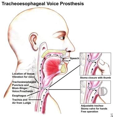
An image depicting laryngectomy rehabilitation can be seen below.
 Diagram of tracheoesophageal puncture and prosthesis placement. Image courtesy of International Healthcare Technologies. Blom-Singer is a registered trademark of Hansa Medical Products.
The 3 basic options for voice restoration after total laryngectomy (TL) are (1) artificial larynx speech, (2) esophageal speech, and (3) tracheoesophageal speech. Selection of a method should be based on input from the surgeon, speech pathologist, and patient. The decision is best made keeping in mind the patient's communicative needs, physical and mental status, and personal preference.
Esophageal speech
See the list below:
-
Principle: Esophageal speech is produced by insufflation of the esophagus and controlled egress of air release that vibrates the pharyngoesophageal (PE) segment for sound production. Anatomic structures for articulation and resonance are usually unaltered.
-
Techniques: The 2 basic approaches to esophageal insufflation are injection and inhalation. Both techniques are based on the pressure differential principle that air flows from areas of higher pressure to areas of lower pressure. Injection involves using the articulators to increase oropharyngeal air pressure, which, in turn, overrides the sphincter pressure of the PE segment, thereby insufflating the esophagus. Inhalation involves decreasing thoracic air pressure below environmental air pressure by rapidly expanding the thorax so air insufflates the esophagus. Proficiency in esophageal speech typically requires several months of speech therapy.
-
Advantages: No apparatus must be purchased or maintained, and no further surgery is required.
-
Disadvantages: Speech acquisition is delayed because of the learning curve, and difficulties with phrasing and loudness are possible.
Artificial larynx speech
See the list below:
-
Principle: An external mechanical sound source is substituted for the larynx. Anatomic structures for articulation and resonance are usually unaltered.
-
Techniques: Two general types of electrolarynges are available, the neck type and the intraoral type. [2] The neck type is placed flush to the skin on the side of the neck, under the chin, or on the cheek. Sound is conducted into the oropharynx and articulated normally. Intraoral devices are used for patients who cannot achieve adequate sound conduction on the skin. A small tube is placed toward the posterior oral cavity, and the generated sound is then articulated. The tube has minimal effect on articulatory accuracy if the patient is taught properly and learns to use it well. A third type of electrolarynx has been developed using an electromyograph (EMG) transducer in the strap muscles to activate a sound source for hands-free use. [3]
-
Advantages: Voice restoration after surgery is immediate, and the maintenance for the electrolarynx is minimal (may last 2-10 y).
-
Disadvantages: The voice quality sounds mechanical.
Tracheoesophageal speech
See the list below:
-
Principle: A surgical fistula is created in the wall separating the trachea and esophagus. This puncture tract can be created primarily, at the time of total laryngectomy (TL), or secondarily, weeks or years following the total laryngectomy (TL). Several days after surgery, a one-way valved prosthesis is placed in the puncture tract, allowing lung air to pass into the esophagus. The lung air induces vibration of the PE segment for sound production. The mechanics of the one-way valve allow lung air to pass into the esophagus without food and liquids passing into the trachea.
-
Technique: During the initial evaluation, a speech pathologist measures the length of the puncture tract and selects a size and style of prosthesis for placement. Once in place, the patient digitally occludes the tracheostoma to direct air through the prosthesis into the esophagus for phonation. Hands-free external airflow valves are also available as accessories.
-
Advantages: The air supply for speech is pulmonary, phonation sounds natural, and voice restoration occurs within 2 weeks of surgery. [4]
-
Disadvantages: Additional surgery is required for secondary punctures, the prosthesis must be maintained, and aspiration may occur if liquids leak through a malfunctioning valve.
Flap reconstruction
The use of autologous free flaps for laryngopharyngeal reconstruction has provided another strategy for voice restoration. A study by Tsao et al compared the use of a J-shaped anterolateral thigh flap with that of a tracheoesophageal puncture plus prosthesis. Patients in the study had undergone either a total laryngectomy (TL) or a laryngopharyngectomy, with partial or complete removal of the surrounding pharynx. The results of objective assessment of phonatory and acoustic outcomes did not significantly differ between the J-flap and tracheoesophageal puncture groups. [5]
However, as evaluated by speech pathologists, consonant pronunciation was considered less accurate in the J-flap patients. In addition, the Voice Handicap Index score indicated that these same individuals had moderate impairment, with the total score being 52.56 for the J-flap group and 18.32 for the tracheoesophageal puncture patients. [5]
Evaluating Tracheoesophageal Speech
Assessing the integrity of the pharyngoesophageal segment
Tracheoesophageal punctures can be created primarily, at the time of total laryngectomy (TL), or secondarily, days to years after surgery. [6] If the plan is for a secondary puncture, a simple insufflation test can be performed preoperatively by the speech pathologist to assess the integrity of the PE segment and potential voice quality. Results indicate whether further surgical intervention is necessary during the puncture procedure. If the puncture is performed primarily, insufflation testing is not an appropriate preoperative assessment because the cricopharyngeus will be reconstructed during the laryngectomy.
Insufflation testing
A catheter is placed through the nose and inserted until the end is just below the PE segment, ie, approximately 25 cm of the catheter length. Air is channeled through the catheter to insufflate the esophagus, simulating tracheoesophageal speech. If insufflation is monitored using manometry, the indication for adequate PE segment integrity is a phonation pressure less than 22 mm Hg. For perceptual assessment, the patient performs speech tasks, such as sustained phonation and/or counting for evaluation of phonatory quality and duration. The patient should be able to sustain phonation of /a/ for at least 10 seconds or produce 10-15 syllables per breath.
If the insufflation test is performed correctly and phonation is not achieved or is of poor quality and duration, the 4 possible conditions of the PE segment that should be considered are (1) hypotonicity, (2) hypertonicity, (3) spasticity upon egress of airflow, or (4) stricture. If perception is uncertain, the PE segment can be further evaluated using fluoroscopy with barium swallows and repeated insufflations.
If insufflation test results indicate failure, several therapies are available. If hypotonicity is present, consider applying digital pressure to the PE segment or an external pressure band around the patient's neck during phonation. If hypertonicity, spasticity, or both is present, consider pharyngeal constrictor myotomy, pharyngeal plexus neurectomy, or botulinum toxin (BOTOX®) injection with electromyographic or radiographic guidance. If stricture is present, dilatation is indicated.
Tracheoesophageal Speech Prostheses
Selecting a Prosthesis
Several sizes and styles of tracheoesophageal prostheses are available. [7, 8] Selecting a valve should be a conscientious decision. The following 4 main issues should be considered when selecting a device:
Phonatory effort
Before any prosthesis is inserted, phonation should be sampled with a patent puncture tract. The perceptual quality and effort of that sample guides decision-making. For example, if the voice quality is effortless, loud, and consistent, then the patient may do well with a higher-resistance device with increased durability. If the voice quality is strained and effortful, a lower-resistance device of greater diameter (20F) may be appropriate.
Candidacy for independent insertion
If the patient and his or her spouse or caregiver appear able and willing to participate in prosthesis management, a valve with no restrictions on placement procedures should be considered. Indwelling devices, although touted for their advanced design, must be inserted by a trained professional. This stipulation creates a situation of patient dependency on the health care professional. Autonomy offered by devices that can be changed without restriction is appealing to many patients. Conversely, if the patient is unable or unwilling to change the valve independently, an indwelling style device offers more security from dislodgement.
Durability
Occasionally, the device that provides the least phonatory effort also has a patient-specific tendency to malfunction rapidly. If the device recurrently leaks in less than a couple of months with no treatable cause (eg, candidal infection), a device with higher resistance and durability should be considered.
Cost
Prices for valves vary from $28 (Inhealth 16F duckbill) to $199 (Provox 2 indwelling, Atos Medical). See the Prosthetic Supply Vendors section for vendor information. Cost issues should be considered when devices are comparable in style and performance. Certain health insurance policies do not cover prosthetic supplies. Patients without prosthesis coverage should be provided cost options when selecting a device.
Prosthesis Choices
Duckbill
-
Size: The prosthesis is 6-28 mm in length and 16F or 20F in diameter.
-
Advantages: It has good durability, can be changed independently, and is inexpensive.
-
Disadvantages: Airflow resistance is increased.
Low resistance/pressure
-
Size: It is 6-28 mm in length and 16F or 20F in diameter.
-
Advantages: It has decreased airflow resistance, has shorter esophageal extension, and can be change independently.
-
Disadvantages: It has decreased durability and is sensitive to esophageal pressure changes.
Indwelling
-
It is 6-22 mm in length and 20F or 22F in diameter.
-
Advantages: It has decreased airflow resistance, increased security from dislodgement, and a removable strap.
-
Disadvantages: It is clinician-dependent and has the potential for gastric distention from excess air insufflation. Also, it is expensive ($130-199).
Steps for Fitting a Prosthesis
See the list below:
Evaluate phonation with a patent puncture tract and stoma occlusion to rule out technique problems.
Measure the length of the puncture tract.
Select and prepare a prosthesis.
Dilate the puncture tract to slightly wider than the prosthesis.
Align the prosthesis with the puncture tract for insertion; alignment is more important than pressure.
Have the patient drink liquid, and watch for any leak through or around the prosthesis.
Assess patient phonation with stoma occlusion.
See the images below.
 Diagram of tracheoesophageal puncture and prosthesis placement. Image courtesy of International Healthcare Technologies. Blom-Singer is a registered trademark of Hansa Medical Products.
 Top photo shows leakage of ingested liquids around the device, which indicates an expanding tracheoesophageal tract possibly due to a prosthesis that is too long. Bottom photo shows an indwelling-style prosthesis in place.
Hands-free tracheostoma valves
Tracheostoma valves provide 2 primary functions: hands-free speech and housing for heat and moisture filters. These external valves are adhered to the neck, with a valve housing directly over the stoma. For speech, the air pressure generated during increased exhalatory effort closes the tracheostoma valve and directs air back through the tracheoesophageal prosthesis. An adequate adhesive seal is essential to generate hands-free speech. Without a tight external seal, stomal air escape reduces the amount of airflow available for speech. Heat-and-moisture–exchange filters are also available to place over, or in lieu of, the tracheostoma valve. These filters modify the inhaled environmental air. Benefits of the filters include decreased airway irritation and maintenance of airway humidification, which may reduce tracheal secretions.
Troubleshooting Tracheoesophageal Punctures
Problems related to tracheoesophageal punctures and prosthetic devices are mentioned, along with typical causes and corresponding solutions. [9]
Leaking through the prosthesis
See the list below:
-
Deteriorated valve: Replace the prosthesis.
-
Candidal infection: Administer antifungal medication.
-
Duckbill tip pressed against esophageal wall: Switch to a low-pressure device.
-
Thoracic pressure changes: Increase the resistance of the valve.
Leaking around the prosthesis
See the list below:
-
Tracheoesophageal puncture tract expansion: Fit a 20F prosthesis.
-
Pistoning of prosthesis: Fit a shorter prosthesis.
-
Radiation effects: Down-stent or perform a repuncture.
Difficult or no phonation
See the list below:
-
Clogged device: Clean the device.
-
Duckbill tip lodged in esophageal wall: Change to a low-pressure prosthesis.
-
Incomplete insertion: Dilate and resize the puncture tract.
-
Closed puncture tract: Perform a repuncture.
Dislodgement of prosthesis
See the list below:
-
Incomplete insertion: Dilate and resize the puncture tract.
-
Inadvertent removal: Fit a more stable prosthesis if this situation is recurrent.
-
Aspiration of device: Remove the device using flexible bronchoscopy.
Granulation tissue
See the list below:
-
Irritation/pistoning of prosthesis: Fit the prosthesis length more securely.
-
Repeated removal/insertion of prosthesis: Decrease the frequency of prosthesis changing, and perform laser removal of the granulation tissue.
Emergent Procedures
When a prosthesis is dislodged, patients are instructed to insert a catheter into the puncture tract as soon as possible to maintain patency and prevent aspiration. If they are unable to place the catheter, they may come to the emergency department for puncture tract stenting. Patients are sometimes unaware that they can phonate without the prosthesis. As long as the puncture tract is patent, phonation is possible. Encouraging tracheoesophageal speech to explain their situation may ease patient anxiety. If the patient cannot speak, have them drink a sip of water, preferably with blue dye. If the water leaks through the puncture tract into the airway, the tract is patent.
The role of the emergency department staff is to stent the puncture tract with a catheter (8-20F). If no catheters are readily available, a Duo Tube or nasogastric tube works. The next step is to dilate the puncture tract. Progressively increase the size of the catheter until a 16F or 20F catheter passes through the tract, depending on the size of the prosthesis. At this point, the prosthesis can be reinserted.
If the patient did not recover the prosthesis, the device may have been aspirated. Some patients report violent coughing after aspirating a valve; however, many patients are asymptomatic. Therefore, diagnostic imaging should be performed. Most prostheses manufactured by InHealth are radiopaque. The indwelling device has only a ring of radiopacity. A chest radiograph should be the first test, followed by a CT scan if a prothesis that is not radiopaque is missing. The final approach should be bronchoscopy. Typically, a prosthesis lodges in the right mainstem bronchi and can be easily retrieved by an otolaryngologist or pulmonologist.
Conclusion
Successful voice restoration for alaryngeal speakers can be attained with any of the 3 speech options. Although, no single method is considered best for every patient, the tracheoesophageal puncture has become the preferred method in the past decade. Perceptual studies have demonstrated listener and speaker advantages of tracheoesophageal speech. Despite the potential facility of voice production with the tracheoesophageal puncture, careful attention must be directed to PE segment integrity and mucosal density, valve selection, and troubleshooting. Voice restoration is a process, not a prosthesis.
Prosthetic Supply Vendors
See the list below:
-
InHealth Technologies : 1110 Mark Avenue, Carpenteria, CA 93013; Phone (800) 477-5969, Fax (888) 371-1530
-
Atos Medical: 11390 W. Theodore Trecker Way, West Allis, WI 53214-1135; Phone (800) 217-0025, Fax (414) 227-9033
-
Diagram of tracheoesophageal puncture and prosthesis placement. Image courtesy of International Healthcare Technologies. Blom-Singer is a registered trademark of Hansa Medical Products.
-
Sizing device used to measure the depth of the tracheoesophageal wall and determine the appropriate prosthesis length.
-
Comparison photos of an aspirated indwelling-style prosthesis on a chest x-ray film versus a CT scan image. Only the outside ring of the esophageal flange is radiopaque.
-
Insertion of a low-pressure prosthesis with a gelatin capsule.
-
Top photo shows leakage of ingested liquids around the device, which indicates an expanding tracheoesophageal tract possibly due to a prosthesis that is too long. Bottom photo shows an indwelling-style prosthesis in place.


