Background
Retinal artery occlusion (RAO) usually presents as painless loss of monocular vision. Ocular stroke commonly is caused by embolism of the retinal artery, although emboli may travel to distal branches of the retinal artery, causing loss of only a section of the visual field. Retinal artery occlusion represents an ophthalmologic emergency, and delay in treatment may result in permanent loss of vision. [1]
Immediate intervention improves chances of visual recovery, but, even then, prognosis is poor, with only 21-35% of eyes retaining useful vision. Although restoration of vision is of immediate concern, retinal artery occlusion is a harbinger for other systemic diseases that must be evaluated immediately.
Pathophysiology
Blood supply to the retina originates from the ophthalmic artery, the first intracranial branch of the internal carotid artery that supplies the eye via the central retinal and the ciliary arteries. The central retinal artery supplies the retina as it branches into smaller segments upon leaving the optic disc. The ciliary arteries supply the choroid and the anterior portion of the globe via the rectus muscles (each rectus muscle has 2 ciliary arteries except the lateral rectus, which has 1).
Anatomical variants include cilioretinal branches from the short posterior ciliary artery, giving additional supply to part of the macular retina. A cilioretinal artery occurs in approximately 14% of the population.
Typical funduscopic findings of a pale retina with a cherry red macula (ie, the cherry red spot) result from obstruction of blood flow to the retina from the retinal artery, causing pallor, and continued supply of blood to the choroid from the ciliary artery, resulting in a bright red coloration at the thinnest part of the retina (ie, macula). These findings do not develop until an hour or more after embolism, and they resolve within days of the acute event. By this time, visual loss is permanent and primary optic atrophy has developed. In those with a cilioretinal artery supplying the macula, a cherry red spot is not observed. [1]
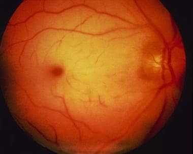 The cherry red spot of central retinal artery occlusion.
An embolism, atherosclerotic changes, inflammatory endarteritis, angiospasm, or hydrostatic arterial occlusion may occlude the retinal artery. The mechanism of obstruction may be obvious from comorbid systemic disease or physical findings. Atrial fibrillation and ipsilateral carotid stenosis are more commonly associated with prolonged visual disturbances. [1]
Animal studies have shown that a retina with completely occluded circulation has irreversible ischemic damage at 105 minutes but may recover at 97 minutes. Complete occlusion of retinal artery circulation in humans is rare with retinal artery disease; thus, retinal recovery is possible even after days of ischemia.
Branch retinal artery occlusion (BRAO) occurs when the embolus lodges in a more distal branch of the retinal artery. BRAO typically involves the temporal retinal vessels and usually does not require ocular therapeutics unless perifoveolar vessels are threatened. The central retinal artery is affected in 57% of occlusions, the branch retinal artery is involved in 38% of occlusions, and cilioretinal artery obstructions occur in 5% of occlusions. [2]
Epidemiology
Frequency
United States
Recent estimates put the incidence of retinal artery occlusion at 0.85 per 100,000 per year, with a 10-year cumulative incidence of retinal emboli of 1.5%. [3]
Mortality/Morbidity
Patients with visualized retinal artery emboli, whether or not obstruction is present, have 56% mortality over 9 years, compared with 27% for an age-matched population without retinal artery emboli. Life expectancy of patients with central RAO (CRAO) is 5.5 years, compared to 15.4 years for an age-matched population without CRAO.
RAO is associated with smoking and cardiovascular disease, with an increased incidence of stroke in patients who have suffered RAO.
Both eyes have an equal incidence of disease, with bilateral involvement in 1-2%.
Sex
Men are affected slightly more frequently than women.
Age
The mean age of presentation of retinal artery occlusion is early in the seventh decade of life, although a few cases have been reported in patients younger than 30 years.
The etiology of occlusion changes, depending on the age at presentation.
Prognosis
Recovery of useful vision is related directly to the rapidity of treatment and presenting visual acuity.
Studies report that 21% of patients exhibited visual improvement of 6 gradients of visual acuity, 35% exhibited improvement of 3 gradients of visual acuity, while 26% showed no improvement in visual acuity.
Patients that showed improvement had presenting visual acuity of counting fingers and a mean duration of visual loss of 21.1 hours; those that did not improve had presenting visual acuity of hand movement and a mean duration of visual loss of 58.6 hours.
The longest delay to treatment that has been associated with significant visual recovery is approximately 72 hours.
Presence of a cilioretinal artery with foveolar sparing increases improvement of visual acuity.
Branch retinal artery occlusions (BRAOs) are associated with a higher recovery rate (80% of eyes improve to 20/40 or better) than central retinal artery occlusions (CRAOs).
Patient Education
Patients must understand that the prognosis for visual recovery is poor and that the visual changes are usually a result of a systemic process that needs treatment.
-
The cherry red spot of central retinal artery occlusion.


