Practice Essentials
More than 16 million people each year receive emergency care for hand injuries. Common emergencies include fractures, ligamentous injuries, and infections. Hand fractures, a frequent emergency department complaint, are the most common fractures of the body and account for about 1.5% of all emergency room visits and 40% of upper extremity fractures. Hand fractures occur in all age groups, although fractures in young children should prompt suspicion of child abuse. [1, 2, 3, 4, 5, 6, 7]
Fractures of the phalanges are the most common hand fractures. Metacarpal and phalangeal fractures are common injuries in athletes who participate in contact and ball-handling sports. Proper management at initial evaluation of hand injuries can prevent a significant amount of morbidity and disability. Emergency physicians, often the first to assess these fractures, must have the skills to properly evaluate and manage these injuries. Most of these injuries are managed nonoperatively with immobilization, but certain displaced and angulated fractures, unstable fractures, and open injuries require surgical intervention. [1, 2, 3, 4, 5, 6, 7]
Basic concepts about bony structures of the hand help to understand injury patterns and manage hand fractures. The hand is a group of gliding bones surrounded by soft tissue. A relatively immobile center consisting of the second and third metacarpal bones provides fixed support on which intrinsic movements of the hand depend. More mobile bones of the hand, such as the first, fourth, and fifth metacarpals, may tolerate a greater degree of angulation without disability, while the less mobile second and third metacarpal bones must have more precise reduction to ensure proper function.
Disability from hand injuries may result in loss of sensation, strength, and flexibility, the chief functions of the hands. Preserving function relies on maintaining the structural relationships of the intrinsic hand structures as well as musculotendinous connections from the forearm. Prevention of disability from hand injuries is the primary goal of treatment. Maintenance of function, rather than cosmesis, is of paramount concern in the management of hand injuries.
In the ED, plain radiography is the diagnostic test of choice to evaluate potential hand fractures. [8] Standard radiographs include AP, lateral, and oblique views (see the images below). CT is sometimes necessary to check for intra-articular displacement and determine need for surgery
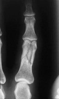 Phalangeal fractures. Complex unstable fracture of the proximal phalanx. Image courtesy of Mark Baratz, MD.
Care for the vast majority of patients with hand fractures is completed on an outpatient basis. Reserve inpatient care for those who must go directly to the operating room for open reduction and internal fixation (ORIF). This is not a common occurrence.
Complications include the following [9, 10, 11, 12] :
-
Malrotation
-
Degenerative arthritis
-
Adhesion of tendon to bone (more likely in open or widely angulated fractures)
-
Joint stiffness from immobilization
-
Boutonniere deformity (may result from improperly treated middle phalanx fracture)
-
Nonunion of fractures resulting in prolonged disability
Prehospital care of most orthopedic injuries consists of administering pain medication, splinting the hand in the position in which it was found, applying ice, and elevating the extremity, if possible.
Obtain as much information about mechanism of injury and conditions at the scene as possible.
Both operative and nonoperative treatment of hand fractures can result in numerous complications, including stiffness, malunion, nonunion, arthritis, infection, and complex regional pain syndrome. [10]
Because most patients have splints applied in the ED, discharge instructions should include signs and symptoms of constrictive splints or casts. Instruct patients of date and time of their follow-up appointment with an orthopedic surgeon or the phone number to call for an appointment.
Instruct patients to rest and elevate the injured hand to reduce swelling and pain. Cold packs may also be recommended to minimize swelling.
-
Assessment of the hand for rotational deformities of the fingers or metacarpals is essential, as such deformities, if untreated, may result in significant functional compromise. With fingers flexed at the metacarpophalangeal and proximal interphalangeal joints and extended at the distal interphalangeal joints, fingers should all point toward the scaphoid bone (see image).
-
Phalangeal fractures. Complex unstable fracture of the proximal phalanx. Image courtesy of Mark Baratz, MD.
-
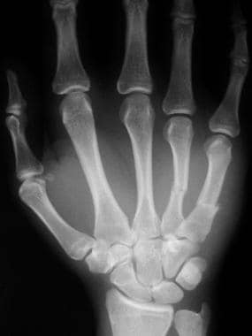
Displaced fourth and fifth metacarpal fractures, anteroposterior view.
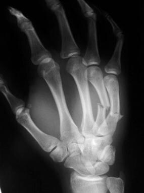
-
Fourth and fifth metacarpal fractures, oblique view.
-
Metacarpophalangeal ligaments.
-
Metacarpophalangeal musculoskeletal structure.


