Practice Essentials
The frontal bone in an adult is an unpaired bone that is part of the bony structure that forms the anterior and superior portions of the skull. At the beginning of life, this bone is separated by a temporary suture called the frontal suture. It then transforms into a singular front bone, absent the suture in most cases. Throughout life, it plays a vital role in protecting important neural structures and forms the superior aspect of the orbit. [1]
Frontal bone injuries in the adult usually occur secondary to trauma. [1] Frontal bone fractures show a low frequency of occurrence at about 5-15% of all maxillofacial fractures occurring as the result of high-velocity injuries such as those seen in motor vehicle accidents, sporting events, and assaults. Successful surgical management revolves around the concept of minimizing cosmetic deformity, maintaining normal sinus function, and avoiding short- and long-term complications. [2]
Clinical symptoms characteristic of a frontal bone fracture include but are not limited to pain, edema, facial laceration, ecchymosis/abrasions, facial asymmetry, facial paresthesia, facial hematoma, emphysema, crepitation, and diplopia. [1]
Management of frontal bone fracture takes into consideration the location and displacement of the fracture as well as the presence or absence of any associated maxillofacial or other head injuries. Common surgical fixation options include the use of titanium plates/screws, titanium mesh/screws, or a combination of these methods to obtain an ideal open reduction internal fixation (ORIF) construct. [1]
The frontal sinuses are located within the frontal bone, superior and medial to the orbits. The frontal sinuses begin to develop at around 5-6 years of age and become fully developed between the ages of 12 and 20 years. Frontal sinus fractures most commonly occur in young males (92%), with a mean age of 20-31 years. [3]
Currently, there is no general consensus on the classification of frontal sinus fractures. Treatment plans for these patients can vary immensely based on their related injuries. [3]
The prognosis for patients with frontal bone fracture is related to the extent of injuries sustained as well as the clinical condition of the patient. If many injuries are present, various specialists and subspecialists may be needed to treat the patient. [3]
In the emergency department (ED), ABCs take priority; reassess the airway frequently.
Do not focus solely on the obvious deformity, thereby failing to perform a complete primary survey.
Rapidly diagnose other life threats, and undertake appropriate resuscitation. Follow with a complete secondary survey.
Diagnosis of frontal bone fracture in the ED is part of the secondary survey.
The support of an interprofessional team should be enlisted for comprehensive management, depending on the extent of injury. Surgical reconstruction will need to be carried out by appropriate specialists. [3]
(See the image below.)
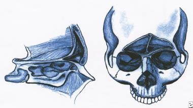 Anterior and lateral views of the frontal sinus. These figures demonstrate the relative thickness of the anterior and posterior tables, as well as the relationship of the frontal sinus to the orbits, ethmoid sinuses, and anterior cranial fossa.
Pathophysiology
Anatomy
The frontal bone borders several bones on its external surface. Anteriorly and medially it joins the nasal bone to create the area called the nasion. Lateral to this, it joins the superior aspect of the maxillary bone bilaterally. Lateral from the maxillary bone, it connects to various bones that form the posterior aspect of the orbit. From medial to lateral, it connects to the lacrimal bone, the sphenoid bone, and the zygomatic bone. Posterior and lateral to the zygomatic bones, it connects again with the temporal surface of the greater wing of the sphenoid, then finally with the parietal bones posteriorly. The frontal bone alone creates the roof of the orbit. It also contains a small zygomatic process to connect with the zygomatic bone just lateral to the lateral wall of the socket. [1]
Two anatomically important landmarks are found on the external surface of the skull that the frontal bone contributes to: the bregma and the pterion. The bregma is the place where the sagittal and coronal sutures meet. The coronal suture is a syndesmosis that joins both parietal bones and the posterior aspect of the frontal bone. The frontal bone has involvement in formation of the pterion through joining of 4 cranial bones: frontal, sphenoidal, parietal, and temporal. Last, the anterior aspect of the frontal bone contains 2 frontal sinuses, each superior to the orbital roof. [1]
Younger patients have more elastic cartilaginous and bony structures in the face and a larger cranium-to-face ratio; thus, fractures in very young children are rare. The growth patterns of the face dictate age-specific fracture patterns that differ from adults. [4]
Frontal cranial bones have greater thickness than the more lateral temporal bones (6.15 cm in males, 7.13 cm in females, compared to 4.33 cm and 4.41 cm, respectively). As a result, these fractures require a more forceful mechanism of injury than other facial bone fractures; they occur less frequently than other forms of skull trauma and often present with concurrent injuries such as naso-orbito-ethmoid fracture, orbital injury, cerebrospinal fluid (CSF) leak, intracranial hemorrhage, and cervical spine fracture, among others. The potential for other potentially devastating injuries to occur along with frontal sinus fractures makes thorough evaluation of these patients imperative. [3]
Object and force
The type of object striking the face and the force behind the object are the main determinants of whether a person sustains soft tissue or bony injury. In automobile accidents, striking a hard dashboard is more likely to cause bony injury than striking a padded dashboard or an airbag. Striking the steering wheel concentrates the force more than striking the flat surface of the dashboard. [5] This also holds true for altercations with a bat, as compared to a bare fist or boxing glove. Penetrating injury from a shotgun at a distance is not likely to cause fractures. Bullets from low-velocity guns are likely to cause fractures; high-velocity bullets cause fractures and extensive soft tissue damage.
The amount of force needed to fracture different bones of the face has been studied; injuries have been divided into those that require high impact to fracture (greater than 50 times the force of gravity [g]) and those that require low impact to fracture (50 g or less). High impact include supraorbital rim, 200 g; symphysis of the mandible, 100 g; frontal-glabellar bone, 100 g; and angle of mandible, 70 g. Low impact include zygoma, 50 g, and nasal bone, 30 g.
Frontal bone and supraorbital ridge fractures require high-energy impact. Forces this strong may indicate intracranial injury. The frontal bone contains the frontal sinus, and fracture of only the anterior (outer) table or of both anterior and posterior (inner) tables is possible. [6] Associated fractures of the supraorbital ridge, the naso-ethmoidal complex, and other facial bones may occur. [7, 8]
Epidemiology
Frontal bone fractures show a low frequency of occurrence at about 5-15% of all maxillofacial fractures, occurring as the result of high-velocity injuries such as those caused by motor vehicle accidents, sporting events, falls, falling objects, assaults, and penetrating trauma. [2] These injuries most commonly occur in young males (92%), with a mean age of 20-31 years. [3]
Among all patients with facial fracture, less than 15% are children. Most facial injuries in children are limited to soft tissues, with only 10-15% of pediatric facial injuries resulting in facial fracture. However, more than half of all facial trauma presentations are associated with concurrent additional severe injuries beyond the face. [4]
Many minor facial traumas are treated at home and may be underreported; facial fractures are likely to cause significant pain and swelling and therefore are more likely to be accurately reported than soft tissue trauma to the face. [4]
Males are more likely to have facial fracture than females, especially during adolescence, when males are approximately twice as likely as females to present with fracture. [4]
Consider domestic violence in women with facial injuries not related to a motor vehicle crash.
Consider child abuse when facial injuries are found in children.
Facial fractures are rare among those younger than 6 years, in whom skull fractures are more likely to be the result of head or facial trauma. When facial fractures occur in children, roughly half are the result of motor vehicle collisions. Beyond motor vehicle collisions, bicycle accidents and sports injuries account for most remaining traumatic events in school-aged children; infants and toddlers are more likely to suffer from falls. Adolescent males are the demographic group most likely to be injured as the result of assault. [4]
Fracture location can be age-dependent due to both activities undertaken affecting the location of likely blunt trauma and differential areas of bone growth and laxity with age. [4]
Nasal fractures are generally thought to be the most common facial fractures, although they are likely underreported, as they do not necessitate evaluation at a trauma center—the source of most pediatric facial trauma data. [4]
Frontal bone fractures are associated with intracranial injury in 35-64% of cases, and with CSF leak in 18-36% of cases (this generally is seen in younger patients). Naso-orbito-ethmoid fractures are uncommon, representing only 1-8% of all pediatric fractures. [4]
In a retrospective review of facial fractures at a level 1 trauma center from 2000 to 2012 in 285 patients aged 18 years or younger, mean patient age was 14.2 years with a male predominance (77.9%). The mechanisms of injury were assault in 108 (37.9%), motor vehicle accident in 68 (23.9%), pedestrian struck in 41 (14.4%), fall in 26 (9.1%), sporting accident in 20 (7.0%), and gunshot injury in 16 (5.6%). The mean Glasgow Coma Scale score (GCS) on arrival to the ED was 13.7. The most common fractures were those of the mandible (29.0%), orbit (26.5%), nasal bone (14.4%), zygoma (7.7%), and frontal bone/frontal sinus (7.5%). Intracranial hemorrhage was present in 70 patients (24.6%). Fractures of the zygoma, orbit, nasal bone, and frontal sinus/bone were significantly associated with intracranial hemorrhage (P< 0.05), and fractures of the zygoma and orbit were significantly associated with cervical spine injury (P< 0.05). [9]
Prognosis
The prognosis for patients with frontal bone fracture is related to the extent of injuries sustained, as well as to the clinical condition of the patient. If many injuries are present, various specialists and subspecialists may be needed to treat the patient. [3]
Ultimately, isolated frontal sinus fractures have a good prognosis, regardless of whether or not the nasofrontal outflow tract or the posterior table is involved. Advancements in surgical technique and in equipment have improved the chances of preservation of the frontal sinuses, as well as patients’ anticipated quality of life. [3]
The prognosis for pediatric patients with facial trauma is generally good, although the more bones that are involved, the greater the chance of long-term deformity and need for surgical repair. Pediatric osteochondral tissues are adept at remodeling, and most patients heal well with minimal later discernible evidence of injury. [4]
Patient Education
The primary method of avoiding or mitigating frontal sinus fracture consists of consistent wearing of helmets during sporting activities involving rapid movement (cycling, driving, skiing, skateboarding, etc.), rapid projectile travel (baseball, cricket, softball, etc.), and full body contact (American football, ice hockey, lacrosse). Helmets should also be worn during activities involving heavy machinery or work in dangerous environments (eg, construction workers, industrial factory workers, soldiers). [3]
Although trauma typically is incidental, some methods remain to reduce the chance of occurrence and the severity of injuries. These involve evaluating potential risk of child abuse, suicidal tendencies, and risky social behaviors (including injurious sports or recreational activities). [4]
Adults and children alike see a decreased rate of injury or death when age- and size-appropriate car seats and restraints are used. Additionally, use of personal protective equipment should be encouraged for participation in recreational and sporting activities. [4]
Patients should be made aware of the high incidence of posttraumatic stress disorder associated with facial injuries and have resources available should symptoms occur. [10]
-
Anterior and lateral views of the frontal sinus. These figures demonstrate the relative thickness of the anterior and posterior tables, as well as the relationship of the frontal sinus to the orbits, ethmoid sinuses, and anterior cranial fossa.


