Practice Essentials
The common forms of esophagitis include reflux esophagitis, infectious esophagitis, pill esophagitis, eosinophilic esophagitis, and radiation and chemoradiation esophagitis. Candida esophagitis (see the image below) is the most common type of infectious esophagitis. The prognosis is good with rapid diagnosis and proper treatment; ultimately, it depends on the severity of the underlying disease. Esophagitis is commonly seen in adults and is uncommon in childhood.
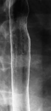 Infectious esophagitis. Candida esophagitis. Double-contrast esophagram shows linear plaquelike lesions in the esophagus, with normal intervening mucosa.
Signs and symptoms
The history and physical examination findings vary according to the type of esophagitis present. Symptoms of reflux esophagitis (the most common type) may include the following:
-
Heartburn, or dyspepsia (the most common symptom)
-
Water brash [1]
-
Regurgitation
-
Other common symptoms include upper abdominal discomfort, nausea, bloating, and fullness
-
Less common symptoms are dysphagia, odynophagia, cough, hoarseness, wheezing, and hematemesis
-
Chest pain indistinguishable from that of coronary artery disease (CAD)
Patients with infectious esophagitis (eg, from Candida, cytomegalovirus [CMV], herpes simplex virus [HSV], or human immunodeficiency virus [HIV]) may be asymptomatic, but typical symptoms include the following:
-
Onset of difficult or painful swallowing (ie, dysphagia or odynophagia)
-
Heartburn
-
Retrosternal discomfort or pain
-
Nausea and vomiting
-
Fever and sepsis
-
Abdominal pain
-
Epigastric pain
-
Hematemesis (occasionally)
-
Anorexia and weight loss
-
Cough
Physical examination usually does not help confirm uncomplicated esophagitis but may reveal other potential sources of pain. The examination should include the following:
-
Rectal examination (to identify the presence of occult bleeding)
-
Examination of the oral cavity (for thrush or ulcers)
-
Search for signs of immunosuppression and skin signs of systemic disease
Complications of esophagitis may include the following:
-
Bleeding and stricture formation
-
Perforation with mediastinitis (rare)
-
Volume depletion and weight loss
-
Laryngitis, aspiration pneumonitis, and bronchospasm
-
In infants, failure to thrive and apnea
See Clinical Presentation for more detail.
Diagnosis
Laboratory tests are usually unhelpful unless complications are present (eg, upper gastrointestinal [GI] hemorrhage). The following may be considered:
-
Complete blood count (CBC) in patients with neutropenia or immunosuppression
-
CD4 count and HIV test in patients with risk factors for HIV
-
Systemic autoimmune disease workup as indicated by the underlying disease
Routine radiography is not indicated unless complications are suspected. Considerations for the use of diagnostic procedures include the following:
-
Double-contrast esophageal barium studies are recommended as the initial imaging study for dysphagia, though a case can be made for an initial upper gastrointestinal (GI) endoscopy (esophagogastroduodenoscopy [EGD])
-
Barium studies are not recommended for patients with absolute dysphagia or odynophagia; upper GI endoscopy is recommended under these circumstances
-
Barium studies and upper GI endoscopy are complementary rather than competing
-
Diagnosis of metastatic cancer is best made by means of barium contrast radiography and computed tomography (CT)
Other studies that may be helpful include the following:
-
Blind brush cytology (now, with the availability of EGD, less commonly used)
-
Electrocardiography (ECG)
-
Troponin or other cardiac markers (if acute coronary syndrome is being considered)
See Workup for more detail.
Management
Treatment includes the following components:
-
Hemodynamic stabilization (eg, in cases of bleeding or perforation)
-
Pain management – Because chest pain of esophageal origin cannot be accurately differentiated from chest pain associated with CAD, prehospital protocols for the latter should be followed
-
Specific therapy, depending on the cause of the esophagitis and any complications
Treatment of reflux esophagitis may include the following:
-
Histamine-2 receptor antagonists (H2RAs)
-
Proton pump inhibitors (PPIs)
-
Cisapride (a gastroprokinetic agent)
-
Sucralfate (a coating agent)
Treatment of infectious esophagitis is directed at the underlying cause, as follows:
-
Fungal esophagitis – Topical, oral, or parenteral antifungals
-
HSV esophagitis – Acyclovir, foscarnet (for acyclovir-resistant cases), or famciclovir
-
CMV esophagitis – Ganciclovir and foscarnet
-
HIV esophagitis – Oral corticosteroids in conjunction with antiretroviral therapy
-
Varicella-zoster virus (VZV) esophagitis – Acyclovir, famciclovir, or foscarnet (for acyclovir-resistant cases)
-
Epstein-Barr virus (EBV) esophagitis – Acyclovir
-
Human papillomavirus (HPV) esophagitis – No treatment, in most cases; systemic interferon alfa, bleomycin, or etoposide
-
Tuberculous esophagitis – Standard antituberculous therapy
-
Bacterial esophagitis – Broad-spectrum beta-lactam antibiotics, usually with an aminoglycoside, adjusted as appropriate
Treatment of nonreflux, noninfectious esophagitis depends on the underlying condition, as follows:
-
Behçet disease esophagitis – Corticosteroids; chlorambucil or azathioprine (long-term therapy)
-
Graft-versus-host disease esophagitis – Dilation and antireflux measures; prednisone, cyclosporine, azathioprine, and thalidomide
-
Inflammatory bowel disease (Crohn disease) esophagitis – Corticosteroids; dilation; occasionally surgery
-
Eosinophilic esophagitis – Diet modification, corticosteroids
-
Metastatic cancer esophagitis – Radiation therapy; palliative stenting
See Treatment and Medication for more detail.
Background
The most common cause of esophagitis is gastroesophageal reflux disease (GERD). Other important, but less common, types of esophagitis include infectious esophagitis (in patients who are immunocompromised), radiation esophagitis, and esophagitis from direct erosive effects of ingested medication or corrosive agents (eg, strong alkalis in liquid and granular forms [3] ) (see the image below). (See Pathophysiology.)
See Pediatric Esophagitis for complete information on this topic.
 Corrosive esophagitis. This is a vinegar-induced esophageal burn. The patient had a fish bone in her throat. She ingested vinegar in an attempt to dissolve the fish bone but to no avail; this led to corrosive esophagitis.
Eosinophilic esophagitis has also emerged as an important cause of esophagitis in both children and adults. [4, 5] Other causes of esophagitis include systemic disease and trauma. (See Etiology.)
The prognosis is good with rapid diagnosis and proper treatment. Ultimately, the prognosis depends on the underlying disease process. (See Prognosis.)
The history findings vary based on the type of esophagitis (eg, reflux or infectious). The physical examination usually is not helpful in confirming the diagnosis of uncomplicated esophagitis. However, the examination may reveal other potential sources of chest or abdominal pain. (See Presentation.)
Laboratory tests are usually unhelpful unless complications are present (eg, upper gastrointestinal [GI] hemorrhage). Routine radiography is not indicated unless complications (eg, perforation, obstruction, bleeding) are suspected. Double-contrast esophageal barium studies and upper endoscopy are the recommended initial diagnostic studies; these tests should be viewed as complementary rather than competing in the evaluation of patients with dysphagia. (See Workup.)
Treatment begins with hemodynamic stabilization and pain management. Subsequent therapy depends on the cause of the esophagitis and on any complications present. [6] Surgery (fundoplication) is sometimes indicated in patients with severe pain who fail to respond to medical management. (See Treatment and Medication.)
Pathophysiology
The pathophysiology of esophagitis depends on its etiology (see Etiology). The common forms of esophagitis include reflux esophagitis, infectious esophagitis, pill esophagitis, eosinophilic esophagitis, and radiation and chemoradiation esophagitis.
Reflux esophagitis
Reflux esophagitis develops when gastric contents are regurgitated into the esophagus. Reflux happens commonly; in most cases, it does not cause major harm, because natural peristalsis of the esophagus clears the refluxate back into the stomach. In other cases, where acid reflux from the stomach is persistent, the result is damage to the esophagus, causing symptoms and macroscopic changes. Gastric acid, pepsin, and bile irritate the squamous epithelium, leading to inflammation, erosion, and ulceration of the esophageal mucosa.
Infectious esophagitis
Infectious esophagitis is most commonly observed in immunosuppressed hosts [7, 8] but has also been reported in healthy adults and children. A wide range of abnormalities in the host defense may predispose an individual to opportunistic infections, such as neutropenia, impaired chemotaxis and phagocytosis, alteration in humoral immunity, and impaired T-cell lymphocyte function.
Patients with systemic diseases (eg, diabetes mellitus, adrenal dysfunction, alcoholism) and those of advanced age can be predisposed to infectious esophagitis because of an altered immune function. Steroids, cytotoxic agents, radiation, and immune modulators can also contribute to the impaired host immune function. Disruption of the mucosal protective barriers and antibiotics that suppress the normal bacterial flora may contribute to the invasive ability of commensal organisms. [9]
Common types of infectious esophagitis include the following:
-
Fungal (eg, candidal) esophagitis
-
Viral (eg, herpes) esophagitis
-
Tuberculous esophagitis
Candida esophagitis results from fungal overgrowth in the esophagus, impaired cell-mediated immunity, or both.
Fungal overgrowth typically occurs in the setting of esophageal stasis resulting from abnormal esophageal motility (eg, achalasia or scleroderma) or mechanical causes (eg, strictures). Impaired cell-mediated immunity can result from immunosuppressive therapy (eg, with steroids or cytotoxic agents, which may suppress both lymphocyte function and granulocyte function), malignancy, or acquired immunodeficiency syndrome (AIDS). Chronic mucocutaneous candidiasis is a congenital immunodeficiency state that is also associated with Candida esophagitis.
Illnesses that interfere with esophageal peristalsis, such as achalasia, progressive systemic sclerosis, and esophageal neoplasias, may contribute to fungal esophagitis.
Initially, herpes esophagitis is manifested by the development of small vesicles that subsequently rupture to form discrete superficial ulcers on the mucosa. In immunocompetent patients, the host response promotes healing of the ulcers, but in patients who are severely immunocompromised, the condition may progress from discrete areas of ulceration to a diffuse hemorrhagic esophagitis. Necrotic herpetic ulcers may become superinfected by candidiasis.
In tuberculous esophagitis, the esophagus is usually involved by erosion of the involved mediastinal lymph nodes abutting the esophagus.
See Cytomegalovirus Esophagitis for complete information on this topic.
Medication-induced esophagitis (pill esophagitis)
Medications associated with pill esophagitis cause damage by local or topical injury. [10, 11, 12] Antibiotics, potassium chloride, nonsteroidal anti-inflammatory drugs (NSAIDs), quinidine, emperonium bromide, and alendronate account for 90% of the reported cases. The following are important pill and patient factors:
-
Chemical nature of drug
-
Solubility
-
Contact time with mucosa
-
Size, shape, and pill coating
-
Amount of water (ie, too little) taken to swallow pill (eg, alendronate)
-
Preexisting esophageal pathology (eg, stricture, achalasia)
Eosinophilic esophagitis
The mechanism of eosinophilic esophagitis remains to be elucidated. However, a corrugated esophagus characterized by fine concentric mucosal rings is commonly observed in patients and is believed to be related to histamine released from sensitized mast cells in the esophageal wall. The release of histamine activates a cascade of reactions, culminating in acetylcholine release that contracts muscle fibers in the muscularis mucosae, resulting in the formation of concentric esophageal rings. [13, 14]
This hypothesis can be tested by performing endoscopic ultrasonography, which will reveal contraction of the muscle layers of the muscularis mucosae and may be related to immunoglobulin E (IgE) activation. [15]
More recent studies have suggested other potential etiopathophysiologic factors, such as an increased susceptibility to eosinophilic esophagitis when genetic predisposition is influenced by environmental factors, [4] or a co-occurrence of potential or probable celiac disease in adults with eosinophilic esophagitis. [16]
Radiation and chemoradiation esophagitis
Radiation dose over 30 Gy to the mediastinum typically causes retrosternal burning and painful swallowing, which is usually mild and limited to the duration of therapy. [17]
-
A dose of 40 Gy causes mucosal redness and edema.
-
A dose of 50 Gy causes a higher incidence and severity of esophageal damage.
-
A dose of 60-70 Gy causes moderate-to-severe esophagitis with strictures, perforations, and fistulas.
Etiology
The various types of esophagitis are associated with differing causative conditions and risk factors.
Reflux esophagitis
Factors or conditions that may increase the risk of reflux esophagitis include the following:
-
Pregnancy
-
Obesity
-
Scleroderma
-
Smoking
-
Alcohol, coffee, chocolate, fatty or spicy foods
-
Certain medications (eg, beta blockers, nonsteroidal anti-inflammatory drugs [NSAIDs], theophylline, nitrates, alendronate, calcium-channel blockers)
-
Intellectual disability requiring institutionalization
-
Spinal cord injury
-
Immunocompromised state
-
Radiation therapy for chest tumors
Helicobacter pylori eradication therapy has been inversely related to reflux esophagitis; it is postulated that the ammonia (alkaline) produced by H pylori reduces the acidity of the stomach and, hence, protects the esophagus from acid spillage.
Infectious esophagitis
Infectious agents known to cause esophagitis include the following:
-
Candida species: Candida albicans is the most common offending pathogen, [7] but other Candida species, such as C tropicalis, C glabrata, and C parapsilosis, have also been implicated as rare causes of esophagitis
-
Noncandidal fungi (eg, Aspergillus, Histoplasma, Cryptococcus, Blastomyces)
-
Herpes simplex virus (HSV)
-
Cytomegalovirus (CMV)
-
Varicella-zoster virus (VZV)
-
Epstein-Barr virus (EBV)
-
In hosts infected with human immunodeficiency virus (HIV), CMV, HSV, Mycobacterium avium-intracellulare, idiopathic
-
Human papillomavirus (HPV) [18]
-
Bacterial species (eg, normal flora, Mycobacterium tuberculosis, M avium-intracellulare, Staphylococcus, Streptococcus, Lactobacillus, Nocardia)
-
Parasitic infections (eg, Chagas disease, Trypanosoma cruzi, Cryptosporidium, Pneumocystis, Leishmania donovani)
Major predisposing factors for Candida esophagitis include antibiotic use, radiation therapy or chemotherapy, hematologic malignancies, and acquired immunodeficiency syndrome (AIDS). Other conditions associated with an increased incidence of Candida esophagitis include esophageal stasis, alcoholism, malnutrition, and advanced age. Occasionally, Candida esophagitis can occur in otherwise healthy individuals with no underlying esophageal or systemic disease. [19, 20, 21, 22, 23, 24, 25]
Other infections of the esophagus are rare and most often develop in patients with neutropenia, AIDS, burns, trauma, or generalized sepsis. Actinomycosis may produce severe esophagitis with deep ulcers and fistulous tracts to the mediastinum, pleural space, tracheobronchial tree, and skin. The diagnosis can be confirmed by the presence of characteristic sulfur granules on endoscopic biopsy specimens.
In persons with HIV, the most significant risk factor for infectious esophagitis is a persistently low CD4 count, but reports exist of individuals who develop fungal esophagitis during the seroconversion phase.
Esophagitis associated with systemic illnesses
Systemic illnesses that can result in esophagitis include the following:
-
Skin disorders, including epidermolysis bullosa, pemphigus vulgaris, bullous pemphigoid, cicatricial pemphigoid, drug-induced skin disorders (eg, erythema multiforme, Stevens-Johnson syndrome, toxic epidermal necrolysis), and others (eg, lichen planus, psoriasis, acanthosis nigricans, leukoplakia)
-
Eosinophilic esophagitis
-
Graft versus host disease (GVHD)
-
Chronic granulomatous disease
-
Metastatic cancer
-
Collagen vascular disease
-
Motility disorders of the esophagus lead to poor acid clearing, with resulting epithelial damage (ie, gastroesophageal reflux disease in scleroderma)
Esophagitis associated with pharmacologic or other therapy
Therapeutic interventions that can cause esophagitis include the following:
-
Medications (eg, pill esophagitis), including alendronate, antibiotics (eg, tetracycline), potassium, NSAIDs, quinidine, and chemotherapeutic agents (eg, dactinomycin, bleomycin, cytarabine, daunorubicin, 5-fluorouracil, methotrexate, vincristine)
-
Radiation esophagitis may occur with radiation treatment of cancers located in the chest (ie, lung, esophagus, mediastinum)
-
Sclerosant and band ligation therapy for varices can cause necrosis of the esophageal tissues and mucosal ulcers; incidence and severity are higher with sclerosant therapy; later, strictures can develop
Pill esophagitis is thought to be secondary to chemical irritation of the esophageal mucosa by certain medications (eg, iron, potassium, quinidine, aspirin, steroids, tetracyclines, NSAIDs), especially when these medications are swallowed with too little fluid.
Epidemiology
Esophagitis is commonly seen in adults and is uncommon in childhood. [26, 27] The most common type of esophagitis is that associated with gastroesophageal reflux disease (GERD) (ie, reflux esophagitis). Candida esophagitis is the most common type of infectious esophagitis. Esophageal reflux symptoms occur monthly in 33%-44% of the general population; as many as 7%-10% of people have daily symptoms.
International statistics
The incidence of symptoms of reflux is up to an order of magnitude higher than the prevalence of esophagitis. In the United Kingdom, patients presenting to a general practitioner with symptoms of reflux esophagitis show rates of esophagitis in the range of 40%-65%. However, a retrospective review of the results of more than 8000 diagnostic endoscopies in Hampshire showed that GERD accounted for 23% of all upper gastrointestinal conditions. [28]
A review of the Swedish National Register estimated the prevalence of esophagitis (diagnosed by endoscopy) to be less than 5% in the 55-year-old group. Other reports have estimated the prevalence to be on the order of 2%.
Prevalence in association with other disorders
The prevalence of symptomatic infectious esophagitis is high in individuals with acquired immunodeficiency syndrome (AIDS), leukemia, and lymphoma and is low (< 5%) in the general medical population.
Candida esophagitis is the most common type of infectious esophagitis. Herpes simplex virus (HSV) type I is the second most common cause of infectious esophagitis. Although obtaining accurate figures regarding the prevalence of herpes esophagitis is difficult, this infection has been reported in approximately 1% of patients who are immunocompromised and in as many as 43% of patients at autopsy. [29, 30, 31, 32, 33, 34]
Cytomegalovirus (CMV) is a recognized cause of esophagitis. Asymptomatic CMV infection is common worldwide, and a large percentage of the world’s population has been exposed to CMV. Before the AIDS epidemic, CMV infections of the esophagus were primarily found on postmortem examinations. The first clinical case of CMV esophagitis was not reported until 1985.
Unlike herpes esophagitis, CMV esophagitis almost never occurs in immunocompetent patients, and the vast majority of affected individuals are found to have AIDS. The incidence of CMV esophagitis—like that of other forms of infectious esophagitis—has declined among AIDS patients since the widespread use of highly active antiretroviral therapy. [35] However, CMV esophagitis has increased among patients with solid organ transplants, [36] in whom delayed-onset disease is typical because of the increasing routine use of early CMV prophylaxis. [37]
Giant esophageal ulcers have been described in patients with AIDS in whom no other infectious etiology for the ulcers can be found. These ulcers have been termed idiopathic or HIV (human immunodeficiency virus) ulcers because they are believed to be caused by HIV. In fact, results of electron microscopy confirm the presence of HIV-like viral particles in these lesions.
Although some patients with HIV ulcers may have undergone recent seroconversion, most are found to have chronic AIDS with CD4 counts lower than 100 cells/μL. HIV ulcers are more common than is generally recognized, accounting for as many as 40% of all esophageal ulcers in patients with AIDS. [23, 38, 39, 40, 41, 42, 43, 44, 45, 46, 47]
Prognosis
The prognosis is good with rapid diagnosis and proper treatment. Ultimately, prognosis depends on the underlying disease process.
Minimal morbidity and mortality result from mild symptoms of esophagitis. Pain from moderate-to-severe symptoms may produce anxiety and lost work and may lead to medical evaluations for more serious causes of pain.
Complicated esophagitis may lead to esophageal strictures (typically long, smooth, tapered areas of narrowing), malnutrition, and, rarely, perforation or bleeding. In addition to strictures, serious gastrointestinal complications of esophagitis include Barrett esophagus and adenocarcinoma. Aspiration of gastric contents is a potentially serious respiratory complication that occurs more often in children. It may be associated with bronchospasm, pneumonitis, and apnea.
Severe esophagitis may lead to dysphagia, pain, odynophagia, and malnutrition. Rarely, life-threatening bleeding occurs and may lead to death. Outcomes and survival in these patients are related to the severity of their underlying systemic illness.
Recurrence is a frequent problem in patients with reflux. Many patients require maintenance therapy to prevent relapse of symptoms.
Candida esophagitis is usually self-limiting, and most patients have a marked response to treatment with antifungal agents. However, necrotic mucosal debris and fungal mycelia in the esophagus occasionally form a mycetoma (ie, fungus ball) that causes obstruction. In other patients, severe Candida esophagitis may lead to the development of strictures. Other complications include ulceration and hemorrhage and, rarely, fistula formation into the bronchial tree. [48]
In immunocompetent patients, herpes esophagitis often resolves spontaneously within 1-2 weeks with conservative treatment involving analgesia and sedation. Rare complications of herpes esophagitis include perforation, tracheoesophageal fistulas, and dissemination to other organs.
Complications
Various complications of esophagitis may be noted, including the following:
-
Bleeding and stricture formation
-
Barrett esophagus occurs when the normal squamous epithelium of the esophagus is replaced with columnar epithelium, and it is linked to the development of esophageal cancer; a systematic review of patients with Barrett esophagus also indicated a link between Barrett esophagus and colonic cancer (7.6% of patients with Barrett esophagus had colonic cancer vs 1.6% in controls) [2]
-
Perforation with mediastinitis, though rare, is a serious complication
-
Volume depletion and weight loss may occur secondary to the inability to swallow
-
Laryngitis, aspiration pneumonitis, and bronchospasm may occur if the gastric contents are refluxed up to the level of the larynx.
-
In infants, apnea and failure to thrive
Patient Education
Lifestyle changes recommended to reduce the frequency and amount of gastric contents that may reflux back into the esophagus include the following:
-
Elevate the head of the bed with 6-inch blocks (sleeping on extra pillows is discouraged because this actually may increase reflux by increasing intra-abdominal pressure caused by the patient bending at the waist)
-
Avoid lying down for several hours after meals.
-
Reduce meal size
-
Lose weight
-
Quit smoking
-
Avoid alcohol and caffeine
-
Avoid citrus, spicy or fatty foods, and chocolate
-
Avoid aggravating medications such as aspirin and other over-the-counter nonsteroidal anti-inflammatory drugs
Educate patients on the disease process and the importance of early medical evaluation at the onset of symptoms.
To prevent pill esophagitis, instruct patients to take medications with plenty of water while sitting upright. Avoid certain medications (eg, alendronate) in patients with known esophageal varices. Alendronate in patients who are cirrhotic could precipitate gastrointestinal bleeding from erosions over an esophageal varix.
For patient education information, see the Heartburn & GERD Center and Digestive Disorders Center, as well as Acid Reflux (GERD), Heartburn FAQs, and GERD and Heartburn Medications.
-
Esophagitis. Location of fungal and viral infections in ulcers.
-
Peptic esophagitis. A rapid urease test (RUT) was performed on the esophageal biopsy sample. The result was positive for Helicobacter pylori.
-
Corrosive esophagitis. This is a vinegar-induced esophageal burn. The patient had a fish bone in her throat. She ingested vinegar in an attempt to dissolve the fish bone but to no avail; this led to corrosive esophagitis.
-
Infectious esophagitis. Candida esophagitis. Double-contrast esophagram shows linear plaquelike lesions in the esophagus, with normal intervening mucosa.
-
Infectious esophagitis. Two examples of advanced Candida esophagitis demonstrate a shaggy esophagus. In both images, the double-contrast esophagram shows a grossly irregular esophageal contour due to innumerable plaques and pseudomembranes, with the trapping of barium between lesions. Patients with this fulminant form of esophageal candidiasis are almost always found to have acquired immunodeficiency syndrome (AIDS).
-
Infectious esophagitis. Candida esophagitis with a foamy esophagus. This patient has a dilated esophagus with beaklike narrowing (arrow) at the gastroesophageal junction as a result of long-standing achalasia. Innumerable tiny bubbles are layering out in the barium column due to infection by the yeast form of candidiasis.
-
Infectious esophagitis. Herpes esophagitis. Double-contrast esophagram shows small, discrete ulcers (arrows) in the mid esophagus on a normal background mucosa. Note the radiolucent mounds of edema surrounding the ulcers. In the appropriate clinical setting, this appearance is highly suggestive of herpes esophagitis, since ulceration in candidiasis almost always occurs on a background of diffuse plaque formation.
-
Infectious esophagitis. Cytomegalovirus esophagitis in a patient with acquired immunodeficiency syndrome (AIDS). Double-contrast esophagram shows a large, flat ulcer in profile (large arrows) in the mid esophagus with a cluster of small satellite ulcers (small arrows). Because HIV esophagitis may produce identical radiographic findings, endoscopy is required to confirm the presence of cytomegalovirus before patients are treated.
-
Infectious esophagitis. Two examples of giant human immunodeficiency virus (HIV) esophageal ulcers (arrows) in patients with acquired immunodeficiency syndrome (AIDS). In A, the ulcer is seen in profile, whereas in B, the ulcer is seen en face. Endoscopy is required to exclude cytomegalovirus as the cause of this finding before treating patients.


