Background
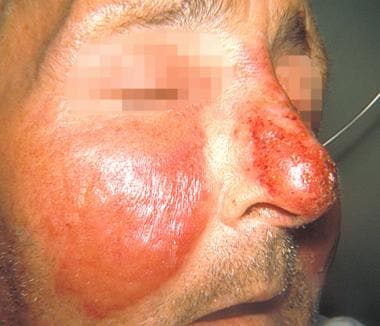
Erysipelas is a bacterial skin infection involving the upper dermis that characteristically extends into the superficial cutaneous lymphatics. It is a tender, intensely erythematous, indurated plaque with a sharply demarcated border. Its well-defined margin can help differentiate it from other skin infections (eg, cellulitis). [1] See the image below. (See Clinical Presentation.)
 Well-demarcated, erythematous plaque of erysipelas. Courtesy of the US Centers for Disease Control and Prevention.
Erysipelas has been traced back to the Middle Ages, where it was referred to as St. Anthony's fire, named after the Christian saint to whom those afflicted would appeal for healing. Around 1095, the Order of St. Anthony, a Roman Catholic congregation, was formed in France to care for those with the ailment. At the time, several diseases were likely grouped under this eponym, including ergotism and herpes zoster (shingles).
Historically, erysipelas occurred on the face, but cases today most often involve the legs. The group A streptococcal bacterium Streptococcus pyogenes causes most of the facial infections; although it can also cause erysipelas on the legs, an increasing percentage of lower extremity infections are now being caused by non–group A streptococci. (See Pathophysiology and Etiology.)
Patient education
Instruct patients to rest, elevate the affected area, and use cold compresses 4 times daily for 48 hours. Patients should return or see a primary care physician if they are experiencing an increase in pain, fever and chills, redness, or other new symptoms. (See Treatment and Medication.)
Pathophysiology and Etiology
Pathophysiology
In erysipelas, the infection rapidly invades and spreads through the lymphatic vessels. This can produce overlying skin "streaking" and regional lymph node swelling and tenderness. Immunity does not develop to the inciting organism.
Etiology
Streptococci are the primary cause of erysipelas. [2] Most facial infections are attributed to group A streptococci, while an increasing percentage of lower extremity infections are caused by non–group A streptococci. Erysipelas in newborns is often caused by group B streptococci, which may also be responsible for perineal and lower-trunk erysipelas occurring in postpartum women. [3] Streptococcal toxins are thought to contribute to the brisk inflammation that is typical of this infection. No clear proof has emerged that other bacteria cause erysipelas, although they coexist with streptococci at sites of inoculation.
The role of Staphylococcus aureus, and specifically methicillin-resistant S aureus (MRSA), remains controversial. No conclusive evidence demonstrates a pathogenic role for staphylococci in typical erysipelas. The infection's predictable response to penicillin, even when S aureus is present, argues against S aureus as an etiologic agent. However, analogous to what occurs in bullous impetigo or staphylococcal scalded skin syndrome, exotoxins from coexisting S aureus may account for the clinical presentation of bullous erysipelas. [4]
Risk factors
Predisposing factors in erysipelas include the following:
-
Lymphatic obstruction or edema
-
Saphenous vein grafting in lower extremities
-
Status postradical mastectomy
-
Immunocompromise: Including patients who are diabetic or alcoholic [5] or who have human immunodeficiency virus (HIV)
-
Arteriovenous insufficiency
-
Paretic limbs
-
Nephrotic syndrome
-
Vagrant lifestyle
Bacterial inoculation into an area of skin trauma is the initial event in developing erysipelas. Thus, local factors, such as venous insufficiency, stasis ulcerations, inflammatory dermatoses, dermatophyte infections, insect bites, and surgical incisions, have been implicated as portals of entry. The source of the bacteria in facial erysipelas is often the host's nasopharynx, and a history of recent streptococcal pharyngitis has been reported in up to one third of cases.
Preexisting lymphedema is a clear-cut risk factor for erysipelas. Recurrent erysipelas complicating the lymphedema from breast cancer treatment is well documented. [6, 7] Lymphoscintigraphy in patients with a first-time episode of lower extremity erysipelas has documented lymphatic impairment in affected and nonaffected legs. Thus, subclinical lymphatic dysfunction is also a risk factor for erysipelas. [8]
Epidemiology
Occurrence in the United States
Isolated cases are the rule with erysipelas, although epidemics have been reported. The incidence of erysipelas declined throughout the mid-20th century, possibly due to antibiotic development, improved sanitation, and decreased virulence. [9] However, an increasing incidence of the condition has been noted since the late 1980s.
The change in distribution from the face to the lower extremities is most likely related to an aging population with risk factors such as lymphedema. Approximately 80% of cases of erysipelas occur on the legs rather than the face.
International occurrence
Erysipelas is somewhat more common in European countries. Isolated cases are still the rule, however, and the distribution and etiology remain similar to those in the United States.
Race-, sex-, and age-related demographics
Erysipelas infections affect persons of all races. The condition has been reported to be more common in females but to occur at an earlier age in males (likely because of a greater incidence of skin injuries in younger males). [10] Other studies indicate that predisposing factors, rather than gender, account for any male/female differences in incidence.
Cases of erysipelas have been reported in all age groups, but it does appear that infants, young children, and elderly patients are most commonly affected. The peak incidence has been reported to be in patients aged 60-80 years, especially in those who are considered to be high-risk and immunocompromised or those with lymphatic drainage problems (eg, after mastectomy, pelvic surgery, bypass grafting).
Prognosis
The prognosis for patients with erysipelas is excellent. Complications of the infection usually are not life threatening, and most cases resolve after antibiotic therapy without sequelae. (The disease may also resolve spontaneously, without treatment.)
Complications of erysipelas may include the following:
-
Gangrene/amputation
-
Chronic edema
-
Scarring
-
Bacteremia sepsis
-
Scarlet fever
-
Pneumonia
-
Embolism
-
Meningitis
-
Death
The most common complications of erysipelas include abscess, gangrene, and thrombophlebitis. [12] Less common complications (< 1%) are acute glomerulonephritis, endocarditis, septicemia, and streptococcal toxic shock syndrome. Rare osteoarticular complications involve joints contiguous with the erysipelas plaques and include bursitis, osteitis, arthritis, and tendinitis. [13]
Local recurrence has been reported in up to 20% of patients with predisposing conditions, and this can lead to disfiguring and disabling sequelae, such as elephantiasis nostras verrucosa. This chronic warty, edematous condition is caused by lymphatic destruction from repeated infection.
Although generally easily and successfully treated with oral antibiotics, with a mortality rate of less than 1% in treated cases, erysipelas can be fatal when associated with bacteremia in very young, elderly, or immunocompromised patients.
-
Well-demarcated, erythematous plaque of erysipelas. Courtesy of the US Centers for Disease Control and Prevention.
-
Facial erysipelas exhibiting classic fiery-red plaque with raised, well-demarcated borders.


