Background
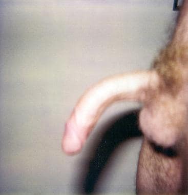
In 1587, Guilio Cesare Aranzi was the first to formally describe Peyronie disease (PD) in his book Tumores praeter naturam. He called Peyronie disease "a rare affection of the genitals in people with excessive sexual intercourse: a little penile tumor palpable like a bean in the flaccid penis causing a deformity similar to a ram horn during erection." The disease was not given its current name until 1743, when Francoise de La Peyronie described the cases of 3 men with fibrous thickening of the penile shaft, painful erections, and penile curvature, as demonstrated in the images below.

Medscape Drugs & Diseases article, Peyronie Disease, also may be helpful.
Pathophysiology
Although the exact etiology of Peyronie disease is not clear, trauma may cause perivascular inflammation. [1] In ordinary tissue, the rupture of blood vessels leads to coagulation and fibrin deposition. Fibrinolysis of the deposited fibrin occurs as tissue cells proliferate to close the site of injury. In Peyronie disease, the tunica albuginea may initially undergo microvascular trauma during sexual intercourse. Adequate postinjury fibrinolysis is prevented because the tunica albuginea is hypovascular.
After multiple microvascular traumas, large quantities of fibrin accumulate in the form of a plaque, generally along the dorsal and ventral midline aspects of the penile shaft. This deposition appears to be immune mediated. Some people may be genetically predisposed to this reaction. The plaque prevents the adequate expansion of the tissue during erection, leading to penile curvature and pain. The relationship of this condition to other fibromatoses suggests a predisposition to fibrous proliferation. Although these microtraumatic events are implicated in the current theory of the pathogenesis of Peyronie disease, no etiology is proven.
Results of animal studies indicate that transforming growth factor-beta (TGF-beta) may be involved in the formation of the plaques via early inflammatory reactions. When TGF-beta analogs are injected into rat tunica albuginea, they form collagen bundles that are morphologically similar to those in Peyronie disease.
Peyronie disease starts in 1 of 2 ways. Most patients report the acute onset of pain accompanied by a lump in the shaft of the penis, followed by gradually increasing curvature with or without pain. Alternatively, a minority of the patients report curvature of the penile shaft that occurs suddenly, seemingly overnight, and remains stable once it occurs.
In the progressive form, the cycle of trauma, fibrin deposition, and attempts at fibrinolysis continue to escalate. Remodeling of the fibrin deposits can take as long as 2 years in the absence of further traumatic events.
Etiology
Although microtraumatic events are implicated in the current theory of the pathogenesis of Peyronie disease, no etiology is proven. No risk factors are known, but associations do exist. Research has demonstrated increased expression of hypoxia-inducible target genes in lesional tissue, suggesting hypoxic injury may be involved in the pathogenesis. [2]
Peyronie disease occurs in men, particularly in older men with weak erections, who engage in frequent and vigorous intercourse (>4 times per wk).
In some studies, HLA-B7 and HLA-DQ5 appeared to correlate with the prevalence of Peyronie disease. Results of other studies have not confirmed this finding. [3]
Peyronie disease may be associated with erectile dysfunction, diabetes mellitus, and hypertension. Resultant partial erections may lead to buckling during intercourse.
Peyronie disease is associated with Dupuytren contracture. Among patients with Peyronie disease, 10% have Dupuytren contracture, and 3% of patients with Dupuytren contracture have Peyronie disease. [4]
Aponeurotic plantar fibrosis (ie, Ledderhose disease) may be found.
Peyronie disease is associated with sporadic and familial knuckle pads, which are circumscribed fibromatous thickenings overlying the finger joints.
Peyronie disease has been seen in systemic sclerosis. [5]
Occasionally, Peyronie disease is associated with fibromatous degeneration of the external ear cartilage.
Antielastin antibodies, anti-DNA antibodies, and elevated antinuclear antibody (ANA) levels have been found in association with Peyronie disease. [6]
Peyronie disease itself is not associated with penile fracture.
Epidemiology
Frequency
United States
The rate for Peyronie disease is 0.3-4% among white men.
International
Peyronie disease is less common in men of African or Asian heritage.
Race
Peyronie disease is well documented in whites. Peyronie disease may occur in black men, often in the presence of preexisting diabetes mellitus and erectile dysfunction.
Age
Peyronie disease predominantly occurs in men aged 40-60 years. The age range of affected persons is 30-80 years.
Prognosis
The spectrum of Peyronie disease ranges from asymptomatic plaques to mild penile curvature or severe curvature that results in a complete inability to have sexual intercourse. Erectile pain can range from none to severe, depending on the site and amount of plaque deposition.
Peyronie disease only occasionally, if ever, disappears completely. The pain may diminish or resolve after the early inflammatory phase, and as many as 13% of patients claim to have some improvement with time. Approximately one half of the remaining individuals have progressive disease, whereas the other half have static disease.
Variable degrees of morbidity may occur. Conditions range from a static painless plaque without penile angulation to painful erections with a curvature significant enough to prevent sexual activity.
Data suggest a combination approach to therapy for Peyronie disease. For instance, the use of partially effective oral therapies with minimally invasive mechanical or intralesional treatments may be more successful than each individual therapy alone.
Patient Education
Patient education in Peyronie disease includes the following:
-
Reassurance that lesions are not cancerous or fatal
-
An explanation that the condition is not curable but that it is treatable and possibly self-resolving
-
An open discussion about the problems with intercourse, the possibility of the resumption of satisfactory sexual activity, and the reduction of pain
-
Lateral view demonstrates vertical curvature.
-
Superior view shows a full erection.
-
Postoperative picture after surgical repair demonstrates a straight erection.
-
Intraoperative picture of an artificial erection demonstrating lateral curvature.
-
Intraoperative picture after penile plication demonstrating a straight erection.
Tables

What would you like to print?
- Overview
- Presentation
- DDx
- Workup
- Treatment
- Medication
- Medication Summary
- Proteinase
- Vitamins
- Nutrients and nutritional agents
- Antihistamines
- Anti-inflammatory agents
- Estrogen receptor antagonists
- Calcium channel blockers
- Nonsteroidal anti-inflammatory agents (NSAIDs)
- Interferon intralesional injections
- Nonselective phosphodiesterase inhibitors
- Show All
- Media Gallery
- References


