Practice Essentials
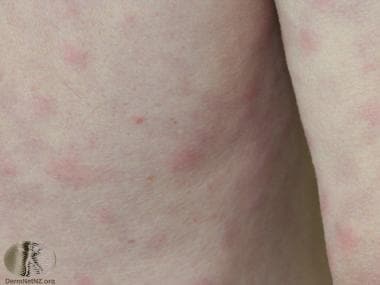
Schnitzler syndrome is an autoinflammatory disease characterized by chronic, nonpruritic urticaria in association with monoclonal gammopathy. Associated sympoms include recurrent fever, bone pain and arthralgia or arthritis. The gammonpathy is most often of the immunoglobulin M (IgM) subtype; those with an immunoglobulin G (IgG) monoclonal gammopathy are considered to be a distinct group.. Approximately 10-15% of patients eventually develop a lymphoproliferative disorder, such as lymphoplasmacytic lymphoma, Waldenström macroglobulinemia, or IgM myeloma. See the image below.
 Rash of Schnitzler syndrome. Courtesy of DermNet New Zealand (http://www.dermnetnz.org/assets/Uploads/systemic/schnitzler.jpg).
Signs and symptoms
The diagnosis of Schnitzler syndrome is made by using the Strausborg criteria [1] :
Major criteria:
Recurrent urticarial eruption
Monoclonal gammopathy. Titers may be low (< 10 g/L).
Minor criteria:
Recurrent fever - present in ~ 90%
Elevated C-reactive protein (CRP) and/or erythrocyte sedimentation rate (ESR)
Leukocytosis
Skin biopsy showing a neutrophilic infiltrate without vasculitis
Evidence of abnormal bone remodeling with or without bone pain ( abnormal MRI, elevated bone alkaline phosphatase or bone scintigraphy)
For a definitive diagnosis:
2 major and 2 minor criteria if the monoclonal gammopathy is IgM.
2 major and 3 minor criteria if the monoclonal gammopathy is IgG.
See Clinical Presentation for more detail.
Imaging studies
Radiologic evaluation shows evidence of hyperostosis in 35% of Schnitzler syndrome patients. Often, the areas of hyperostosis coincide with areas of symptomatic bone pain, such as the iliac bone, tibia, femur, and vertebral column.
See Workup for more detail.
Management
Ibrutinib, a blocker of Bruton's tyrosine kinase (BTK), has been shown to be successful in treating Schnitzler sybdrome, with or without an accompaying hematologic malignancy. [2]
Inhibitors of interleukin (IL)–1 (anakinra, rilonacept, and canakinumab) are often effective. [3, 4]
See Treatment and Medication for more detail.
Background
Schnitzler syndrome was first described in a 1972 case report by French dermatologist Liliane Schnitzler, [5] who further described it 2 years later with several cases. The diagnostic criteria were established in 1999 by Lipsker et al. [6] Most cases have been reported in Europe, although more and more are being described in North America and elsewhere.
Pathophysiology
Schnitzler syndrome is an autoinflammatory disease for which the exact pathophysiology remains unclear but seems to involve the innate immune system. Current data suggests activation of the inflammasome, leading to overproduction of proinflammatory cytokines, is central to the development of Schnitzler syndrome.
Interleukin 1-beta(IL-1beta) appears to play an important role, and inhibitors of IL-1 beta are often helpful in treatment. Increased levels of several other members of the cytokine IL-1 family have been found, including IL-18. [7] Neutrophil extracellular traps (NETs) are net-like structures released by neutrophils to combat pathogens but also have immune-modulating properties that may be important in the pathogenesis of neutrophil mediated disorders including Schnitzler, Sweet syndrome and pyoderma gangrenosum [8] Hypersensitivity to lipo-polysaccharide IL-1 production has been found in peripheral blood cells, particularly mononuclear cells.
Elevated levels of interleukin 6 (IL-6), granulocyte-macrophage colony-stimulating factor (GM-CSF), and granulocyte colony-stimulating factor (G-CSF) have been found in the serum of some patients. [9] .
Several genetic mutations have been found in Schnitzler syndrome patients, though not consistently. Mutations in the NLRP3 gene (nucleotide-binding oligoisomerization domain [NOD]-like [NLR] family pyrin domain containing 3) that are also found in cryopyrin-associated periodic syndrome (CAPS) has been found in a few Schnitzler syndrome patients. Mutations in MYD88 L265P have also been observed in a few patients.
Patients have shown deposition of IgM in the involved tissue, and using anti-idiotype antibodies, IgM monoclonal antibodies were demonstrated to react with epidermal antigens. [10] In one case, monoclonal IgM was found to target 50-, 31-, and 17-kd proteins within epidermal extracts. [11] These findings suggest that the IgM deposits may be involved in the pathogenesis, perhaps via the formation of immune complexes and activation of the complement system.
Disturbances in mitochondrial function have also been reported. [12]
Epidemiology
United States
Only a few cases of Schnitzler syndrome have been reported from the United States.
International
Schnitzler syndrome is relatively rare, though more and more cases are being reported in the literature. The original case was from France, with the greatest number of cases originating from Western Europe.
Race
Occurs in all races.
Sex
Males have a slight predominance with a ratio of 1.76:1.
Age
Patients with Schnitzler syndrome have ranged from age 13-71 years at the time of diagnosis. Itis generally considered to be of "late onset" with the average age of onset approximately 52 years, [13, 14] although the average delay to diagnosis is more than 5 years.
Prognosis
Most Schnitzler syndrome patients have a chronic benign course, though quality of life may be lowered due to symptoms. No spontaneous complete remissions have been reported. Neuropathy, both sensory and motor, has been reported as well as hearing loss and anemia [15] .
Approximately 10-15% of patients eventually develop a lymphoproliferative disorder, including lymphoplasmacytic lymphoma, Waldenström macroglobulinemia, or IgM myeloma. Schnitzler's original patient died at age 88 years, with a diffuse lymphoplasmacytic infiltration of his liver and bone marrow. Thus, the initial workup of a Schnitzler syndrome patient should include an examination of the bone marrow, immunoelectrophoresis of serum, and a urinary protein level. A lymph node biopsy should be performed if the nodes are enlarged. Other associated diseases include AA amyloidosis.
Kidney involvement has been described as a rare complication, but it improved with treatment in the cases reported. [16]
-
Rash of Schnitzler syndrome. Courtesy of DermNet New Zealand (http://www.dermnetnz.org/assets/Uploads/systemic/schnitzler.jpg).


