Practice Essentials
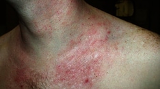
Nummular (meaning round or "coin shaped") dermatitis or eczema is an inflammatory skin condition characterized by the presence of well-demarcated round-to-oval erythematous plaques. The term nummular dermatitis has been used both as an independent disease and as a description of lesion morphology that can be found in many different diseases, including atopic dermatitis, contact dermatitis, and asteatotic eczema. This discussion focuses on the disease entity that has been described in the literature. Other names have included discoid eczema or orbicular eczema.
Signs and symptoms
Especially in the earlier phases, the plaques may be studded with small vesicles. Nummular eczema is usually extremely pruritic and may occur in all ages.
Diagnostics
Tinea corporis should be excluded by scraping and microscopically analyzing a potassium hydroxide preparation of a lesion.
For lesions that have erythema spreading away from the lesions, suggesting cellulitis, swab culture of the exudate may be helpful. First-generation cephalosporins are still usually effective first-line treatment. As methicillin-resistant Staphylococcus aureus becomes more common in a community, the culture results help choose appropriate antibiotics in treatment-resistant cases of documented secondarily infected lesions.
Consider a workup for infection, such as with H pylori and for giardiasis, if appropriate history is suggestive of infection. [1]
Management
Treatment includes moisturizers and topical steroids.
Background
Nummular eczema was first described by Deverigie in 1857 [2] as coin-shaped lesions on the upper extremities. Since then, it has been reported in all age groups and in all body areas but is most commonly found on the upper and lower extremities. [3] Lesions most often start as papules, which coalesce into plaques; they are usually scaly. Early lesions may be studded with vesicles containing serous exudate. Nummular eczema is usually very pruritic. Many precipitating factors have been reported, including dry skin, contact allergies, weather (particularly winter), nutritional issues, and emotional stress.
Pathophysiology
Nummular dermatitis is a condition confined to the skin.
Little is known about the pathophysiology of nummular eczema, but it is frequently accompanied or preceeded by xerosis. Like most other forms of dermatitis, the cause is likely to be a combination of epidermal lipid barrier dysfunction and an immunologic response. Dryness of the skin results in "leaking" of the epidermal lipid barrier; this allows environmental allergens and bacteria to penetrate the skin and induce an allergic or irritant immune response. [4, 5] This is supported by one study that showed that elderly patients with nummular dermatitis had increased sensitivity to environmental aeroallergens compared with age-matched controls. This impaired cutaneous barrier in the setting of nummular eczema may also lead to increased susceptibility to allergic contact dermatitis to materials such as metals, soaps, and chemicals. [6]
Nummular eczema has been associated with medications. Any medication that induces dryness of the skin can theoretically initiate nummular eczema, particularly diuretics and statins. Onset of severe, generalized nummular lesions has been reported in association with interferon and ribavirin therapy for hepatitis C. [7, 8] Other medications that influence immune response have also been reported to induce lesions of nummular eczema, including inhibitors of tumor necrosis factor [9] and guselkumab. [10] A review of 1662 Japanese women found that nummular eczema developed in almost 3% of patients undergoing breast reconstruction, assumed to be due to surgical cleansers or bandage tape. [11]
Onset has also been described in association with mercury in dental amalgams. Hypersensitivity to the metals in the mouth is posulated to be sufficient to drive an immune response that results in cutaneous nummular plaques.
Because of the intense pruritus associated with nummular eczema, the potential role of mast cells in the disease process has been investigated. Increased numbers of mast cells have been observed in lesional compared with nonlesional samples in persons with nummular dermatitis.
One study identified neurogenic contributors to inflammation in both nummular eczema and atopic dermatitis by investigating the association between mast cells and sensory nerves and identifying the distribution of neuropeptides in the epidermis and upper dermis of patients with nummular eczema. Researchers hypothesized that release of histamine and other inflammatory mediators from mast cells may initiate pruritus by interacting with neural C-fibers. The research showed that dermal contacts between mast cells and nerves were increased in number in both lesional and nonlesional samples of nummular eczema compared with normal controls. In addition, substance P and calcitonin gene-related peptide fibers were prominently increased in lesional samples compared with nonlesional samples from patients with nummular eczema. These neuropeptides may stimulate release of other cytokines and promote inflammation. [12, 13, 14]
Other research has demonstrated that mast cells present in the dermis of patients with nummular eczema may have decreased chymase activity, imparting reduced ability to degrade neuropeptides and protein. This dysregulation could lead to decreased capability of the enzyme to suppress inflammation.
Colonization of the skin with Staphylococcus aureus has been described both on lesional skin and in the nares of patients and their close contacts. [15] Whether this is important in the precipitation of disease has yet to be determined.
Etiology Nummular Dermatitis
The etiology of nummular eczema is unknown and likely multifactorial. Most patients with nummular eczema also have very dry (xerotic) skin. Local trauma, such as arthropod bites, contact with chemicals, or abrasions, may precede an outbreak.
Contact dermatitis may play a role in some cases. Contact dermatitis may be irritant or allergic in nature. Sensitivity to nickel, cobalt, or chromates has been reported in patients with nummular dermatitis. In one study, the most frequent sensitizers were colophony, nitrofurazone, neomycin sulfate, and nickel sulfate. In the past, cases of nummular eczema–like eruptions have been caused by ethyl cyanoacrylate–containing glue, thimerosal, [16] mercury-containing dental amalgams, [17] and depilating creams containing potassium thioglycolate. [18] .
Venous insufficiency (and varicosities), stasis dermatitis, and edema may be related to the development of nummular eczema on the affected lower extremities. [19]
Autoeczematization (ie, lesional spread from the initial focal site) may account for the presence of multiple plaques.
Onset of severe, generalized nummular lesions has been reported in association with interferon therapy for hepatitis C as well as exposure to mercury.19. Various types of eczematous eruptions, including nummular eczema, have been observed following tumor necrosis factor-alpha–blocking therapy. [9] and it has been reported after treatment of psoriasis with guselkumab as well. [20]
Nummular eczema of the breast has reported in breast cancer patients undergoing mastectomy with subsequent breast reconstruction. [21, 22] . The majority of these patients developed it after the insertion of tissue expanders or breast implants; the authors suggested a role in stretching of the skin as a cause.
Nummular eczema has been found in association with infection in rare cases. Giardiasis has been reported. [1] One study reported that in patients with Helicobacter pylori infection and nummular eczema, eradication of H pylori caused clearance of the skin lesions in 54% of the patients. [23] Another case report noted nummular eczema in association with a dental infection that cleared after the treatment of the infection. [24]
In children, it occurs most often in association with atopic dermatitis [25] .
Epidemiology
Frequency
The prevalence of nummular eczema is two cases per 1000 people. Dermatitis in general (eg, atopic, asteatotic, dyshidrotic, nummular, hand) is one of the most common of dermatologic conditions.
Race
No racial predilection has been observed. In children, it appears to be more common in those of skin of color [25] .
Sex
Nummular eczema is more common in males than in females (see Age below).
Age
Nummular eczema has two peaks of age distribution. The most common is in the sixth to seventh decade of life and is most often seen in males. A smaller peak occurs in the second to third decade of life, which is most often seen in association with atopic dermatitis. This is more often seen in females, by two thirds in one study. [26] It is uncommon in children. [27] . Nummular eczema also occurs in children.
Prognosis
Nummular eczema tends to be a chronic condition that remits and relapses. Patients need to be informed that once nummular eczema develops, it is often recurrent. Avoidance of exacerbating factors and close attention to moisturizing the skin may help reduce the frequency.
Pruritus, often worst at night, may cause irritability, insomnia, or both.
Secondary infection may result in lesions that ooze serosanguineous exudate. The most common organism revealed by culture is Staphylococcus aureus.
Generalized flares may require systemic antibiotics and/or systemic steroids.
Increased contact sensitivity to environmental antigens (especially metals) could limit ability to tolerate those antigens, especially clothing, metal snaps, jewelry, dental amalgams or occupational exposure.
Patient Education
Patients must be educated about the most important predisposing condition to nummular eczema—dry skin. Use of gentle soaps and copious application of moisturizers, especially while the skin is still damp after bathing, is imperative. Once the lesions develop, use of topical steroids or calcineurin inhibitors helps with the itch and hastens resolution.
For patient education resources, visit the Skin Conditions and Beauty Center. Also, see the patient education article Eczema.
-
Dry, scaling plaque of nummular dermatitis (size, 3 X 5 cm) on the shin.