Background
In 1895, Kofmann [1] described the first case of nevus comedonicus. It manifests as groups of closely set, dilated follicular openings with dark keratin plugs resembling comedones. The majority of cases are isolated. However, nevus comedonicus may be part of nevus comedonicus syndrome in association with skeletal or central nervous system anomalies, ocular abnormalities, and cutaneous defects. [2, 3, 4]
See the image below.
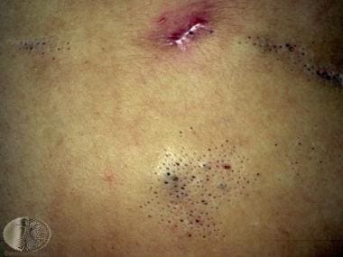 Nevus comedonicus. Courtesy of DermNet New Zealand (http://www.dermnetnz.org/assets/Uploads/lesions/com-naev1.jpg).
Pathophysiology
Many consider nevus comedonicus to be a hamartoma deriving from a failure of the mesodermal part of the folliculosebaceous unit to develop properly, with subsequent abnormal differentiation of the epithelial portion. The follicular structures that result are unable to form terminal hair or sebaceous glands and are capable only of producing soft keratin, which accumulates in the adnexal orifices and produces the comedonelike lesions observed in persons with this condition. Another view is that nevus comedonicus is an epidermal nevus involving hair follicles or an appendageal nevus of sweat ducts. Lesions that extend onto a palm or sole typically demonstrate sweat duct dilatation with keratin in the volar portion of the lesion. See Epidermal Nevus Syndrome for more information.
The etiology of nevus comedonicus is unclear. Why some nevus comedonicus patients present late in life is not known, although a genetic mosaicism has been proposed. While the majority of cases are sporadic, several families with this condition have been documented. Only one report has described nevus comedonicus occurring in homozygous twins. [5]
Etiology
The etiology is unknown. However, whole-exome sequencing in nevus comedonicus identified somatic NEK9 mutations, each affecting highly conserved residues within its kinase or RCC1 domains. [6] All mutations were gain of function, resulting in increased phosphorylation at Thr210, a hallmark of NEK9 kinase activation. The authors found that comedo formation in nevus comedonicus is marked by loss of follicular differentiation markers, expansion of keratin-15‒positive cells from localization within the bulge to the entire sub-bulge follicle and cyst, and ectopic expression of keratin 10, a marker of interfollicular differentiation not present in normal follicles. These findings suggest that NEK9 mutations in nevus comedonicus disrupt normal follicular differentiation and identify NEK9 as a potential regulator of follicular homeostasis. [6]
Another study of two families with nevus comedonicus found an up-regulation of ABCA12 in the sebaceous glands of affected individuals compared with controls. This same gene is altered in keratosis pilaris. [7]
Epidemiology
Frequency
Exact figures are lacking. Nevus comedonicus is considered relatively rare. One dermatology department found 12 cases in 100,000 skin biopsy specimens. Another department reported an incidence of 1 case per 45,000 dermatology visits. The incidence of nevus comedonicus syndrome is even more difficult to estimate; it is considered less common than nonsyndromal nevus comedonicus.
Race
No racial predilection is recognized.
Sex
Males and females are equally affected.
Age
Approximately 50% of cases of nevus comedonicus are evident at birth, with the other 50% developing during childhood, usually before age 10 years. A few case reports describe onset later in life, including in the seventh decade. These cases usually occur after some form of trauma [8] or a rash.
Prognosis
Most patients are asymptomatic. Uncommonly, the lesions become repeatedly inflamed and infected, leading to painful cysts, abscesses, fistula formation, and scarring. Additionally, patients may be distressed over the cosmetic appearance of the lesions. Spontaneous resolution has not been described. All lesions persist unless treated. They often enlarge during puberty.
-
Nevus comedonicus. Courtesy of DermNet New Zealand (http://www.dermnetnz.org/assets/Uploads/lesions/com-naev1.jpg).


