Practice Essentials
Klippel-Trenaunay-Weber syndrome (KTWS) is characterized by a triad of port-wine stain, varicose veins, and bony and soft tissue hypertrophy involving an extremity. Note the image below.
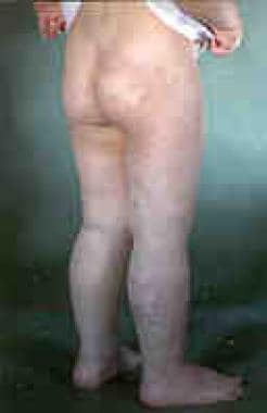 Klippel-Trenaunay syndrome in a young person. Note the port-wine stain extending to the buttocks. These lesions can be associated with venous malformations involving the rectum and bladder.
In 1900, noted French physicians Klippel and Trenaunay first described a syndrome in 2 patients presenting with a port-wine stain and varicosities of an extremity associated with hypertrophy of the affected limb's bony and soft tissue. They termed the syndrome "naevus vasculosus osteohypertrophicus." In 1907, Parkes Weber, unaware of Klippel and Trenaunay's report, described a patient with the 3 aforementioned symptoms as well as an arteriovenous malformation of the affected extremity. He termed the process hemangiectatic hypertrophy.
Today, conflicting opinion exists in the literature as whether to separately designate the original triad as Klippel-Trenaunay syndrome and the triad with the addition of arteriovenous malformation as Parkes Weber syndrome. Making the distinction is probably wise given the increased morbidity associated with arteriovenous malformations. For this discussion, the 2 types are considered together.
Prognosis
Hand or foot malformations in Klippel-Trenaunay-Weber syndrome (KTWS) may predict the presence of deep venous system anomalies. [1]
Signs and symptoms
Klippel-Trenaunay-Weber syndrome (KTWS) generally affects a single extremity, although cases of multiple affected limbs have been reported. The leg is the most common site followed by the arms, the trunk, and rarely the head and the neck. One report describes only upper limb involvement. [2]
Most patients demonstrate all 3 signs of the clinical syndrome: port-wine stain, varicose veins, and bony and soft tissue hypertrophies.
Also see Presentation.
Diagnostics
In many instances, a thorough history and physical examination are all that is required to diagnose Klippel-Trenaunay-Weber syndrome (KTWS). However, when complications are present, imaging studies can be useful. Color Doppler sonography is an accurate, reliable, and noninvasive way to evaluate patients with possible KTWS. [3]
Multidetector row computed tomography arteriography may be of value in the preoperative assessment of patients with KTWS. [4]
Evaluation of the deep venous system can be completed with duplex scanning contrast venography, ultrasonography, contrast venography and arteriography, and nuclear MRI studies. Arteriography is especially helpful in the diagnosis of an arteriovenous fistula.
MRI is also helpful in imaging the soft tissue hypertrophy. In addition, magnetic resonance angiography can be very helpful in identifying and defining vascular malformations.
In the case of major limb length discrepancies, serial radiographic studies, including but not limited to scanograms, orthoroentgenograms, and CT scans, for measurement of limb length are necessary. Clinical measurements can only guarantee measurement within 0.5-1.0 cm, whereas radiographic examinations can be used to determine the exact differences to within 0.1 cm. These studies help to determine how fast a limb is growing and may help in determining proper timing for limb length equalization procedures.
Prenatal diagnosis by ultrasonography has been reported.
In lesions extending onto the perineum or abdomen, performing imaging studies can be helpful to rule out internal involvement. Vascular malformations have been reported throughout the gastrointestinal tract. Although this typically does not cause symptoms, gastrointestinal bleeding has been reported. Similarly, vascular malformations have also been reported within the genitourinary tract.
Lymphoscintigraphy may be used to evaluate potential KTWS candidates for thoracic duct decompression if needed. [5]
See Treatment and Medication.
Pathophysiology
The exact cause of Klippel-Trenaunay-Weber syndrome (KTWS) remains to be elucidated, although several theories exist. Bliznak and Staple suggested intrauterine damage to the sympathetic ganglia or intermediolateral tract leading to dilated microscopic arteriovenous anastomoses as the cause. [6] Servelle believes that deep vein abnormalities, with resultant obstruction of venous flow, lead to venous hypertension, the development of varices, and limb hypertrophy. [7] Baskerville et al contend that a mesodermal defect during fetal development causes maintenance of microscopic arteriovenous communications. [8] Finally, McGrory and Amadio believe that an underlying mixed mesodermal and ectodermal dysplasia is likely responsible for the development of KTWS. [9]
Most cases KTWS are sporadic, although a few cases in the literature report an autosomal dominant pattern of inheritance. [10] A case report of KTWS in a monozygotic twin with an unaffected twin advances the theory of a paradominant inheritance pattern. [11] This theory suggests that KTWS is produced by a single gene defect lethal in individuals who are homozygous for this gene. Heterozygotes carry the gene but are unaffected. The disease manifests in individuals who demonstrate loss of heterozygosity from a somatic mutation during embryogenesis. In these individuals, only the skin region harboring this cell population demonstrates the KTWS mutation.
The association between the angiogenic factor gene AGGF1 and KTS appears to be significant. [12] Common AGGF1 variants may confer risk of KTWS.
Kihiczak et al report that KTWS may result from a pathogenic gene for vascular and tissue overgrowth. [13]
Epidemiology
No racial predilection is documented for Klippel-Trenaunay-Weber syndrome (KTWS).
KTWS affects females and males equally. [14] KTWS presents at birth or during early infancy or childhood.
-
Klippel-Trenaunay syndrome in a young person. Note the port-wine stain extending to the buttocks. These lesions can be associated with venous malformations involving the rectum and bladder.


