| Patient Education |
|
Click here for patient education.
|
|
You are in: eMedicine Specialties >
Sports Medicine > Knee
Jumper's Knee
Article Last Updated: Sep 6, 2006
AUTHOR AND EDITOR INFORMATION
Section 1 of 11
Author: Garrett Hyman, MD, MPH, Clinical Assistant Professor, University of Washington; Consulting Staff, Department of Rehabilitation Medicine, Northwest Spine & Sports Physicians, PC
Garrett Hyman is a member of the following medical societies: American Academy of Physical Medicine and Rehabilitation, American College of Sports Medicine, American Medical Association, and North American Spine Society
Coauthor(s):
Gerard A Malanga, MD, Associate Professor, Department of Physical Medicine and Rehabilitation, New Jersey Medical School; Director of Pain Management, University of Medicine and Dentistry at New Jersey, Overlook Hospital; Director of Sports Medicine, Mountainside Hospital;
Irfan Alladin, MD, Department of Physical Medicine and Rehabilitation, Staff Physician, University of Medicine and Dentistry at New Jersey
Editors: Leslie Milne, MD, Department of Emergency Medicine, Assistant Clinical Instructor, Harvard University School of Medicine; Francisco Talavera, PharmD, PhD, Senior Pharmacy Editor, eMedicine; Russell D White, MD, Professor of Medicine, Department of Community and Family Medicine, University of Missouri-Kansas City School of Medicine, Truman Medical Center Lakewood; Jon Whitehurst, MD, Consulting Staff, Rockford Orthopedic Associates; Wylie D Lowery, Jr, MD, Department of Orthopedic Surgery, Associate Professor, George Washington University
Author and Editor Disclosure
Synonyms and related keywords:
jumper's knee, patellar tendinitis, patellar tendinosis, patellar tendinopathy, patellar apicitis, quadriceps tendinitis, infrapatellar tendinopathy
Background
Although Sinding-Larson, Johansson, and Smillie once described this condition, Blazina et al first used the term jumper's knee in 1973 to describe an insertional tendinopathy seen in skeletally mature athletes. Jumper's knee usually affects the attachment of the patellar tendon to the inferior patellar pole. The definition was subsequently widened to include tendinopathy of the attachment of the quadriceps tendon to the superior patellar pole or tendinopathy of the attachment of the patellar tendon to the anterior tuberosity of the tibia. The term jumper's knee implies functional stress overload due to jumping.
Frequency
United States
Jumper's knee is certainly one of the more common tendinopathies affecting skeletally mature athletes, occurring in as many as 20% of jumping athletes. With regard to bilateral tendinopathy, males and females are equally affected. With regard to unilateral tendinopathy, the male-to-female ratio is 2:1.
Functional Anatomy
The rectus femoris and 3 vasti muscles (ie, the vastus medialis, vastus lateralis, and vastus intermedius muscles) join in a common quadriceps tendon that inserts on the patella, the largest sesamoid bone in the human body. This same tendon is known as the patellar tendon from the inferior pole of the patella to its distal insertion at the tibial tuberosity.
Radiologic and histologic studies have shown that the posterior proximal fibers of the patellar tendon appear to be most commonly affected in jumper's knee. However, biomechanical research has demonstrated that these posterior fibers can withstand greater tensile strains before failing, compared with anterior fibers.
Sport Specific Biomechanics
Jumper's knee is believed to be caused by repetitive stress placed on the patellar or quadriceps tendon during jumping. It is an injury specific to athletes, particularly those participating in jumping sports such as basketball, volleyball, or high or long jumping. Jumper's knee is occasionally found in soccer players, and in rare cases, it may be seen in athletes in nonjumping sports such as weight lifting and cycling. Investigators have implicated sex, greater body weight, genu varum and genu valgum, an increased Q angle, patella alta and patella baja, and limb-length inequality as intrinsic risk factors. However, the only biomechanical impairment prospectively linked to jumper's knee is poor quadriceps and hamstring flexibility. Vertical jump ability, as well as jumping and landing technique, are believed to influence tendon loading. Overtraining and playing on hard surfaces have been implicated as extrinsic risk factors. Interestingly, the patellar tendon experiences greater mechanical load during landing than during jumping, because of the eccentric muscle contraction of the quadriceps. Therefore, eccentric muscle action during landing, rather than concentric muscle contraction during jumping, may exert the tensile loads that lead to injury.
History
Jumper's knee commonly occurs in athletes involved in jumping sports such as basketball and volleyball. Patients report anterior knee pain, often with an aching quality. Symptom onset is insidious. Rarely is a discrete injury described. Usually, involvement is infrapatellar at or near the infrapatellar pole, but it may also be suprapatellar.
Depending on the duration of symptoms, jumper's knee can be classified into 1 of 4 stages:
- Stage 1 - Pain only after activity, without functional impairment
-
- Stage 2 - Pain during and after activity, although the patient is still able to perform satisfactorily in his or her sport
-
- Stage 3 - Prolonged pain during and after activity, with increasing difficulty in performing at a satisfactory level
-
- Stage 4 - Complete tendon tear requiring surgical repair
Physical
Physical examination may reveal the following findings:
- Point tenderness at the inferior patellar pole, superior patellar pole, or tibial tuberosity
-
- Hamstring and quadriceps tightness
-
- Normal ligamentous stability of the knee during testing
-
- Normal knee range of motion
-
- Normal neurovascular examination
-
- Normal hip and ankle examination
-
- Intra-articular effusion of the knee (rare)
Causes
The cause of jumper's knee remains unclear.
Histologic specimens are devoid of inflammatory cells; therefore, the disease process probably rarely involves a true tendinitis.
Biomechanical research has shown that a greater tensile load is borne by the anterior fibers. This observation contradicts the theory that jumper's knee is caused by repetitive tensile loading given that the proximal posterior patellar tendon is routinely affected.
The degenerative "tendinosis" rather than inflammatory "tendinitis" model has prevailed since the 1970s. Hamilton and Purdam have proposed an adaptive model, whereby compressive rather than tensile loads occur at the proximal posterior aspect of the tendon, resulting in structural changes from altered biomechanical forces. This model is said to account for the presence of the routinely found asymptomatic lesions, and perhaps the absence of inflammatory cells in histologic specimens.
In 2002, Almekinders et al suggested that stress shielding by the anterior fibers may lead to degenerative change or tendon wear of the posterior ones due to compressive forces. They also proposed that such stress shielding and/or compressive forces, rather than repetitive tensile loads, may be more important etiologic factors in insertional tendinopathy.
Knee Osteochondritis Dissecans
Meniscus Injuries
Osgood-Schlatter Disease
Patellar Injury and Dislocation
Patellofemoral Joint Syndromes
Pes Anserine Bursitis
Quadriceps Injury
Other Problems to be Considered
Infrapatellar bursitis Pes anserine bursitis Tibial plateau fracture infrapatellar fat pad syndrome Sinding-Larsen-Johansson syndrome Tumor Knee-joint infection
Lab Studies
- The diagnosis of jumper's knee is based on the history and clinical findings. Laboratory and imaging tests are rarely necessary.
-
- Laboratory studies are not indicated unless other potential causes, such as systemic, inflammatory, or metabolic disease, must be ruled out.
Imaging Studies
- Radiographic imaging is not necessary to make the diagnosis. Such imaging may be helpful for excluding other potential maladies.
- Certain radiography findings have been reported to support the diagnosis. For instance, plain images may show a radiolucency at or elongation of the involved pole.
- Ultrasonography and MRI are both highly sensitive for detecting tendon abnormalities in both symptomatic and asymptomatic athletes. Therefore, a significant number of false-positive results makes routine testing with these techniques impractical.
- Ultrasonography combined with color or power Doppler may demonstrate peritendinous neovascularization. Some investigators advocate sclerosing injections targeting this neovascularization as a treatment for jumper's knee.
Other Tests
Procedures
Acute Phase
Rehabilitation Program
Physical Therapy
Most patients respond to a conservative management program such as the one suggested below.
- Activity modification: Decrease activities that increase patellofemoral pressure (eg, jumping, squatting). Possibly initiate gentle eccentric loading activities.
- Cryotherapy: Apply ice for 20-30 minutes, 4-6 times per day, especially after activity.
- Joint motion and kinematics assessment: Evaluate hip, knee, and ankle joint range of motion.
- Stretching: Stretch (1) flexors of the hip and knee (hamstrings, gastrocnemius, iliopsoas, rectus femoris, adductors), (2) extensors of the hip and knee (quadriceps, gluteals), (3) the iliotibial band, and (4) the patellar retinaculum.
- Strengthening: Strengthen using closed kinetic chain and eccentric exercise (ie, single-leg squat descents). Jonsson and Alfredson found eccentric quadriceps strengthening on a decline board superior to concentric strengthening in terms of pain, treatment satisfaction, and return to play (Jonsson, 2005). Kongsgaard et al demonstrated that eccentric squats on a decline board of 25 degrees increases patellar tendon loading versus standard eccentric squats on a level surface (Kongsgaard, 2006).
- Sport-specific proprioceptive training and plyometrics
Ultrasound or phonophoresis may decrease pain symptoms. A patellofemoral brace with a patellar cutout and lateral stabilizer or McConnell taping may improve patellar tracking and provide stability through augmentation of proprioception. Provide arch supports or orthotics to correct foot malalignments. Stage I The treatment of jumper's knee is often specific to the degree of involvement. Stage I, which is characterized by pain only after activity and no undue functional impairment, is often treated with cryotherapy. The patient should use ice packs or ice massage after terminating the activity that exacerbates the pain and later again that evening. If aching persists, a course of regularly prescribed anti-inflammatory medications should be administered for 10-14 days. Often, cryotherapy and anti-inflammatories suffice in the first stage, though some cases relapse once the course of anti-inflammatories is completed. Long-term use of anti-inflammatories should be avoided in the young athlete. Local corticosteroid injections are not advised in stage I treatment. In addition, most competitive athletes do not agree to rest and immobilization, given that the condition does not affect their performance at this point. For this reason, use of a counterforce strap has been suggested to relieve some of the tension on the affected area and thereby ameliorate the symptoms. A comprehensive physical therapy program should include aggressive quadriceps and hip flexor stretching along with progressive strengthening. Begin with closed kinetic strengthening exercises, such as lunges, leg presses, and squats. Focus is given to the eccentric phase with each activity. Strict attention to proper technique is important to reduce stress on the patellofemoral joint and patellar tendon and to allow for progressive strengthening. The strengthening program should progress to proprioceptive and plyometric exercises. Plyometrics include activities, such as jump roping, within the patient's pain tolerance. A good warm-up activity prior to stretching and strengthening is important to increase blood flow and tissue compliance. An exercise bicycle can be used to perform aerobic warm-up exercises. If significant anterior knee pain occurs, the seat should be adjusted slightly higher to eliminate excessive and repetitive knee flexion loading. Stage II In stage II, the patient has pain both during and after activity but is still able to participate in the sport satisfactorily. The pain may interfere with sleep. At this point, activities that cause increased loading of the patellar tendon (eg, running, jumping) should be avoided. A comprehensive physical therapy program, as discussed above, should be implemented. For pain relief, the knee should be protected by avoiding high loads to the patellar tendon, and cryotherapy should continue. The athlete should be instructed in alternative conditioning. Once the pain improves, therapy should focus on knee, ankle, and hip joint range of motion, flexibility, and strengthening. Eccentric strengthening (perhaps on a decline board) should be emphasized because eccentric contractions allow for greater generation of force and simulate landing in many sports. Advanced therapy should include plyometrics and sport-specific training as tolerated. If the pain becomes increasingly intense and if the athlete becomes more concerned about his or her performance, a local corticosteroid injection may be considered. The physician should explain that the steroid could cause further tendon degeneration, and perhaps even rupture, if the athlete begins loading the tendon too quickly once the symptoms improve. Stage III In stage III, the patient's pain is sustained, and performance and sport participation are adversely affected. Though discomfort increases, therapeutic measures similar to those described above should be continued, along with abstinence from the inciting activity. Relative rest for an extended period (eg, 3-6 wk) may be necessary in stage III. The athlete should avoid only those activities that incite the pain to prevent deconditioning and should be encouraged to continue an aggressive alternative cardiovascular and strength-training program. If the pain is refractory to these measures, options are limited. One can either abandon participation in jumping sports and/or consider surgery. In a randomized controlled trial, Bahr et al demonstrated no difference in outcome between surgical and nonsurgical treatment for patients with grade III tendinopathy. The authors advocated 12 weeks of eccentric quadriceps strengthening before considering tenotomy. Only approximately half of athletes in both surgical and nonsurgical treatment groups were well enough to return to sport within 1 year (Bahr, 2006).
Stage IV Tendon rupture requires surgical repair.
Medical Issues/Complications
Knee immobilization is contraindicated because it results in stiffness and may lead to muscle or joint contracture, further prolonging an athlete's return to play.
Surgical Intervention
Three principal surgical procedures include drilling of the involved pole, tendon excision, and resection of the involved pole. The goal of drilling is to increase the vascular supply to the affected area. This should allow for healing of the affected tendon and improve pain and tenderness. However, this procedure has yielded disappointing results. The second procedure involves longitudinal excision of the involved tendon with subsequent restructuring of the residual tendon. Excision need not be limited to the tendon, as the inferior pole of the patella may be removed as well. The advantage of this procedure is that it does not disrupt the continuity of the quadriceps mechanism and allows for relatively rapid return to play. On the other hand, visualization of the area is rather poor, and the clinician may overlook degradation of the patellar or peripatellar area or at the intercondylar portion of the femur, or they may even overlook degenerative changes in the tendon itself. Results have been mixed; some patients improve while others have been unable to return to their prior level of performance despite prolonged rest. The third procedure calls for resection of the involved pole of the patella after inspection of the undersurface of the patella, the corresponding patellar face of the femur, and the involved tendon. Reattachment of the involved tendon and reinforcement of the medial patellar retinaculum are also parts of this procedure. As reported by Blazina et al in 1973, the principal benefit is that this resection allows complete assessment of the extensor mechanism and provides the most suitable bed for favorable reattachment of the involved tendon into a raw bony surface. In addition, a tendon with degradation in multiple locations is best treated with such a procedure, as it enhances visualization of the affected area. Although this is the most involved procedure, it is effective in select patients. Complete overhaul of the entire extensor mechanism, however, is not required in many cases.
Open tenotomy was compared with an eccentric strengthening regime in a randomized, controlled trial (Bahr, 2006). The authors found both surgery and the eccentric strengthening provided equivalent benefit in terms of pain reduction and functional improvement and return to sport at 1 year.
Consultations
Consultation with a physical medicine and rehabilitation specialist or an orthopedic specialist is recommended.
Other Treatment
- Ultrasonographic-guided sclerosing injections into the neovascularized patellar tendon have shown promise in reducing pain and improving function in elite Norwegian basketball, volleyball, and handball players (Hoksrud, 2006).
- Peritendinous corticosteroid/anesthetic injection:
- Consider this injection for patients with tendonitis in whom conservative therapy has failed.
- The usual dose is triamcinolone (Kenalog, Aristospan) at 40 mg/1 mL. Inject 0.5-1 mL with or without 0.5-1 mL of a local anesthetic agent.
- Use aseptic technique; prepare the area with alcohol or povidone-iodine solution (Betadine).
- Direct the injection into the most symptomatic peritendinous area (usually at or near the infrapatellar pole).
- Fredberg and colleagues demonstrated efficacy of ultrasound-guided peritendinous steroid injections.
- Never inject into the patellar tendon directly because of the potential for patellar tendon rupture after the injection of corticosteroid.
- Avoid repetitive corticosteroid injections in any site, as well as injection directly into a tendon, because of the risk of tendon rupture.
- Following the injection, the patient should ice the injection site 3 times per day, 20 minutes at a time, for 48 hours after the injection.
- The patient should avoid any running or jumping activities for 10-14 days after the injection.
- Extracorporeal shock wave therapy (ESWT) provides a comparable functional outcome compared with surgery, according to Peers and colleagues. To the author's knowledge, ESWT has not been studied against placebo for patellar tendinopathy; thus, its efficacy for this condition remains in question.
Recovery Phase
Rehabilitation Program
Physical Therapy
An in-depth, stage-specific description of a conservative therapy program is described above (see Acute Phase). In brief, in the recovery phase, the athlete and therapist should work to restore pain-free joint range of motion and muscle flexibility, symmetric strength in the lower extremities, and joint proprioception. Sport-specific training, including high-level plyometric exercises, should then be initiated.
Surgical Intervention
Surgical intervention is indicated for stage IV, and refractory stage III tendinopathy. See Acute Phase above.
Maintenance Phase
Rehabilitation Program
Physical Therapy
An in-depth, stage-specific description of a conservative therapy program is described above (see Acute Phase). Briefly, once in the maintenance phase, the athlete should complete a sport-specific training program before returning to competition. The physician and physical therapist can assist the athlete in determining when to return to competition based on the patient's symptoms, current physical examination findings, and functional test results. Once the athlete returns to play, he or she must work to maintain gains in flexibility and strength.
Surgical Intervention
Surgical intervention is indicated for stage IV disease. See Acute Phase above.
Nonsteroidal anti-inflammatory drugs may be used judiciously in the acute phase in conjunction with non-pharmacologic modalities for pain relief and anti-inflammatory effects.
Drug Category: Nonsteroidal anti-inflammatory drugs
These agents have analgesic, anti-inflammatory, and antipyretic activities. Their mechanism of action is not known, but NSAIDs may inhibit cyclo-oxygenase activity and prostaglandin synthesis. Other mechanisms may exist as well, such as inhibition of leukotriene synthesis, lysosomal enzyme release, lipoxygenase activity, neutrophil aggregation and various cell-membrane functions.
| Drug Name | Naproxen (Aleve, Anaprox, Naprelan, Naprosyn) |
| Description | For relief of mild to moderate pain; inhibits inflammatory reactions and pain by decreasing activity of cyclo-oxygenase, which results in decrease of prostaglandin synthesis. |
| Adult Dose | 500 mg PO bid for 5-7 d; not to exceed 1.5 g/d |
| Pediatric Dose | <2 y: Not established >2 y: 2.5 mg/kg/dose PO; not to exceed 10 mg/kg/d |
| Contraindications | Documented hypersensitivity; peptic ulcer disease; recent GI bleeding or perforation; renal insufficiency |
| Interactions | Coadministration with aspirin increases risk of inducing serious NSAID-related adverse effects; probenecid may increase concentrations and, possibly, toxicity of NSAIDs; may decrease effect of hydralazine, captopril, and beta-blockers; may decrease diuretic effects of furosemide and thiazides; may increase PT when patient is taking anticoagulants (instruct patients to watch for signs of bleeding); may increase risk of methotrexate toxicity; phenytoin levels may be increased when administered concurrently |
| Pregnancy | B - Usually safe but benefits must outweigh the risks.
|
| Precautions | Category D in third trimester of pregnancy; acute renal insufficiency, interstitial nephritis, hyperkalemia, hyponatremia, and renal papillary necrosis may occur; patients with preexisting renal disease or compromised renal perfusion risk acute renal failure; leukopenia occurs rarely, is transient, and usually returns to normal during therapy; persistent leukopenia, granulocytopenia, or thrombocytopenia warrants further evaluation and may require discontinuation of drug |
| Drug Name | Ibuprofen (Motrin, Advil, Excedrin IB, Ibuprin) |
| Description | DOC for patients with mild to moderate pain. Inhibits inflammatory reactions and pain by decreasing prostaglandin synthesis. |
| Adult Dose | 400-600 mg PO q8h for 5-7 d; not to exceed 3.2 g/d |
| Pediatric Dose | <6 mo: Not established 6 mo to 12 y: 4-10 mg/kg/dose PO tid/qid >12 y: Administer as in adults |
| Contraindications | Documented hypersensitivity; peptic ulcer disease, recent GI bleeding or perforation, renal insufficiency, or high risk of bleeding |
| Interactions | Coadministration with aspirin increases risk of inducing serious NSAID-related adverse effects; probenecid may increase concentrations and, possibly, toxicity of NSAIDs; may decrease effect of hydralazine, captopril, and beta-blockers; may decrease diuretic effects of furosemide and thiazides; may increase PT when patient is taking anticoagulants (instruct patients to watch for signs of bleeding); may increase risk of methotrexate toxicity; phenytoin levels may be increased when administered concurrently |
| Pregnancy | B - Usually safe but benefits must outweigh the risks.
|
| Precautions | Category D in third trimester of pregnancy; caution in congestive heart failure, hypertension, and decreased renal and hepatic function; caution in coagulation abnormalities or during anticoagulant therapy |
Return to Play
Return to play should be based on an athlete's ability to safely and skillfully perform sport-specific activities. When symptoms persist despite exhaustive conservative or even surgical treatment, the athlete must weigh the benefits and the consequences of playing in pain.
Functional testing at the end of the recovery phase of rehabilitation, administered by a physical therapist, athletic trainer, or physician, is helpful in determining the athlete's readiness to return to his or her sport.
Complications
The most common complication is persistent pain during jumping that may result in activity avoidance.
Prevention
Sport-specific training and physical fitness prior to competition may help prevent jumper's knee. To the author's knowledge, no research validates any particular preventive training regimen more so than any other.
Prognosis
The prognosis for jumper's knee stage I or II is typically excellent with conservative treatment. Stage III carries a guarded prognosis for a full-recovery, while those few with stage IV injury (complete tendon rupture) require surgical repair of the tendon and are least likely to return to competitive play.
Education
Jumper's knee affects jumping athletes. It is nearly always amenable to conservative treatment with a comprehensive rehabilitation program. The persistence of pain during and after play guides the staging and treatment of this disorder. Judicious use of relative rest, reducing pain and inflammation, and alternative conditioning methods help to expedite an athlete's return to competition.
Medical/Legal Pitfalls
- The only medicolegal pitfall is a failure to diagnose another condition, such as a joint infection or tumor, that can cause significant morbidity.
Special Concerns
- Competitive or elite athletes may choose to play in severe pain rather than seek medical attention until the disease has progressed to later stages and their level of play is affected.
| Media file 1:
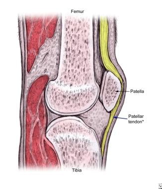
The proximal patellar tendon is most commonly affected in jumper's knee. |
 | View Full Size Image | |
Media type: Image
|
- Al-Duri ZA, Aichroth PM. Surgical aspects of patellar tendonitis: technique and results. Am J Knee Surg. 2001;14(1):43-50. [Medline].
- Alfredson H, Ohberg L. Neovascularisation in chronic painful patellar tendinosis--promising results after sclerosing neovessels outside the tendon challenge the need for surgery. Knee Surg Sports Traumatol Arthrosc. Mar 2005;13(2):74-80. [Medline].
- Almekinders LC, Vellema JH, Weinhold PS. Strain patterns in the patellar tendon and the implications for patellar tendinopathy. Knee Surg Sports Traumatol Arthrosc. Jan 2002;10(1):2-5. [Medline].
- Bahr R, Fossan B, Loken S, Engebretsen L. Surgical Treatment Compared with Eccentric Training for Patellar Tendinopathy (Jumper's Knee) A Randomized, Controlled Trial. J Bone Joint Surg Am. Aug 2006;88(8):1689-98. [Medline].
- Basso O, Amis AA, Race A, Johnson DP. Patellar tendon fiber strains: their differential responses to quadriceps tension. Clin Orthop Relat Res. Jul 2002;(400):246-53. [Medline].
- Blazina ME, Kerlan RK, Jobe FW, et al. Jumper's knee. Orthop Clin North Am. Jul 1973;4(3):665-78. [Medline].
- Busch MT. Sports medicine in children and adolescents. In: Lovell and Winter's Pediatric Orthopaedics. Philadelphia, Pa: Lippincott-Raven; 1990.
- Cook JL, Khan KM, Harcourt PR, et al. A cross sectional study of 100 athletes with jumper's knee managed conservatively and surgically. The Victorian Institute of Sport Tendon Study Group. Br J Sports Med. Dec 1997;31(4):332-6. [Medline].
- Cook JL, Khan KM, Kiss ZS, et al. Prospective imaging study of asymptomatic patellar tendinopathy in elite junior basketball players. J Ultrasound Med. Jul 2000;19(7):473-9. [Medline].
- Cook JL, Khan KM, Kiss ZS, et al. Reproducibility and clinical utility of tendon palpation to detect patellar tendinopathy in young basketball players. Victorian Institute of Sport tendon study group. Br J Sports Med. Feb 2001;35(1):65-9. [Medline].
- Cook JL, Kiss ZS, Khan KM, et al. Anthropometry, physical performance, and ultrasound patellar tendon abnormality in elite junior basketball players: a cross-sectional study. Br J Sports Med. Apr 2004;38(2):206-9. [Medline].
- Ferretti A. Epidemiology of jumper's knee. Sports Med. Jul-Aug 1986;3(4):289-95. [Medline].
- Ferretti A, Ippolito E, Mariani P, Puddu G. Jumper's knee. Am J Sports Med. Mar-Apr 1983;11(2):58-62. [Medline].
- Gisslén K, Alfredson H. Neovascularisation and pain in jumper's knee: a prospective clinical and sonographic study in elite junior volleyball players. Br J Sports Med. Jul 2005;39(7):423-8; discussion 423-8. [Medline].
- Hamilton B, Purdam C. Patellar tendinosis as an adaptive process: a new hypothesis. Br J Sports Med. Dec 2004;38(6):758-61. [Medline].
- Hoksrud A, Ohberg L, Alfredson H, Bahr R. Ultrasound-Guided Sclerosis of Neovessels in Painful Chronic Patellar Tendinopathy: A Randomized Controlled Trial. Am J Sports Med. Jul 10 2006;[Medline].
- Johnson DP, Wakeley CJ, Watt I. Magnetic resonance imaging of patellar tendonitis. J Bone Joint Surg Br. May 1996;78(3):452-7. [Medline].
- Jonsson P, Alfredson H. Superior results with eccentric compared to concentric quadriceps training in patients with jumper's knee: a prospective randomised study. Br J Sports Med. Nov 2005;39(11):847-50. [Medline].
- Khan KM, Bonar F, Desmond PM, et al. Patellar tendinosis (jumper's knee): findings at histopathologic examination, US, and MR imaging. Victorian Institute of Sport Tendon Study Group. Radiology. Sep 1996;200(3):821-7. [Medline].
- Kongsgaard M, Aagaard P, Roikjaer S, Olsen D, Jensen M, Langberg H. Decline eccentric squats increases patellar tendon loading compared to standard eccentric squats. Clin Biomech (Bristol, Avon). Aug 2006;21(7):748-54. [Medline].
- Kujala UM, Osterman K, Kvist M, et al. Factors predisposing to patellar chondropathy and patellar apicitis in athletes. Int Orthop. 1986;10(3):195-200. [Medline].
- Lian O, Refsnes PE, Engebretsen L, Bahr R. Performance characteristics of volleyball players with patellar tendinopathy. Am J Sports Med. May-Jun 2003;31(3):408-13. [Medline].
- Matheson GO. Long-term prognosis for jumper's knee. Clin J Sport Med. May 2003;13(3):196. [Medline].
- Medlar RC, Lyne ED. Sinding-Larsen-Johansson disease. Its etiology and natural history. J Bone Joint Surg Am. Dec 1978;60(8):1113-6. [Medline].
- Ogden JA, Southwick WO. Osgood-Schlatter's disease and tibial tuberosity development. Clin Orthop Relat Res. May 1976;(116):180-9. [Medline].
- Peers KH, Lysens RJ, Brys P, Bellemans J. Cross-sectional outcome analysis of athletes with chronic patellar tendinopathy treated surgically and by extracorporeal shock wave therapy. Clin J Sport Med. Mar 2003;13(2):79-83. [Medline].
- Purdam CR, Jonsson P, Alfredson H, et al. A pilot study of the eccentric decline squat in the management of painful chronic patellar tendinopathy. Br J Sports Med. Aug 2004;38(4):395-7. [Medline].
- Roels J, Martens M, Mulier JC, Burssens A. Patellar tendinitis (jumper's knee). Am J Sports Med. Nov-Dec 1978;6(6):362-8. [Medline].
- Witvrouw E, Bellemans J, Lysens R, et al. Intrinsic risk factors for the development of patellar tendinitis in an athletic population. A two-year prospective study. Am J Sports Med. Mar-Apr 2001;29(2):190-5. [Medline].
- Lian O, Refsnes PE, Engebretsen L, Bahr R. Performance characteristics of volleyball players with patellar tendinopathy. Am J Sports Med. May-Jun 2003;31(3):408-13. [Medline].
Jumper's Knee excerpt Article Last Updated: Sep 6, 2006
|