Practice Essentials
Although sports injuries to the knee, ankle, and shoulder have been well documented, injuries to the pelvis, hip, and thigh get little attention because of their low prevalence. Unfortunately, severe consequences may result if these injuries are improperly managed. [1, 2, 3, 4]
Femoral neck stress fractures were mainly seen in military recruits due to a triad of activity that is new, strenuous, and highly repetitive. However, as a result of self-imposed fitness regimens of recreational athletes, over the last 20 years the number of these injuries has been increasing in nonmilitary populations. Stress fractures of the hip and pelvic region have been increasingly reported in long-distance runners. [5] Factors such as muscle fatigue (which leads to abnormal gait patterns and altered stress distribution), training errors, improper footwear, and poor training surfaces can predispose an athlete to the development of stress fractures.
In contrast, contact sports such as football, rugby, and soccer are usually the cause of most fractures of the hip. Stress fractures occur in normal bone undergoing repeated submaximal stress. As the bone attempts to remodel, osteoclastic activity occurs at a greater rate than osteoblastic activity. When these cumulative forces exceed the structural strength of bone, stress fractures occur. [6, 7, 8]
Stress fractures occur mainly at the femoral neck and are classified as either tension (at the superior aspect of the femoral neck) or compression (at the inferior aspect of the femoral neck). See the images below.
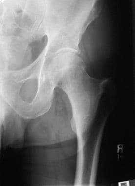 A subcapital femoral neck fracture. Slight compression of the femoral head onto the femoral neck can be seen. Note the cortical break medially. This fracture could be missed if not closely evaluated.
Hip fractures are classified as intracapsular, which includes femoral head and neck fractures, or extracapsular, which includes trochanteric, intertrochanteric, and subtrochanteric fractures. The location of the fracture and the amount of angulation and comminution play integral roles in the overall morbidity of the patient, as does the preexisting physical condition of the individual. Fractures of the proximal femur are extremely rare in young athletes and are usually caused by high-energy motor vehicle accidents or significant trauma during athletic activity. Other causes may be an underlying disease process such as Gaucher disease, fibrous dysplasia, or bone cysts.
Identification and initiation of treatment is imperative in attempts to avoid complications, such as avascular necrosis (AVN). AVN is more common in patients in the pediatric and adolescent age groups. This outcome is due to the precarious nature of the blood supply to the subchondral region of the femoral head, which does not stabilize until years after skeletal maturity, after which collateral flow develops.
Epidemiology
United States statistics
An estimated 340,000 hip fractures occur each year. Estimates indicate that in 2040, approximately 500,000 hip fractures will occur.
Nine of 10 hip fractures occur in patients aged 65 years and older, and 3 of 4 occur in women.
White females have been reported to be twice as likely to fracture their hips than Black and Hispanic females. This frequency has been associated with a metropolitan setting, increased caffeine use, alcohol use, sedentary lifestyle, psychotropic drug use, and dementia.
The rate of fractures is low in adolescent and young athletic populations, estimated to be less than 2% of all hip fractures (one hundredth of adult hip fractures).
Functional Anatomy
The hip is a ball-and-socket joint composed of the acetabulum and the head of the femur. The femoral head is connected to the shaft by the femoral neck. These are supported by a network of trabecular bone.
Two other important landmarks on the proximal femur are the greater and lesser trochanters. These 2 structures are the main muscle attachment sites for the proximal bone. The iliopsoas muscle is connected to the lesser trochanter, and the abductors and short rotator muscles act through their insertion on the greater trochanter. In addition, many additional muscles attach along the intertrochanteric line, and, along with the muscles, they bring with them an abundant and redundant blood supply, which is conducive to healing. This is in contrast to the intercapsular femoral neck, which is prone to healing complications.
The blood supply to the femoral head has been studied extensively and has been found to change substantially during development. Until the cartilaginous growth plate forms a barrier at age 4 years, the major blood supply comes from the medial and lateral circumflex arteries (metaphyseal arteries), which arise from the deep femoral artery. After age 4 years, the posterosuperior and posteroinferior arterial branches of the medial femoral circumflex bypass the growth plate and form the main blood supply to the femoral head. During adolescence, the growth plate fuses and the metaphyseal vessels again become significant, traveling along the femoral neck. Fractures in this area can disrupt this delicate blood supply, leading to AVN, the most severe complication of this fracture.
Sport-Specific Biomechanics
The ball-and-socket joint provides most of the inherent stability of the hip joint, while allowing for a large range of motion. Additional stability is provided by the thick capsule and strong ligamentous structures that actually enforce the capsule, namely the iliofemoral, pubofemoral, and ischiofemoral ligaments. These ligaments are taut with internal rotation, which limits motion, and become lax with external rotation.
Motion about the hip occurs in the sagittal, frontal, and transverse planes. During normal gait, motion occurs in all 3 planes, and normal activities occur within the range of 120 º flexion, 20 º extension, 40 º abduction, 25 º adduction, and 45 º external and internal rotation.
The biomechanics of the neck-shaft angle, which averages 135 º and 10-15 º of anteversion, allows for a unique arrangement. This permits angular movements of the thigh to be converted to rotatory hip motion.
Prognosis
The prognosis for hip fractures is dependent on the age and condition of the patient and on the location and type of fracture. Athletes who sustain femoral neck stress fractures may or may not be able to return to their sport. Tension stress fractures are generally unstable and have an unfavorable prognosis. On the other hand, compression fractures are usually successfully treated with conservative measures and have a good prognosis for recovery. Hip fractures in elderly individuals have a mortality rate of 14-36% one year following surgery.
Complications
Complications related to poorly treated or misdiagnosed stress fractures are considerable. Avascular necrosis, nonunion, varus deformity, chronic pain, and completely displaced femoral neck fractures may occur and may lead to serious life-altering changes in function and the patient's ability to ambulate efficiently. [1]
Patient Education
Patient education is a very important aspect to the rehabilitation process following hip fracture, regardless of the patient's age. Patients must be thoroughly informed about treatment options following their diagnosis, and they must understand the benefits and risks of treatment. If conservative treatment is an option, the patient may need instruction in the use of crutches initially to restrict weight bearing. A physical therapist should be involved in the patient's care for instructions in mobility training and reconditioning of the affected lower extremity. Patients are usually instructed in a home exercise program for continuing strengthening of the hip so that they are able to return to their previous level of activity.
-
A subcapital femoral neck fracture. Slight compression of the femoral head onto the femoral neck can be seen. Note the cortical break medially. This fracture could be missed if not closely evaluated.
-
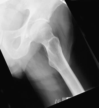
A view of the contralateral hip for comparison.
-
Intraoperative x-ray film (fluoroscopic view) of placement of the lag screw.
-
Addition of a superior derotational screw to maintain alignment and allow compression.
-
Internal fixation of the subcapital femoral neck fracture with a screw and short side plate with an additional derotational screw above. Final anteroposterior view.
-
Garden I femoral neck fracture. Note the valgus impaction with compression of the superior femoral head-neck junction.
-
Lateral view of a Garden I femoral neck fracture. Compression of the head-neck junction inferiorly.
-
Anteroposterior view of the pelvis with a displaced femoral neck fracture.
-
Lateral view of a displaced femoral neck fracture.
-
Displaced femoral neck fracture treated with a conventional, noncemented monopolar hemiarthroplasty.
-
Lateral view of a unipolar hemiarthroplasty.
-
An example of a calcar replacement hemiarthroplasty. A low femoral neck fracture extending into the calcar femoralis, not amenable to internal fixation or conventional hemiarthroplasty, requiring a calcar replacement prosthesis.
-
A lateral x-ray film of a calcar replacement hemiarthroplasty.


