Continually Updated Clinical Reference
 
 
  All Sources     eMedicine     Medscape     Drug Reference     MEDLINE
 
eMedicine - Hamate Fracture : Article by

Quick Find
Authors & Editors
Introduction
Clinical
Differentials
Workup
Treatment
Medication
Follow-up
Multimedia
References


Patient Education
Hand, Wrist, Elbow, and Shoulder Center

Breaks, Fractures, and Dislocations Center

Sports Injury Center

Wrist Injury Overview

Wrist Injury Causes

Wrist Injury Symptoms

Wrist Injury Treatment

Broken Hand Overview


Author: Amy Powell, MD, Assistant Clinical Professor, Department of Orthopedics, University of Utah

Amy Powell is a member of the following medical societies: American College of Physicians, American College of Sports Medicine, and American Medical Society for Sports Medicine

Coauthor(s): Emily Harold, MD, Staff Physician, Department of Internal Medicine, University of Utah Hospital; Janos P Ertl, MD, Clinical Assistant Professor, Department of Orthopedic Surgery, University of California at Davis; Director of Amputee Clinic, Chief of Orthopedic Trauma, Kaiser Hospital; Warren S Theis, MD, Staff Physician, Department of General Surgery, Carilion Roanoke Memorial Hospital; Nancy J Taubenheim, DPT, Staff Physical Therapist, Clinical Instructor, Department of Rehabilitation Services, Bryan LGH Medical Center

Editors: Gerard A Malanga, MD, Founder and Director, New Jersey Sports Medicine Institute; Director of Pain Management, Overlook Hospital; Director of Sports Medicine, Sports Medicine Fellowship Director, Mountainside Hospital; Clinical Chief, Rehabilitation Medicine and Electrodiagnosis, St Michael's Medical Center; Medical Director, Consultant, Horizon Healthcare Worker's Compensation Services, Blue Cross and Blue Shield Worker's Compensation; Francisco Talavera, PharmD, PhD, Senior Pharmacy Editor, eMedicine; Henry T Goitz, MD, Chief, Sports Medicine, Associate Professor, Department of Orthopaedic Surgery, Medical College of Ohio; Jon B Whitehurst, MD, Clinical Instructor of Surgery, University of Illinois College of Medicine; Partner and Executive Board Member, Rockford Orthopedic Associates; Orthopedic Chairman, Rockford Memorial Hospital; Sherwin SW Ho, MD, Associate Professor, Department of Surgery, Section of Orthopedic Surgery and Rehabilitation Medicine, University of Chicago

Author and Editor Disclosure

Synonyms and related keywords: fracture of the hook, hook fracture, fracture of the hamate, hook of hamate fracture, fracture of the hook of hamate, wrist fracture, wrist trauma, hamate trauma, broken wrist, wrist injury

Background

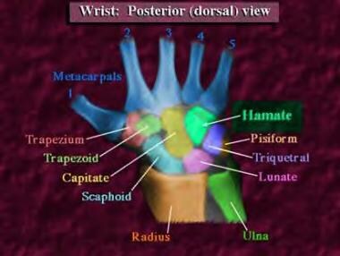
Although hamate fractures are increasing in incidence secondary to the popularity of sports activities involving racquets, bats, and clubs, these injuries remain relatively rare. Estimates suggest hamate fractures constitute 2% of all carpal fractures. The hamate bone is a roughly triangular-shaped bone composed of both a body and a hook (see Images 1-2). Hamate fractures are thus classified as type I fractures involving the hook and type II fractures involving the body. Type I fractures are more common than type II fractures.1, 2

For excellent patient education resources, visit eMedicine's Hand, Wrist, Elbow, and Shoulder Center, Breaks, Fractures, and Dislocations Center, and Sports Injury Center. Also, see eMedicine's patient education articles Wrist Injury and Broken Hand.

Related eMedicine topics:
Carpal Fractures
Fracture, Wrist
Hand, Fractures and Dislocations: Wrist
Metacarpal Fractures

Related Medscape topics:
Resource Center Exercise and Sports Medicine
CME Physical Exercise May Help Reduce Fatigue During and After Cancer Treatment
CME Regular Exercise Through Middle Age May Delay Biological Aging
CME/CE Risks and Benefits of Exercise Reviewed in AHA Statement

Frequency

United States

Hamate fractures account for 2% of all carpal fractures. Of the 2%, one third are hamate hook fractures due to repetitive swinging by golfers.

Functional Anatomy

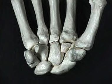
The hamate is a triangular bone located in the distal carpal row farthest to the ulnar side (see Images 1-2). The hamate is bordered proximally by the pisiform and the lunate in the proximal carpal row, radially by the capitate, and distally by the bases of the fourth and fifth metacarpals.

A roughly circular projection or hook on the volar surface of the hamate is the inferolateral border of the Guyon canal. The roof (superficial) of the canal is formed by the palmar carpal ligament, and the floor (deep) is formed by the flexor retinaculum. The canal carries the ulnar artery and nerve, and, for this reason, hook fractures should suggest a high probability of ulnar artery and nerve damage.3 In addition, the hamate hook has a dual blood supply, with vessels entering from both the ulnar tip and radial base. These vessels often have a poor anastomosis, which clinically can result in nonunion due to insufficient blood supply.

Sport-Specific Biomechanics

Type I fractures involving the hook of the hamate are the most common and can occur via several different mechanisms.1, 2, 4, 5, 6, 7 First, repeated microtrauma to the hook during sports involving swinging clubs, bats, or racquets can result in a hook stress fracture. These usually occur in the nondominant hand and account for approximately one third of hamate fractures. Second, direct trauma can be applied during sports when the butt of the club rests on the hamate and the force of the swing is then transmitted directly to the bone. In addition, indirect trauma can be applied to the hook through its muscular and ligamentous attachments. This can occur either when falling on a hyperextended wrist or during power grips.

Type II fractures involving the body of the hamate are less common than type I fractures and always require direct force.4 Most commonly, these fractures occur with a punch-press injury or dorsopalmar compression of the wrist between heavy weights.

Related Medscape topic:
Resource Center Exercise and Sports Medicine


History

Hamate hook fractures are usually seen in individuals who participate in sports involving a racquet, bat, or club or in individuals who have a history of falling on an outstretched hand.1, 2, 4, 5, 6, 7 Because most patients with this injury seek medical advice only after persistent symptoms, they often present weeks to months after the initial injury. Most report palmar pain aggravated by grasp, pain with dorsoulnar deviation, and pain with flexion of the fourth and fifth digits.

In the case of a hamate body fracture or direct trauma, persons may present immediately. Fractures involving the body of the hamate are typically associated with high-energy, direct-force trauma or crushing injuries. External evidence of these forces is evident in these individuals.

Related eMedicine topics:
Carpal Fractures
Fracture, Wrist
Hand, Fractures and Dislocations: Wrist
Metacarpal Fractures

Related Medscape topics:
Resource Center Exercise and Sports Medicine
Resource Center Fracture
Resource Center Trauma

Physical

Physical examination findings are usually nonspecific and may even be absent. If symptoms are present, physical examination typically reveals discrete point tenderness with palpation over the hook of the hamate, diminished grip strength, and, secondary to the proximity of hamate fractures to the ulnar nerve, paresthesia may be present in the fourth and fifth fingers.

Resisted distal interphalangeal flexion of the fourth and fifth fingers with the wrist in ulnar deviation causes pain over the fractured hook, whereas testing in radial deviation does not. In the case of more severe injury, brief examination for neurologic and vascular competency, accompanied by basic radiographs, are all that is appropriate in an emergency department setting. More detailed exploration and radiographic studies should be performed later, while the patient is under anesthesia.

Causes

Hamate fractures are generally associated with sports activities that use a racquet, bat, or club. For a more complete discussion of the causes and mechanisms of hamate fractures, see Sport-Specific Biomechanics.


Other Problems to Be Considered

Consider the following when evaluating hamate fractures:

  • Tears to any of the ligaments surrounding the hamate (eg, palmar carpal ligament, flexor retinaculum, pisohamate ligament, palmar carpometacarpal ligament, dorsal carpometacarpal ligament)
  • Injury to the ulnar flexor profundus tendon, such as rupture or tenosynovitis


Lab Studies

  • Laboratory studies are not necessary for the diagnosis of hamate fractures.

Imaging Studies

  • Radiographs: Fractures to the hamate may not be readily evident on radiographic images (see Images 3-5).8, 9 For this reason, multiple views of the wrist, including a carpal tunnel view, supination oblique view (hook of hamate view), and flexion and extension films, should be ordered. Even when appropriate radiographs are obtained, some studies demonstrate 72% sensitivity and 88% specificity for detecting hamate fractures.9
  • Computed tomography (CT) scan: In cases in which clinical findings suggest a fracture but the radiographic evidence is questionable, a CT scan should be ordered (see Images 6-8).8 CT scanning is considered the criterion standard, with sensitivity and specificity approaching 100%. In addition, this imaging modality determines the degree of fracture displacement, which aids in therapeutic decision making.
  • Magnetic resonance imaging (MRI): This study can be performed instead of CT scanning if the patient lacks neurologic and/or vascular competency in order to better view soft-tissue structures. Sensitivity and specificity approach 100% for diagnosing fractures.


Acute Phase

Medical Issues/Complications

Complications include ulnar nerve compression at the level of the Guyon canal. The hook of the hamate is the distal lateral border of the Guyon canal and is close to the motor branch of the ulnar nerve and ulnar artery as they pass through the canal. The ulnar nerve then turns around the hook of the hamate and travels deep to innervate the intrinsic musculature. When surgical treatment is indicated, care must be taken to prevent damage to the motor branch of the ulnar nerve. Additionally, rupture of the small- and ring-finger flexor tendons may occur if injured by the irregular fracture edges.

Surgical Intervention

In the past, hamate hook fractures were treated conservatively with lower arm cast immobilization for 6 weeks, provided the fracture was diagnosed within 1 week of the injury.10 Retrospective analyses have demonstrated nonunion rates greater than 50% and as high as 80-90% with conservative treatment. These nonunions are likely multifactorial, involving poor blood supply, delayed diagnosis, and fragment displacement with continuous movement of the fourth and fifth digits while casted. Therefore, all hamate hook fractures should be referred to a hand surgeon for possible surgical intervention.

Two types of surgeries are commonly performed for hamate hook fractures.4, 10 One involves excision of the hook itself. The other is an open reduction and internal fixation (ORIF) procedure. Of the 2 procedures, the former (excision of the hook) is considered the criterion standard.

Complications that may arise from hook excision include decreased grip strength secondary to removal of the attachment for the transverse carpal ligament, pisohamate ligament, and flexor and opponens digiti minimi muscles. Because of this concern, 2 retrospective studies compared grip strength in patients who underwent excision versus those who underwent ORIF.4, 10 Both studies failed to show any statistically significant difference in grip strength up to 3 years post procedure.

Hamate body fractures are commonly associated with dislocation of the fourth and fifth fingers.11, 12, 13 Shearing forces from the metacarpals can cause either a dorsal cortical hamate fracture or a coronal body fracture.14, 15 These fractures can be reapproximated by reduction of the carpometacarpal joint; however, they usually require internal fixation because of the high incidence of instability in these injuries.

Consultations

Consultation with an orthopedist or hand surgeon is recommended for all patients with hamate fractures secondary to the high risk of nonunion with conservative treatment (see Surgical Intervention). If an ulnar nerve injury is suspected, an electrodiagnostic evaluation should be performed by a physiatrist or neurologist with electrodiagnostic expertise before surgical exploration and treatment in order to determine the degree of axonal injury.

Related eMedicine topics:
Electrophysiology
Physical Assessment for Electrodiagnostic Medicine

Other Treatment

Data are emerging that suggest ultrasound is useful in promoting fracture healing. Fujioka et al published a case report of a hamate nonunion in an 18-year-old woman treated with low-intensity ultrasound that was performed 20 min/d for 4.5 months.16 During her ultrasound treatments, the patient was allowed routine activities and was not immobilized. CT scanning confirmed union of her fracture.

The role ultrasound will play in the future remains unclear; however, it will likely be limited because this imaging modality is both more time consuming and requires longer activity limitations than current treatments.

Recovery Phase

Rehabilitation Program

Physical Therapy

Recovery from injury occurs in 4 stages (ie, wound healing, recovery of motion, recovery of strength and power, and recovery of endurance). Although these divisions are arbitrary, they provide a progressive sequence for management and a target for physical therapy. Early motion, instituted before collagen cross-linking and scarring occur, favors functional recovery. Collagen has more elasticity in the earlier stages of formation; and this elasticity decreases with time and immobilization.

The goals of physical therapy are tailored to the desires of the patient and the demands of his or her occupation or lifestyle. In all phases of recovery, special attention is directed to any development of edema. Fibrin contained in the exudate has the potential to evolve into scar tissue and limit function. For this reason, recommend elevation, compression, and motion in order to mobilize excess fluid from around the wound until maximum medical recovery is achieved.

The ability to begin physical therapy and increase the intensity of exercises varies according to the initial treatment of the hamate fracture. Conservative treatment requires immobilization with casting for 6 weeks, followed by an additional 4-6 weeks of physical therapy. If the injury is treated surgically with hook excision, the patient can start physical therapy immediately, without limitations, and can return to full activity within 6-8 weeks. If the injury is treated surgically with ORIF, the patient requires casting for 2 weeks, followed by an additional 4 weeks of physical therapy without placing strain on the affected wrist, before progressing to full activity in 6-8 weeks (see Return to Play).

Maintenance Phase

Rehabilitation Program

Physical Therapy

The maintenance phase is the final phase of the rehabilitation process. The goals for therapy are focused on specific functional activities, whether related to work or sports, to enable the patient to safely return to his or her premorbid level of functioning. In addition to working on specific strengthening activities with a physical therapist, the patient should also be independently engaging in a home exercise program for continued range-of-motion therapy and strengthening of the wrist and hand if optimum mobility and strength has not been attained.


Pain control following surgery is the most common medication concern. Usually, 5-7 days of a low-strength narcotic analgesic, followed by over-the-counter (OTC) nonsteroidal anti-inflammatory drugs (NSAIDs), is sufficient to control pain in most patients. Sufficient amounts of pain medication should be used in the early phases of physical therapy to allow maximum movement with minimal discomfort. Pain control encourages the patient to continue in the program and speeds recovery of the wrist.

Antibiotic coverage has proven to have little value in the full spectrum of hand injuries; however, its use in open fractures is of definite value.17, 18 Although only a short course, 1-5 days of cephalosporin therapy must be administered. The speed of administration is of primary concern. Continued therapy more than 48 hours after definitive wound closure has been achieved is not necessary. No antibiotic therapy can compensate for a lack of adequate debridement.19

Drug Category: Analgesics

Pain control is essential to quality patient care. Analgesics ensure patient comfort, promote pulmonary toilet, and have sedating properties, which are beneficial for patients who have sustained trauma.

Related Medscape topics:
Resource Center Pain Management: Advanced Approaches to Chronic Pain Management
Resource Center Pain Management: Pharmacologic Approaches

Drug NameHydrocodone/Acetaminophen (Lortab, Vicodin, Norcet),
DescriptionIndicated for moderate to severe pain.
Adult Dose1-2 tab PO q4-6h prn; not to exceed 6-8 tab/24 h (60 mg hydrocodone/4 g acetaminophen)
Pediatric Dose<2 years: Not established

2-12 years: 0.27 mL/kg (0.135 mg/kg hydrocodone and 9 mg/kg acetaminophen) PO q4-6h prn; not to exceed 6 doses/d

>12 years: Administer as in adults
ContraindicationsDocumented hypersensitivity to acetaminophen/hydrocodone products
InteractionsToxicity of hydrocodone increases with CNS depressants, TCAs, MAOIs, neuromuscular blockers, CNS depressants, phenothiazines, and narcotic analgesics; rifampin can reduce analgesic effects; coadministration with barbiturates, carbamazepine, hydantoins, and isoniazid may increase hepatotoxicity.
PregnancyC - Fetal risk revealed in studies in animals but not established or not studied in humans; may use if benefits outweigh risk to fetus
PrecautionsThe tablet formulation contains metabisulfite, which may cause hypersensitivity; caution in patients who are dependent on opiates because this substitution may result in acute opiate-withdrawal symptoms; caution in the presence of severe renal or hepatic dysfunction

Drug Category: Nonsteroidal anti-inflammatory drugs

Although most NSAIDs are used primarily for their anti-inflammatory effects, they are effective analgesics and are useful for mild to moderate pain.

Related eMedicine topic:
Toxicity, Nonsteroidal Anti-inflammatory Agents

Drug NameIbuprofen (Motrin, Ibuprin)
DescriptionDOC for mild to moderate pain. Inhibits inflammatory reactions and pain by decreasing prostaglandin synthesis.
Adult Dose400-800 mg PO tid with food
Pediatric Dose<6 months: Not established

6 months to 12 years: 10 mg/kg PO tid with food

>12 years: Administer as in adults
ContraindicationsDocumented hypersensitivity; peptic ulcer disease, recent GI bleeding or perforation, renal insufficiency, or high risk of bleeding
InteractionsCoadministration with aspirin increases the risk of inducing serious NSAID-related adverse effects; probenecid may increase the concentrations and, possibly, the toxicity; may decrease the effect of hydralazine, captopril, and beta-blockers; may decrease the diuretic effects of furosemide and thiazides; may increase PT duration when taking anticoagulants (instruct patients to watch for signs of bleeding); may increase the risk of methotrexate toxicity; phenytoin levels may be increased when administered concurrently.
PregnancyB - Fetal risk not confirmed in studies in humans but has been shown in some studies in animals
D - Fetal risk shown in humans; use only if benefits outweigh risk to fetus
PrecautionsCaution in patients with congestive heart failure, hypertension, and decreased renal and hepatic function; caution in the presence of coagulation abnormalities or during anticoagulant therapy

Drug Category: Antibiotics

Antibiotic therapy must be comprehensive and cover all likely pathogens in the context of this clinical setting.

Related Medscape topic:
Resource Center Sepsis: Pathophysiology and Treatment

Drug NameCefazolin sodium (Ancef, Kefzol, Zolicef)
DescriptionFirst-generation cephalosporin. Bactericidal, binds to bacterial membranes, and inhibits cell wall synthesis. Has a half-life of 1.4-1.8 h, which is increased in the presence of renal dysfunction. Excreted primarily unchanged in urine.
Adult Dose500 mg PO q6h or 1 g IV q8h
Pediatric Dose<1 month: Not established

>1 month: 25-100 mg/kg/d IV divided tid/qid
ContraindicationsDocumented hypersensitivity
InteractionsProbenecid prolongs effect; coadministration with aminoglycosides may increase renal toxicity; may yield false-positive urine-dip test results for glucose
PregnancyB - Fetal risk not confirmed in studies in humans but has been shown in some studies in animals
PrecautionsAdjust dose in patients with severe renal insufficiency (high doses may cause CNS toxicity); superinfections and promotion of nonsusceptible organisms may occur with prolonged use or repeated therapy; adverse effects include antibiotic-associated colitis, nephrotoxicity, and superinfections.


Return to Play

Return to full activity depends on the patient's activity level and desires. Return to full activity is also dependent on the initial fracture treatment (see Physical Therapy). Typically, if treated conservatively, simple fractures of the hamate are unified within 6-8 weeks of injury.

Patient participation in full-contact sports, such as football, usually requires bracing or protection for the wrist until full musculature and flexibility have returned. This improvement should be achieved within 12 weeks with a diligent physical therapy program. In contrast, if the injury is treated surgically with either ORIF or excision, return to play occurs much sooner. Although no evidence-based guidelines have been developed, the general consensus is that return to play takes 6-8 weeks after either surgery, but this is very individualized and often depends on the level of the athlete.

Complications

The most frequent complication is nonunion.3, 20, 21 This can follow conservative treatment in more than 50% of patients. Often, these patients present with continued palmar pain, especially with grip. Conventional radiographs can miss this diagnosis in 30-50% of patients. Therefore if the clinical suspicion is high and radiographic findings are negative, CT scanning should be performed. The treatment of nonunion involves either excision of the hamate hook or ORIF (see Surgical Intervention).

In cases in which internal fixation has been tried and has failed, excision of the fragment is the recommended treatment. These fragments may be small, and full range of motion is often preserved. Pathologic fractures due to cyst formation in the hamate may also occur. These types of fractures are treated best with bone packing, using tissue from the iliac crest, and external fixation. In addition, there has been one case report that described avascular necrosis occurring in the hamate hook.21

Prevention

Having good strength and flexibility of both the wrist flexors and extensors can aid in the prevention of some wrist injuries. If participating in sports activities in which diving or falling is not an uncommon occurrence (eg, rollerblading, skiing, ice skating), a protective wrist guard may be recommended to prevent injury to the wrist and hand. Athletes who golf may have increased risk for fracturing the hook of the hamate secondary to repetitive wrist extension. One good method of prevention in this population is to ensure that a proper length of club is always used.

Related eMedicine topics:
Carpal Fractures
Fracture, Wrist
Hand, Fractures and Dislocations: Wrist
Metacarpal Fractures

Related Medscape topics:
Resource Center Exercise and Sports Medicine
Resource Center Fracture
Resource Center Trauma

Prognosis

The prognosis of hamate fractures depends on the degree of injury encountered and the patient's effort in the physical therapy program. In a retrospective review of 29 cases, the patient's functional recovery was indirectly related to the degree of soft-tissue damage at the time of the injury (an increase in soft-tissue damage results in a decrease in functional recovery).4 For most isolated hamate fractures treated soon after the injury, the prognosis is excellent.

Education

Patient education is an important part of the rehabilitation program for patients recovering from hamate fractures. Patients need to have a good understanding of the healing process and must adhere to recommendations provided by their physician and physical therapist to recover full strength and functional abilities.


Media file 1:  Posterior (dorsal) view of the wrist.
Click to see larger pictureClick to see detailView Full Size Image
Media type:  Image

Media file 2:  Anterior palmar view.
Click to see larger pictureClick to see detailView Full Size Image
Media type:  Photo

Media file 3:  Anteroposterior view of the wrist.
Click to see larger pictureClick to see detailView Full Size Image
Media type:  X-RAY

Media file 4:  Lateral view of the wrist.
Click to see larger pictureClick to see detailView Full Size Image
Media type:  X-RAY

Media file 5:  Oblique view of the wrist.
Click to see larger pictureClick to see detailView Full Size Image
Media type:  X-RAY

Media file 6:  Computed tomography scan of the wrist.
Click to see larger pictureClick to see detailView Full Size Image
Media type:  CT

Media file 7:  Lateral computed tomography scan of the wrist.
Click to see larger pictureClick to see detailView Full Size Image
Media type:  CT

Media file 8:  Reconstruction of the hamate fracture.
Click to see larger pictureClick to see detailView Full Size Image
Media type:  CT


  1. Lister G. The Hand: Diagnosis and Indications. 3rd ed. Philadelphia, Pa: Churchill Livingstone; 1993:88-92.
  2. Dobyns JH, Linscheid RL, Cooney WP 3rd. Fractures and dislocations on the wrist. In: Rockwood CA, Green DP, eds. Fractures in Adults. Vol 1. Philadelphia, Pa: JB Lippincott; 1984:411-51.
  3. Failla JM. Hook of hamate vascularity: vulnerability to osteonecrosis and nonunion. J Hand Surg [Am]. Nov 1993;18(6):1075-9. [Medline].
  4. Hirano K, Inoue G. Classification and treatment of hamate fractures. Hand Surg. 2005;10(2-3):151-7. [Medline].
  5. Guha AR, Marynissen H. Stress fracture of the hook of the hamate. Br J Sports Med. Jun 2002;36(3):224-5. [Medline][Full Text].
  6. Boulas HJ, Milek MA. Hook of the hamate fractures. Diagnosis, treatment, and complications. Orthop Rev. Jun 1990;19(6):518-29. [Medline].
  7. Bishop AT, Beckenbaugh RD. Fracture of the hamate hook. J Hand Surg [Am]. Jan 1988;13(1):135-9. [Medline].
  8. Welling RD, Jacobson JA, Jamadar DA, et al. MDCT and radiography of wrist fractures: radiographic sensitivity and fracture patterns. AJR Am J Roentgenol. Jan 2008;190(1):10-6. [Medline].
  9. Andresen R, Radmer S, Sparmann M, Bogusch G, Banzer D. Imaging of hamate bone fractures in conventional X-rays and high-resolution computed tomography. An in vitro study. Invest Radiol. Jan 1999;34(1):46-50. [Medline].
  10. Scheufler O, Andresen R, Radmer S, et al. Hook of hamate fractures: critical evaluation of different therapeutic procedures. Plast Reconstr Surg. Feb 2005;115(2):488-97. [Medline].
  11. Valente L, Sousa A, Gonçalves AM, Loureiro M, Almeida L. [Fracture of the hamate with carpometacarpal dislocation] [Portugese, English]. Acta Med Port. Mar-Apr 2007;20(2):179-84. [Medline][Full Text].
  12. Marck KW, Klasen HJ. Fracture-dislocation of the hamatometacarpal joint: a case report. J Hand Surg [Am]. Jan 1986;11(1):128-30. [Medline].
  13. Kapickis M, Looi KP, Khin-Sze Chong A. Combined fractures of the body and hook of hamate: a form of ulnar axial injury of the wrist. Scand J Plast Reconstr Surg Hand Surg. 2005;39(2):116-9. [Medline].
  14. Gillespy T 3rd, Stork JJ, Dell PC. Dorsal fracture of the hamate: distinctive radiographic appearance. AJR Am J Roentgenol. Aug 1988;151(2):351-3. [Medline][Full Text].
  15. Freeland AE, Finley JS. Displaced dorsal oblique fracture of the hamate treated with a cortical mini lag screw. J Hand Surg [Am]. Sep 1986;11(5):656-8. [Medline].
  16. Fujioka H, Tsunoda M, Noda M, Matsui N, Mizuno K. Treatment of ununited fracture of the hook of hamate by low-intensity pulsed ultrasound: a case report. J Hand Surg [Am]. Jan 2000;25(1):77-9. [Medline].
  17. Peacock KC, Hanna DP, Kirkpatrick K, et al. Efficacy of perioperative cefamandole with postoperative cephalexin in the primary outpatient treatment of open wounds of the hand. J Hand Surg [Am]. Nov 1988;13(6):960-4. [Medline].
  18. Antrum RM, Solomkin JS. A review of antibiotic prophylaxis for open fractures. Orthop Rev. Apr 1987;16(4):246-54. [Medline].
  19. Freeland AE, Jabaley ME. Stabilization of fractures in the hand and wrist with traumatic soft tissue and bone loss. Hand Clin. Aug 1988;4(3):425-36. [Medline].
  20. Scheufler O, Radmer S, Erdmann D, et al. Therapeutic alternatives in nonunion of hamate hook fractures: personal experience in 8 patients and review of literature. Ann Plast Surg. Aug 2005;55(2):149-54. [Medline].
  21. Failla JM. Osteonecrosis associated with nonunion of the hook of the hamate. Orthopedics. Feb 1993;16(2):217-8. [Medline].
  22. Dahlin LB, Ljungberg E, Esserlind AL. Injuries of the hand and forearm in young children caused by steam roller presses in laundries. Scand J Plast Reconstr Surg Hand Surg. 2008;42(1):43-7. [Medline].
  23. Failla JM, Amadio PC. Recognition and treatment of uncommon carpal fractures. Hand Clin. Aug 1988;4(3):469-76. [Medline].

Hamate Fracture excerpt

Article Last Updated: May 13, 2008