| Patient Education |
|
Click here for patient education.
|
|
You are in: eMedicine Specialties >
Sports Medicine > Wrist and Hand
Gamekeeper's Thumb
Article Last Updated: Nov 1, 2007
AUTHOR AND EDITOR INFORMATION
Section 1 of 11
Author: Matthew Hannibal, MD, Staff Physician, Department of Orthopedics, St Mary's Medical Center
Coauthor(s):
Daniel Roger, MD, Assistant Professor, Department of Orthopedics, Catholic Medical Center of Brooklyn and Queens, New York Medical College
Editors: Gerard A Malanga, MD, Associate Professor, Department of Physical Medicine and Rehabilitation, New Jersey Medical School; Director of Pain Management, University of Medicine and Dentistry at New Jersey, Overlook Hospital; Director of Sports Medicine, Mountainside Hospital; Francisco Talavera, PharmD, PhD, Senior Pharmacy Editor, eMedicine; Jon B Whitehurst, MD, Clinical Instructor of Surgery, University of Illinois College of Medicine; Partner and Executive Board Member, Rockford Orthopedic Associates; Orthopedic Chairman, Rockford Memorial Hospital; Craig C Young, MD, Professor, Departments of Orthopedic Surgery and Community and Family Medicine, Medical Director of Sports Medicine, Sports Medicine Fellowship Director, Medical College of Wisconsin
Author and Editor Disclosure
Synonyms and related keywords:
instability of the thumb, skier's thumb, Stener lesion, ulnar collateral ligament tear, UCL tear, proper collateral ligament tear, thumb injury, thumb pain
Background
Gamekeeper's thumb is an insufficiency of the ulnar collateral ligament (UCL) of the metacarpophalangeal (MCP) joint of the thumb. Campbell originally coined the term in 1955 because the condition was most commonly associated with Scottish gamekeepers (especially rabbit keepers) as a work-related injury.1 The injury occurred as the gamekeepers sacrificed game such as rabbits by breaking the animals' necks between the thumb and index finger of the gamekeeper and the ground. As a result, a valgus force was placed onto the abducted metacarpophalangeal (MCP) joint, leading to a ruptured ulnar collateral ligament (UCL) injury and chronic attritional injury that resulted in instability, which was accompanied by pain and weakness of the pinch grasp. (See also the eMedicine article Gamekeeper Thumb.) In the present day, this type of injury is typically more acute. The most common cause is a skier's hand landing on a ski pole, causing a valgus force on the thumb.2 The term "skier's thumb" represents the more acute nature of the injury. Because stability of the thumb is important for prehension, treatment is directed toward optimizing ligament healing to restore full function. (See also the eMedicine article Skier's Thumb.)
For excellent patient education resources, visit eMedicine's Hand, Wrist, Elbow, and Shoulder Center and Sports Injury Center. Also, see eMedicine's patient education article Skier's Thumb and Repetitive Motion Injuries.
Frequency
United States
Gamekeeper's thumb is a fairly common injury, with an increased incidence in skiers that does not depend on the type of ski pole used. No known sex predilection is associated with this condition.
International
No apparent difference exists in the international population with regard to the frequency or incidence of gamekeeper's thumb.
Functional Anatomy
The MCP joint is a diarthrodial joint that is primarily involved in flexion and extension. The static restraints and some dynamic stabilizers provide joint stability. The static restraints include the proper collateral ligament (mostly in flexion), the accessory collateral ligament (mostly in extension), the palmar plate (mostly in extension), and the dorsal capsule (limited, in flexion). The dynamic stabilizers include the thumb intrinsic and extrinsic muscles. The adductor mechanism is particularly important here, because it inserts onto the extensor expansion through its aponeurosis, which lies superficial to the UCL. 
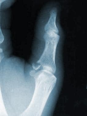
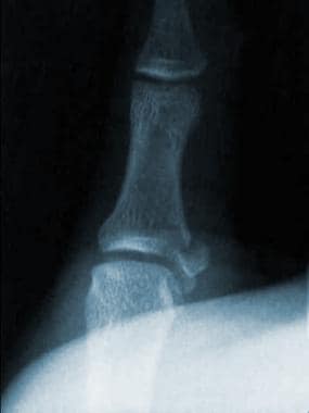
The UCL is a 4- to 8-mm X 12- to 14-mm band that originates from the metacarpal head and inserts into the medial aspect and base of the proximal phalanx of the thumb. Occasionally, when the UCL is strained, it avulses the bone at its insertion and leads to a gamekeeper's fracture (see Images 1-2). A Stener lesion occurs when the adductor aponeurosis becomes interposed between the ruptured UCL and its site of insertion at the base of the proximal phalanx. Thus, the distal portion of the ligament retracts and points superficially and proximally. A rupture of the proper and accessory collateral ligaments must occur for this injury to happen. The UCL no longer contacts its area of insertion and cannot heal. This lesion can also be associated with a gamekeeper's fracture,3 which can be subtle or obvious (see Images 1-2). However, a lump or mass over the ulnar aspect of the MCP joint of the thumb does not necessarily imply a fracture; it may be the result of the Stener lesion. (See also the eMedicine article Stener Lesion.)
Sport-Specific Biomechanics
Considerable variation may be observed in the range of flexion and extension of the thumb MCP joint. The variation of normal joints can include ranges of motion (ROMs) from 5-115° of flexion and extension. In full extension, valgus laxity averages 6° and increases to an average of 12° in 15° of flexion.
History
The gamekeeper's thumb injury may be caused by a valgus stress of any kind to the thumb. The most common history is a fall onto an outstretched arm with an abducted thumb. This commonly occurs in skiers with a ski pole in the hand, which prevents adduction of the thumb when the skier falls.
Physical
- The contralateral, unaffected thumb should be evaluated first for ROM and valgus stability in both extension and 30° of flexion. Then, evaluate the injured thumb for swelling and pain at the ulnar aspect of the MCP joint. Ecchymosis is frequently seen.
- Proper collateral ligament rupture
- The accessory collateral ligament may remain intact, and gross instability may not be present.
- The thumb should be placed in 30° of flexion and tested for valgus instability in this position. This should only be done after radiographs have been obtained that rule out a gamekeeper's fracture. Although a gamekeeper's fracture is a contraindication to stress testing, a nondisplaced avulsion fracture is not. If the patient's pain is too severe, the joint may be anesthetized with a lidocaine injection before stress testing.
- Thirty degrees of laxity or one that is 15° more than that present on the uninjured side represents a ruptured proper collateral ligament in this position.4, 5
- A supination deformity of the MCP joint, which may also be visualized, can be associated with volar subluxation of the MCP joint and suggests instability.
- Accessory collateral ligament rupture
- A palpable mass present on the ulnar aspect of the MCP joint may represent the retracted UCL stump, which is displaced proximally and dorsally to the adductor aponeurosis.
- A Stener lesion can only be present when both the proper and accessory collateral ligaments are ruptured. In more than 80% of complete ruptures of the UCL, a Stener lesion is present, whether it is palpable or not.
- Stress testing with the thumb in the extended position is the best test for determining the competence of the accessory collateral portion of the UCL. Again, valgus laxity of more than 30º or a laxity that is 15º more than that on the uninjured side suggests rupture of this portion of the ligament.4, 5
- If valgus laxity of the thumb's MCP joint is present in both the flexed and extended positions, complete UCL rupture should be suspected.
Causes
- Skiing with poles
- Falling onto an outstretched arm with the thumb abducted
- Valgus-producing force on the thumb MCP joint
- Rheumatoid arthritis (See also Medscape's Rheumatoid Arthritis Resource Center and the eMedicine articles Rheumatoid Arthritis [in the Physical Medicine and Rehabilitation section] and Rheumatoid Arthritis [in the Rheumatology section].)
- Generalized ligamentous laxity
Metacarpophalangeal Joint Dislocation
Phalangeal Fractures
Skier's Thumb
Lab Studies
- No laboratory tests are necessary for the diagnosis of gamekeeper's thumb.
- In cases that require surgical intervention, routine preoperative laboratory workup is indicated.
Imaging Studies
- Radiographs
- Before any manipulation of the thumb, obtain standard anteroposterior (see Image 2), lateral (see Image 1), and oblique radiographs to exclude metacarpal fractures and gamekeeper's fractures.
- Small, nondisplaced avulsion fractures that are associated with rupture of the insertion point of the UCL are not contraindications to manipulation. If displacement of these fractures did not take place at the time of injury and greatest stress, it is believed that they are stable enough for the manipulation of stress testing.
- Three millimeters of volar subluxation of the phalanx on the metacarpal is suggestive of complete UCL rupture and instability.
- Instability is also indicated in cases in which there is a radial deviation of >40º in extension and >20º in flexion.
- Stress radiographs: Radiographs obtained with the thumb in the flexed and extended positions and with valgus stress at the MCP joint can help the physician to determine the degree of instability of partial tears of the UCL (see Image 3).
- Arthrography, ultrasound, and magnetic resonance imaging (MRI) have been used to identify complete tears; however, these tests are not particularly cost effective.2, 6, 7 Careful stress examination is still the optimal method for determining ligament integrity.
Procedures
- Stress testing under local anesthesia (see Images 4-5)
- The patient often has considerable pain in the thumb; stressing the MCP joint leads to patient guarding and, thus, misleading findings on examination.
- The thumb is best examined under local anesthesia, which can be administered in the emergency department (ED) or office setting. Often, 2-3 mL of 1% lidocaine administered into the MCP joint of the thumb is sufficient to relieve the pain and relax the patient's guarding.
- If more anesthesia is required, perform a metacarpal or digital block. Some authors recommend the use of an ulnar or median nerve block to negate the effects of the intrinsic muscles. If the injection into the joint relieves the pain, no further anesthesia is necessary.
Acute Phase
Medical Issues/Complications
- Delayed repair of the UCL
- Complete ruptures of the UCL require surgical intervention for repair within the first 3 weeks of the injury (preferably in the first week while the anatomic position is maintained).
- Complete tears that are repaired after 3 weeks have an increased incidence of weakness and pain on pinch grasp. An associated increase in MCP joint arthritis is noted in the long term.
- Closed treatment of a completely torn UCL
- A 50% failure rate is reported with complete tears that are treated with bracing and early motion. If a patient is unable to tolerate or refuses surgery, then bracing is the treatment of choice. A thumb spica splint or functional brace can be attempted, but it must be understood that full stability of the thumb may not occur.
- Soft-tissue interposition, which is caused by the adductor aponeurosis holding the torn UCL in a displaced position, prevents healing. Surgical correction is necessary to reconstruct the UCL and allow healing.
- ROM is limited in the thumb at the MCP joint if the joint is immobilized longer than 4 weeks.
Surgical Intervention
All complete UCL tears require operative intervention. An incision is made over the ulnar border of the MCP joint of the thumb. The adductor aponeurosis is incised longitudinally and retracted distally. The dorsal capsule is then exposed, and the proper and accessory collateral ligaments are assessed. If the joint is subluxed and if the soft-tissue repair seems insufficient to hold the reduced joint, a small-gauge Kirschner wire (ie, K-wire) can be inserted to maintain the MCP joint in position. The UCL can then be repaired. If a small piece of avulsed bone is present, remove it; a large bone fragment should be reduced and preserved.
Other Treatment
For small, nondisplaced avulsion fractures of the proximal phalanx that are found to be stable on stress testing, nonoperative treatment by a spica-type cast for 4 weeks can be completed with good results. - Closed treatment
- Nonoperative treatment can be considered for partial tears (grade I or grade II) of the UCL, which usually involve an isolated rupture of the proper collateral portion of the ligament. This may be treated with immobilization in a thumb spica-type cast for 4 weeks. The cast should be well-molded around the MCP joint, and the interphalangeal joint can be left free. With appropriate closed treatment, expect 90% good to excellent results.8
- Nonoperative treatment can also be considered in patients that either refuse surgery or who are too infirm to tolerate an operative procedure despite a complete UCL tear. In these patients a functional brace or well-molded spica splint can be applied,8, 9 but it must be understood by the patient that complete healing of the UCL is not expected if the tear was complete.
Recovery Phase
Rehabilitation Program
Physical Therapy
Postoperatively, the patient is immobilized in a thumb spica cast for 4 weeks. At 4 weeks, the cast and any pins that were placed may be removed. The patient is then placed in a hand-based splint that immobilizes the MCP joint for 2 weeks. The splint is removed for therapy during this 2-week period, and active motion of the MCP joint is begun. Unrestricted usage is allowed at 3 months.
Medical Issues/Complications
- Radial sensory nerve injury
- Even when the radial sensory nerve is isolated and protected during surgery, a neuropraxia may still occur.
- The neuropraxia usually resolves spontaneously. Only if the neuropraxia persists after 6 months to 1 year should surgical re-exploration be considered.
- Postsurgical stiffness of the MCP and interphalangeal joints is a common complication (although it is usually not a functional problem) and tends to improve with time.
Maintenance Phase
Medical Issues/Complications
- Chronic instability is a long-term complication of UCL rupture.
- The most common cause is the patient's failure to seek medical attention in a timely fashion for diagnosis and treatment.
- The longer a complete rupture of the UCL exists, the more likely it is to progress to chronic instability, even after its repair. Success has been limited with repairing the tissues after 6 weeks from the time of injury.
- The dorsal capsule, as well as the extensor pollicis brevis and extensor pollicis longus muscles, become attenuated, adding to the dorsal instability of the MCP joint. The thumb then tends to displace volarly and to rotate into supination.
Surgical Intervention
Chronic instability is difficult to treat. Limited success has been associated with repair of gamekeeper's thumb injuries using the capsuloligamentous structures on the ulnar border of the MCP joint.
- If no degenerative changes are present at the MCP joint, consider a ligament reconstruction. A free tendon, usually the palmaris longus, can be woven through the metacarpal neck and the base of the proximal phalanx.
- If arthritis is present or if the patient is a manual laborer, consider an arthrodesis of the MCP joint. Arthrodesis does not lead to significant impairment, if motion of the interphalangeal and carpometacarpal joints is maintained.
Nonsteroidal anti-inflammatory drugs (NSAIDs), which decrease pain and swelling, are the drugs of choice (DOC) in the acute phase of a gamekeeper's thumb injury. The anti-inflammatory effects decrease the acute swelling that is associated with this condition, allowing for a better examination at follow-up (if necessary). NSAIDs are nonopioid analgesics that are made up of different compounds but classed together based solely on their clinical effects. They typically work by inhibiting the formation of prostaglandins through the cyclooxygenase pathway. NSAIDs do not promote dependence like the opioid agents, but they can be associated with renal, liver, and gastrointestinal (GI) toxicities.
Drug Category: Nonsteroidal anti-inflammatory drugs (NSAIDs)
Pain control and anti-inflammatory effects are essential to the quality of patient care. They are beneficial to patients who have a painful injury.
| Drug Name | Ibuprofen (Ibuprin, Advil, Motrin) |
| Description | DOC for mild to moderate pain. Inhibits inflammatory reactions and pain by decreasing prostaglandin synthesis. |
| Adult Dose | 400 mg q4-6h, 600 mg q6h, or 800 mg q8h PO while symptoms persist; not to exceed 3.2 g/d |
| Pediatric Dose | 6 months to 12 years: 20-40 mg/kg/d divided tid or qid >12 years: Administer as in adults |
| Contraindications | Documented hypersensitivity; avoid in patients with peptic ulcer disease, recent GI bleeding or perforation, renal insufficiency, and high risk of bleeding |
| Interactions | May decrease the effects of loop diuretics with coadministration; coadministration with anticoagulants may increase PT duration (monitor and watch for signs of bleeding); may increase serum lithium levels and increase the risk of methotrexate toxicity; probenecid may increase toxicity of NSAIDs |
| Pregnancy | B - Fetal risk not confirmed in studies in humans but has been shown in some studies in animals
|
| Precautions | Caution in patients with congestive heart failure, hypertension, and decreased renal and hepatic function; caution in the presence of anticoagulation abnormalities or during anticoagulant therapy |
| Drug Name | Naproxen (Anaprox, Naprelan, Naprosyn) |
| Description | For relief of mild to moderate pain; inhibits inflammatory reactions and pain by decreasing the activity of cyclooxygenase, which is responsible for prostaglandin synthesis. |
| Adult Dose | 500 mg PO, followed by 250 mg q6-8h; not to exceed 1.25 g/d; may increase to 1.5 g/d for limited time periods |
| Pediatric Dose | < 2 years: Not established > 2 years: 2.5 mg/kg/dose PO; not to exceed 10 mg/kg/d |
| Contraindications | Documented hypersensitivity; peptic ulcer disease; recent GI bleeding or perforation; renal insufficiency |
| Interactions | Probenecid may increase the toxicity of NSAIDs; coadministration with ibuprofen may decrease the effects of loop diuretics; coadministration with anticoagulants may prolong PT duration (watch for signs of bleeding); NSAIDs may increase serum lithium levels and increase the risk of methotrexate toxicity (eg, stomatitis, bone marrow suppression, nephrotoxicity) |
| Pregnancy | B - Fetal risk not confirmed in studies in humans but has been shown in some studies in animals
|
| Precautions | Acute renal insufficiency, interstitial nephritis, hyperkalemia, hyponatremia, and renal papillary necrosis may occur; patients with preexisting renal disease or compromised renal perfusion risk acute renal failure; leukopenia occurs rarely, is transient, and usually returns to normal during therapy; persistent leukopenia, granulocytopenia, or thrombocytopenia warrant further evaluation and may require discontinuation of the drug |
Drug Category: Cyclooxygenase-2 (COX-2) inhibitors
Although increased cost can be a negative factor in the use of COX-2 inhibitors, the incidence of costly and potentially fatal GI bleeds is clearly less with COX-2 inhibitors than with traditional NSAIDs. Ongoing analysis of the cost avoidance of GI bleeds will further define the populations that will find these agents the most beneficial.
| Drug Name | Celecoxib (Celebrex) |
| Description | Celecoxib inhibits primarily COX-2. COX-2 is considered an inducible isoenzyme, induced during pain and inflammatory stimuli. Inhibition of COX-1 may contribute to NSAID GI toxicity. At therapeutic concentrations, the COX-1 isoenzyme is not inhibited, thus GI toxicity may be decreased. Seek the lowest dose of celecoxib for each patient. |
| Adult Dose | 200 mg/d PO qd; alternatively, 100 mg PO bid |
| Pediatric Dose | Not established |
| Contraindications | Documented hypersensitivity to sulfonamides |
| Interactions | Coadministration with fluconazole may cause an increase in celecoxib plasma concentrations because of inhibition of the celecoxib metabolism; coadministration of celecoxib with rifampin may decrease celecoxib plasma concentrations |
| Pregnancy | C - Fetal risk revealed in studies in animals but not established or not studied in humans; may use if benefits outweigh risk to fetus
|
| Precautions | May cause fluid retention and peripheral edema; caution in patients with compromised cardiac function, hypertension, conditions predisposing to fluid retention; caution in the presence of severe heart failure and hyponatremia because circulatory hemodynamics may deteriorate; NSAIDs may mask the usual signs of infection; caution in the presence of existing controlled infections; evaluate symptoms and signs that suggest liver dysfunction, or in abnormal liver laboratory results |
Return to Play
After surgical repair, the MCP joint is immobilized in a cast for 4 weeks. After this period, the cast is removed and replaced with a removable thumb spica splint so that MCP motion can begin. This is continued for 2 weeks; then the splint is removed completely so the MCP joint can be fully mobilized. Unrestricted usage and return to sports can begin at 3 months. Patients are not recommended to return to sports that may stress the hand before 3 months have passed. Premature return to full activity can stress the repair or reconstruction and cause chronic joint instability, which is then very difficult to treat. Chronic pain and degeneration can then develop at the MCP joint of the thumb.
Complications
Chronic instability of the MCP joint can occur despite a good surgical repair, especially if motion and return to play are resumed prematurely. This instability is difficult to treat and can lead to arthritic changes in the MCP joint, as well as weak pinch grasp in the long term.
Prevention
Currently, there are no proven accessories for the prevention of Gamekeeper's thumb injuries. Ski gloves are being designed to protect the thumbs during skiing, but these have not yet been proven effective.
Prognosis
Early diagnosis of Gamekeeper's thumb injuries is one of the most important factors that determines functional outcome. In thumbs with partial ligament injuries, nonoperative treatment by immobilization will yield a stable, painless thumb with nearly normal motion in most cases. In thumbs with a complete rupture that are treated operatively within 3 weeks of injury, a good to excellent result can be expected in >90% of cases. Pain and stiffness in the affected thumb can be expected to be mild or absent, and pinch and grip strength should be nearly normal. The rate of return to former activities, including recreational sports, has been reported as high as 96%.
Medical/Legal Pitfalls
- Failure to diagnose this injury or failure of the patient to seek medical treatment is the most common cause for a poor outcome for Gamekeeper's thumb injuries.
- Significant future morbidity in the affected hand can result, if the diagnosis is delayed or missed.
- Ligamentous testing should be conducted in all patients with a thumb injury and who have normal radiographs.
Special Concerns
- Pediatric gamekeeper's thumb10
- Unlike the adult type, this injury usually involves a Salter Harris III fracture of the proximal phalanx of the thumb.
- Stress testing is not indicated in these injuries, because the stability is determined by the displacement of the fracture fragment.
- If the fragment is displaced <2 mm, then nonoperative management is indicated. For greater displacement, the fracture should be treated with open reduction and fixation.
| Media file 1:
Lateral radiograph displaying a gamekeeper's fracture. |
 | View Full Size Image | |
Media type: X-RAY
|
| Media file 2:
Anteroposterior radiograph displaying a gamekeeper's fracture. |
 | View Full Size Image | |
Media type: X-RAY
|
| Media file 3:
Radiographic stress test view of the thumb, showing an ulnar collateral ligament tear. |
 | View Full Size Image | |
Media type: X-RAY
|
| Media file 4:
Ulnar collateral ligament stress test in full extension. |
 | View Full Size Image | |
Media type: Photo
|
| Media file 5:
Ulnar collateral ligament stress test in a flexed position to isolate the proper portion of the ligament. |
 | View Full Size Image | |
Media type: Photo
|
| Media file 6:
Anterior view of a hand placed in a thumb spica splint. |
 | View Full Size Image | |
Media type: Photo
|
| Media file 7:
Lateral view of a hand placed in a thumb spica splint. |
 | View Full Size Image | |
Media type: Photo
|
- Campbell CS. Gamekeeper's thumb. J Bone Joint Surg Br. Feb 1955;37-B(1):148-9. [Medline]. [Full Text].
- Newland CC. Gamekeeper's thumb. Orthop Clin North Am. Jan 1992;23(1):41-8. [Medline].
- Bekler H, Gokce A, Beyzadeoglu T. Avulsion fractures from the base of phalanges of the fingers. Tech Hand Up Extrem Surg. Sep 2006;10(3):157-61. [Medline].
- Heyman P. Injuries to the ulnar collateral ligament of the thumb metacarpophalangeal joint. J Am Acad Orthop Surg. Jul 1997;5(4):224-9. [Medline].
- Baskies MA, Tuckman D, Paksima N, Posner MA. A new technique for reconstruction of the ulnar collateral ligament of the thumb. Am J Sports Med. Aug 2007;35(8):1321-5. [Medline].
- Shinohara T, Horii E, Majima M, et al. Sonographic diagnosis of acute injuries of the ulnar collateral ligament of the metacarpophalangeal joint of the thumb. J Clin Ultrasound. Feb 2007;35(2):73-7. [Medline].
- Peterson JJ, Bancroft LW. Injuries of the fingers and thumb in the athlete. Clin Sports Med. Jul 2006;25(3):527-42, vii-viii. [Medline].
- Pichora DR, McMurtry RY, Bell MJ. Gamekeepers thumb: a prospective study of functional bracing. J Hand Surg [Am]. May 1989;14(3):567-73. [Medline].
- Sollerman C, Abrahamsson SO, Lundborg G, Adalbert K. Functional splinting versus plaster cast for ruptures of the ulnar collateral ligament of the thumb. A prospective randomized study of 63 cases. Acta Orthop Scand. Dec 1991;62(6):524-6. [Medline].
- Maheshwari R, Sharma H, Duncan RD. Metacarpophalangeal joint dislocation of the thumb in children. J Bone Joint Surg Br. Feb 2007;89(2):227-9. [Medline].
- Abrahamsson SO, Sollerman C, Lundborg G, Larsson J, Egund N. Diagnosis of displaced ulnar collateral ligament of the metacarpophalangeal joint of the thumb. J Hand Surg [Am]. May 1990;15(3):457-60. [Medline].
- Gerber C, Senn E, Matter P. Skier's thumb. Surgical treatment of recent injuries to the ulnar collateral ligament of the thumb's metacarpophalangeal joint. Am J Sports Med. May-Jun 1981;9(3):171-7. [Medline].
- Stener B. Displacement of the ruptured ulnar collateral ligament of the metacarpo-phalangeal joint of the thumb. J Bone Joint Surg [Br]. July 1962;44:869-79. [Full Text].
Gamekeeper's Thumb excerpt Article Last Updated: Nov 1, 2007
|