Continually Updated Clinical Reference
 
 
  All Sources     eMedicine     Medscape     Drug Reference     MEDLINE
 
eMedicine - Athletic Foot Injuries : Article by

Quick Find
Authors & Editors
Introduction
Clinical
Differentials
Workup
Treatment
Medication
Follow-up
Multimedia
References

Related Articles
Achilles Tendonitis

Ankle Sprain

Contusions

Plantar Fasciitis


Patient Education
Breaks, Fractures, and Dislocations Center

Sports Injury Center

Broken Foot Overview

Broken Foot Causes

Broken Foot Symptoms

Broken Foot Treatment


Author: Timothy J Rupp, MD, FACEP, Assistant Professor, Associate Residency Director, Division of Emergency Medicine, University of Texas Southwestern Medical Center; Consulting Staff, Department of Emergency Medicine, Children's Medical Center of Dallas

Timothy J Rupp is a member of the following medical societies: American College of Emergency Physicians, American Medical Association, Gay and Lesbian Medical Association, Society for Academic Emergency Medicine, and Texas Medical Association

Coauthor(s): Steven Karageanes, DO, Director of Primary Care Sports Medicine Fellowship, Department of Orthopedics, Henry Ford Hospital Center for Athletic Medicine; Dean E Schanen, MD, Consulting Staff, Emergency Service Partners, Seton Medical Center

Editors: David T Bernhardt, MD, Director of Primary Care Sports Medicine Fellowship, Professor, Department of Pediatrics, University of Wisconsin; Francisco Talavera, PharmD, PhD, Senior Pharmacy Editor, eMedicine; Russell D White, MD, Professor of Medicine, Department of Community and Family Medicine, University of Missouri-Kansas City School of Medicine, Truman Medical Center Lakewood; Jon Whitehurst, MD, Consulting Staff, Rockford Orthopedic Associates; William Jay Bryan, MD, Clinical Professor, Department of Orthopedic Surgery, Baylor University College of Medicine

Author and Editor Disclosure

Synonyms and related keywords: sesamoiditis, turf toe, Sever disease, posterior tibial tendonitis, peroneal tendon subluxation/dislocation, peroneal tendonitis, flexor hallucis longus tenosynovitis, FHL tenosynovitis, fifth metatarsal fractures, Morton neuroma, Jones fracture, stress fractures, Lisfranc sprain

Background

Athletic foot injuries can be difficult to properly diagnose and treat. Bearing the weight of the entire body, the foot is under tremendous stress. In many sports, the foot absorbs tremendous shearing and loading forces, sometimes reaching over 20 times the person's body weight. Physicians who treat these disorders must have a good understanding of the anatomy and kinesiology of the foot.

Although foot injuries can occur from a variety of causes, the most common cause is trauma. Other etiologies include (1) rapid or improper warm-up, (2) overuse, (3) intense workouts, (4) improper footwear, and (5) playing on hard surfaces.

Physicians who evaluate and treat common foot problems should have a working knowledge of the individual sports and the injuries that are commonly associated with them. An understanding of the basic treatment approaches for these injuries also is imperative.

Frequency

United States

Estimates indicate that 15% of sports-related injuries affect the foot alone.

Functional Anatomy

Foot and ankle

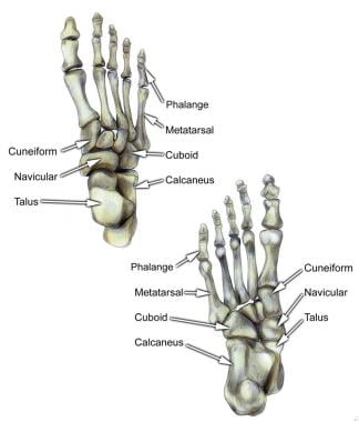
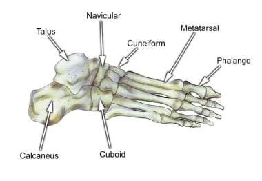
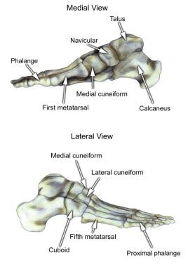
The foot is composed of 26 major bones, which can be divided into 3 regions: the forefoot, midfoot, and hindfoot. The forefoot is comprised of the 5 metatarsals and the 14 phalanges. The 3 cuneiforms (ie, lateral, intermediate, medial), the cuboid, and the navicular represent the midfoot. The hindfoot is composed of the talus and the calcaneus (see Image 1).

  • The talus is oriented so as to transmit forces from the foot through the ankle to the leg.


  • The calcaneus is the largest bone in the foot. The Achilles tendon inserts on the posterior aspect of the calcaneus.


  • The navicular lies anterior to the talus and medial to the cuboid.


  • The cuboid articulates with the calcaneus proximally, with the fourth and fifth metatarsals distally, and with the lateral cuneiform medially.


  • Each of the cuneiform bones is wedge-shaped. The medial, intermediate, and lateral cuneiform bones articulate with the first 3 metatarsals distally and the navicular proximally. The cuboid articulates with the lateral cuneiform.


  • The 5 metatarsals articulate with the proximal phalanges.


  • The great toe is composed of 2 phalanges, with 3 for each lesser toe.


  • Although variation exists in the number and location of sesamoid bones, 2 constant sesamoids are present beneath the metatarsal head. The sesamoids usually are present within tendons juxtaposed to articulations.
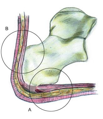
Select tendons of the foot (see Image 2)

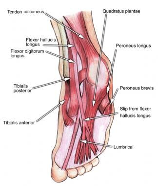
  • The flexor hallucis longus (FHL) tendon is one of 3 structures that lie in the tarsal tunnel. Running behind the medial malleolus, the FHL is the most posterolateral. The FHL runs anterior to insert onto the distal phalanx of the great toe. The FHL acts as a flexor of the great toe, elevates the arch, and assists with plantar flexion of the ankle.


  • The flexor digitorum longus (FDL) tendon passes between the FHL and tibialis posterior tendon. The FDL inserts onto the distal phalanges of the 4 lateral digits and acts to flex the distal phalanges.


  • The tibialis posterior tendon is the most anteromedial of the tarsal tunnel tendons. This tendon inserts on the navicular tuberosity; the 3 cuneiforms; the cuboid; and the second, third, and fourth metatarsals. The tibialis posterior muscle flexes, inverts, and adducts the foot.


  • Laterally, the peroneus longus and peroneus brevis tendons share the common peroneal tunnel running behind and around the lateral malleolus. The peroneus longus plantar flexes the first metatarsal, flexes the ankle, and abducts the foot. The peroneus brevis flexes the ankle and everts the foot.

Other important structures

  • The plantar aponeurosis or fascia is a deep span of connective tissue extending from the anteromedial tubercle of the calcaneus to the proximal phalanges of each of the toes. Medial and lateral fibrous septa originate from the medial and lateral borders to attach to the first and fifth metatarsal bones.


  • Nerve innervation of the foot runs along the medial and lateral metatarsals and phalanges in a neurovascular bundle. These nerves are vulnerable to compressive forces that, in time, can generate the painful Morton neuroma, which most commonly affects the interspace between the third and fourth metatarsals. Four nerves supply the forefoot: the sural nerve (most lateral), branches of the superficial peroneal nerve, the deep peroneal nerve, and the saphenous nerve.

Sport Specific Biomechanics

The 3 planes in which the foot and ankle function are the transverse plane, the sagittal plane, and the frontal plane. Movement is possible in all 3 planes.

  • Plantar flexion and dorsiflexion occur in the sagittal plane. Plantar flexion involves the foot moving from the anterior leg distally. Dorsiflexion is the opposite motion.

  • Inversion and eversion occur in the frontal plane of motion. Eversion occurs when the bottom of the foot turns away from the midline of the body. Inversion is the opposite action.

  • The 2 transverse plane motions are abduction and adduction. Adduction involves the foot moving toward the midline of the body, while abduction is the opposite action.


History

  • Sesamoiditis is manifested by pain beneath the first metatarsal head with weight bearing on the ball of the foot or with motion at the first metatarsophalangeal (MTP) joint. Common complaints include pain with jumping and pushing off to run.
  • Turf toe is an acute injury involving forced hyperdorsiflexion of the first MTP joint as the classic mechanism of injury. This results in a sprain of the first MTP joint. Symptoms include pain and decreased range of motion (ROM) at the MTP joint and difficulty running or changing directions.
  • Sever disease (eg, calcaneal apophysitis) is a common cause of acute or chronic heel pain in children during early adolescence. Athletes typically complain of heel pain or soreness that improves with rest and worsens with prolonged running.
  • Posterior tibial tendonitis occurs most commonly as an idiopathic condition in middle-aged females. Athletes with this condition may present with planovalgus deformity and often play sports with sudden stop-start or push-off activity, such as soccer, football, and basketball. Patients typically complain of pain inferior to the medial malleolus and decreased ROM.
  • Patients with peroneal tendon subluxation/dislocation typically present with acute pain and swelling centered behind the lateral malleolus with extension proximally over the tendons. These symptoms are caused by a dorsiflexion-inversion stress injury that pulls the peroneal retinaculum off the lateral malleolus. Athletes usually complain of snapping and sudden sharp pain when changing directions or pushing off with the foot.
  • Patients with peroneal tendonitis present with pain and swelling on the lateral aspect of the ankle, usually posterior to the lateral malleolus. Patients also may complain of either a "giving way" or "sharp pinching" sensation of the lateral ankle. Long-distance running and any activity that requires repetitive cutting and pushing off can aggravate this condition.
  • Patients with FHL tenosynovitis usually present with pain in the posteromedial aspect of the ankle. The pain improves with rest and increases in sports requiring push-off and extended running.
  • Fifth metatarsal fractures are a common complication with ankle sprains, so physicians must always address this when obtaining the patient's history. Three types of fractures occur in the fifth metatarsal.
    • Avulsion fractures off the base commonly occur with ankle sprains, particularly the plantar flexion-inversion variety.

    • Proximal diaphyseal fractures result from repetitive cyclical stress to the foot and typically have a prodromal presentation.

    • Transverse fractures occurring within 1.5 cm from the tuberosity at the metaphyseal-diaphyseal junction are the definitive Jones fracture. Contrary to popular belief, true Jones fractures primarily occur traumatically. Pain may be diffuse and difficult to localize, depending on the type and location of the fracture.

  • Morton neuroma causes pain over the ball of the foot, followed by radiation of pain to the affected toes. The patient may complain of numbness, tingling, burning, or a sensation similar to an electrical shock. Pain usually eases upon removal of the offending shoes and rubbing the ball of the foot near the affected web space. The information obtained in the history usually reveals wearing of tight-fitting, high-heeled, or pointed-toed shoes, which are commonly worn by females who are young to middle-aged. Athletes who use a repetitive step-off motion (eg, sprinters, jumpers, those who regularly use stair steppers or treadmill machines) may complain of these symptoms.
  • Most athletes with stress fractures complain of progressively increasing pain correlating with a change in activity, footwear, training, playing surface, or equipment. Trauma is not part of the history. Pain is exacerbated by impact loading and is ameliorated with rest.
  • The tarsometatarsal (TMT) fracture-dislocation, or Lisfranc fracture-dislocation, is named after a field surgeon in Napoleon's army who described amputations through the TMT joint. Injury to the TMT joint was common when a soldier's boot became caught in the stirrup during a fall from horseback. More recently, Lisfranc injuries have been observed in snowboarders and windsurfers, as well as in football and rugby players. Typically, the Lisfranc fracture-dislocation occurs when one player falls onto the heel of another while the foot is plantar flexed and fixed, resulting in axial loading. The clinical presentation depends on the degree of displacement. Almost universally, patients complain of pain in the midfoot with the inability to bear weight. Edema and ecchymosis usually are present. Gross deformity of the forefoot may be seen in severe cases. Vascular compromise may manifest as absence of the dorsalis pedis pulse.

Physical

  • Sesamoiditis: Pain on dorsiflexion of the hallux, restricted motion of the first MTP joint, or pain on dorsal palpation of a sesamoid bone occurs.


  • Turf toe: The first MTP joint is red, swollen, tender, and stiff. Pain usually is greatest with end-range dorsiflexion of the foot. Collateral ligaments are stable, but there may be laxity with anterior-posterior translation. Consider gout if a patient presents with this particular clinical picture. If there is no history of repetitive motion or if in doubt, the joint may need to be aspirated. The resultant fluid should be analyzed for the culture and the presence of negative birefringent crystals.


  • Sever disease: Pain is provoked by palpation along the posterior portion of the heel and the Achilles tendon insertion (see Image 3). Restricted ankle dorsiflexion and knee extension may contribute to symptoms.


  • Posterior tibial tendonitis: Tenderness is revealed at the posterior tibial insertion, often with a swollen erythematous navicular prominence, or along the distal path around the posterior aspect of the medial malleolus. Patients may have pain and/or weakness with resisted inversion with a tendency to have a flexible flatfoot, genu valgum, and tibia varum.


  • Peroneal tendon subluxation/dislocation: Palpation reveals direct tenderness over the peroneal tendons. Subluxation, dislocation, or tear of the peroneal tendon results in weakness of eversion and dorsiflexion. Snapping is palpated through a ROM, occasionally only when bearing weight.


  • Peroneal tendonitis: Examination usually reveals swelling and tenderness along the tendons at the lateral aspect of the ankle. Eversion of the foot against resistance may elicit pain.


  • FHL tenosynovitis: Usually no palpable tenderness is present due to the deep location of the tendon. Pain and weakness are noted with resistance to plantar flexion of the first MTP joint. Pain also may be present in the tarsal tunnel.


  • Jones fracture: Tenderness may be difficult to localize specifically, but focal pain on the proximal fifth metatarsal indicates a fracture until proven otherwise. Passive inversion or resisted eversion also may be painful with a fifth metatarsal base fracture.


  • Morton neuroma: Perform a compression test by squeezing the metatarsal heads together with one hand while palpating and compressing the involved web space with the other hand. If a Morton neuroma is present, the interspaces between the metatarsals will be tender to palpation.


  • Stress fractures: Physical examination usually is unremarkable, but it may reveal minor swelling and warmth over the forefoot, while point tenderness may be elicited by applying pressure under the affected metatarsal in a dorsal direction. Maneuvers such as walking on toes or running in place can reproduce symptoms.


  • Lisfranc sprain: The physical examination may reveal prominence of the first metatarsal or shortening of the forefoot. The patient complains of pain and may note paresthesias of the forefoot and digits. Passive range of motion (PROM) and palpation over the metatarsals, with attention to the base of the second metatarsal, will likely reveal tenderness, which mandates that careful radiographic examination be performed. In a dislocation-fracture, the foot may have more swelling and deformity. Radiographs can demonstrate a fracture, seen most at the base of the second metatarsal.

Causes

  • Sesamoiditis usually is caused by overuse of the foot in a plantar flexed position. Cleats with little insole padding can focus excess stress on the first MTP or sesamoid and can trigger this condition. A depressed first ray, forefoot valgus, or pes cavus deformity can lead to sesamoiditis as well.
  • Turf toe is caused by hyperextension of the first MTP joint beyond the normal 60° of dorsiflexion. Incidence of the injury has increased in recent years with the widespread use of artificial surfaces. Lightweight, poorly supported, flexible shoes can predispose the athlete to injury.
  • Sever disease is caused by excessive traction on the calcaneal apophysis by the Achilles tendon, particularly during running and jumping. Inflexibility of the gastrocnemius, hamstring, quadriceps, and hip flexor muscles can exacerbate this condition.
  • Posterior tibial tendonitis is caused by repetitive trauma during the pronation phase of cutting, jumping, or running. Pes planus is a risk factor.
  • Peroneal tendon subluxation/dislocation most commonly occurs with powerful contraction of the peroneal muscles, usually in maximal dorsiflexion. The peroneal tendons are retained in place by the superior and inferior peroneal retinaculum. If enough stress is applied, these may rupture, resulting in subluxation or dislocation of the peroneal tendons.
  • Peroneal tendonitis may be related to acute inversion injury or chronic overuse secondary to hindfoot varus. Iliotibial band restriction increases force on the peroneal tendons.
  • FHL tenosynovitis typically is associated with repeated push-off maneuvers, such as those executed by ballet dancers or sprinters.
  • A Jones fracture usually occurs when a load is applied to the lateral forefoot in the absence of inversion. A Jones fracture also can occur with an acute inversion injury. Jones fractures usually occur in sports involving running and jumping. Fifth metatarsal proximal diaphyseal fractures can occur with chronic overuse and poor biomechanics. Fifth metatarsal base avulsion fractures commonly occur via inversion sprain mechanisms.
  • Morton (intermetatarsal) neuroma is a biomechanically induced neuropathy of the common digital nerve of the foot. Repetitive microdamage to the nerve fibers produces degeneration and reparative fibrosis, significantly increasing the nerve diameter. Restrictive toe boxes combined with overuse can cause this condition.
  • Stress fractures
    • Stress fractures are defined as spontaneous fractures of normal bone that result from the summation of stresses, any of which by themselves would be harmless. Stress fractures of the foot were first described by Breihaupt in 1855. Serving as a physician in the Prussian Army, Breihaupt observed fractures of the metatarsals in otherwise healthy military recruits after long marches. These became know as March fractures.

    • Of all sports-related injuries, it is estimated that 5-10% involve stress fractures. Nine of the 24 members of the 1994 United States National World Cup Soccer Team were diagnosed with stress fractures. The incidence of stress fractures is increasing for the following reasons: (1) increasing numbers of persons participating in sporting activities, (2) increasing awareness and suspicion of stress fractures, and (3) change in the nature and type of sporting activities (ie, rollerblading). Stress fractures of the second, third, and fourth metatarsals account for 90% of metatarsal injuries

  • Lisfranc fracture-dislocation
    • The transverse ligaments connect the second, third, fourth, and fifth metatarsal bases. The second metatarsal is recessed and bound to the medial and intermediate cuneiform bones by ligaments. The Lisfranc ligament joins the first (medial) cuneiform bone to the base of the second metatarsal. The base of the second metatarsal, recessed tightly into the mortise formed by the cuneiform bones and attached by multiple ligaments, forms the keystone of the midfoot and, thus, is the primary stabilizing structure of the TMT joint complex.

    • Lisfranc fracture-dislocations are classified as homolateral or divergent. A homolateral dislocation involves displacement of 4 or 5 metatarsals in the same direction. A divergent dislocation involves a split between the first and second metatarsals.


Achilles Tendonitis
Ankle Sprain
Contusions
Plantar Fasciitis

Other Problems to be Considered

Sesamoiditis

Sesamoid fracture, congenital bipartite, or tripartite sesamoids that result from coalition of the primordial calcification center of the ossicle

Sever disease

Achilles bursitis

Peroneal tendon subluxation/dislocation

Tenosynovitis

Flexor hallucis longus tenosynovitis

Symptomatic os trigonum that may become impinged with extreme plantar flexion

Jones fracture

Fracture of the base of the fifth metatarsal
Ankle sprain
Peroneal apophysitis (ie, Iselin disease)


Imaging Studies

  • Sesamoiditis: Stress radiographs, axial silhouette views, bone scans, and CT scan are helpful diagnostic aids. Radiographic examination of the contralateral foot is useful. Radiographs often are normal. A bipartite sesamoid bone is a common normal variant that can be mistaken for a stress fracture. Additional testing may be needed to help evaluate this possibility further, depending on the patient's symptoms.
  • Turf toe: Plain radiographs may reveal a small avulsion fracture from the plantar metatarsal head.
  • Sever disease: Plain radiographs often are not necessary. However, plain radiographs should be obtained if symptoms are not alleviated with relative rest or if the clinical picture is somewhat atypical in order to rule out a fracture or tumor.
  • Posterior tibial tendonitis: A weight-bearing plain radiograph may help determine the degree and type of flatfoot abnormality. MRI is the imaging modality of choice for imaging posterior tibial tendon tenosynovitis and degenerative tears. Ultrasound (US) imaging can provide a dynamic picture of the tendon.
  • Peroneal tendon subluxation/dislocation: The diagnosis is made on clinical grounds. Most peroneal dislocations reduce spontaneously; therefore, most of these injuries are unrecognized.
  • Peroneal tendonitis: Plain radiographs may reveal hindfoot varus. US imaging and MRI usually are not necessary but may reveal synovitis, peroneal retinaculum, or tendon tearing.
  • FHL tenosynovitis: Plain radiographs of the foot are helpful with the differential diagnosis, and US imaging or MRI can help rule out a tear.
  • Jones fracture: Standard foot radiographs demonstrate most metatarsal base fractures. Note any intra-articular fractures, and determine the percentage of articular surface that is involved, as this percentage is essential to determining management. Avulsion of the fifth metatarsal apophysis should not be confused for a Jones fracture as the apophyseal avulsion is parallel to the shaft of the fifth metatarsal compared to the perpendicular fracture of the standard Jones fracture.
  • Morton neuroma: Radiographic imaging may be employed if the diagnosis is in question. US can help the physician make a reliable estimate of the size of the neuroma.
  • Stress fractures
    • Plain radiography is the first imaging study recommended to help confirm the diagnosis of stress fracture. However, radiographic changes may not be evident for 2-3 weeks following the onset of symptoms. Periosteal and endosteal callus formation typically is seen within 2 weeks of injury, while callus formation reaches its maximum at 6 weeks. Moreover, only 50% of stress fractures are seen on plain radiographs. Findings of radionuclide bone scanning or MRI help confirm the diagnosis.

    • The criterion standard for the diagnosis of stress fractures is a technetium bone scan. The osteoblast incorporates the isotopes in new bone formation and may be positive as early as 48-72 hours following clinical signs of injury. MRI has been found to be just as sensitive and more specific. However, MRI may be cost prohibitive or may not be available, depending on the medical center.

  • Lisfranc fracture-dislocation
    • Meticulous evaluation of weight-bearing foot radiographs is essential for confirmation of suspected Lisfranc fracture-dislocation. A fracture of the base of the second metatarsal is virtually pathognomonic for TMT joint disruption.

    • The medial aspects of the first, second, and third metatarsals should align evenly with the medial borders of the first, second, and third cuneiform bones, respectively. The medial border of the fourth metatarsal should align evenly with the medial border of the cuboid bone. Comparison views of the feet may reveal a widening of the space between the first and second metatarsals or between the second and third metatarsals. Disruption of these anatomic relationships suggests a Lisfranc fracture-dislocation. US imaging and MRI can indicate ligamentous disruption by showing fluid in the ligaments and intertarsal joint spaces.


Acute Phase

Rehabilitation Program

Physical Therapy

Physical therapy is effective in treating inversion injuries and tendonitis of the foot, particularly in athletes who are continuing competition. Most athletes with fractures rehabilitate around the injury to minimize joint restriction and to maintain fitness levels. Acute phase treatment includes relative rest, ice, electrical stimulation, phonophoresis, and iontophoresis.

  • Sesamoiditis: Treatment consists of wearing cushioned-soled shoes with total contact inserts to relieve first metatarsal head stress; taking NSAIDs; and implementing rest, ice, compression, and elevation (RICE). An orthotic device should be worn for at least 6 months.


  • Turf toe: Acute treatment consists of a period of RICE, taping, and strapping the toe in a plantar-flexed position to avoid further hyperextension. Rigid turf-toe orthotics may be helpful as well. Ambulating is well tolerated in a hard-soled shoe. Mild-to-moderate sprains may require rest from the activity from days to weeks. Severe sprains may necessitate relative rest for up to 6 weeks.


  • Sever disease: Treatment consists of implementing RICE, wearing protective heel inserts or prefabricated arch supports, performing stretching and strengthening exercises, and, occasionally, taking NSAIDs (see Image 2).


  • Posterior tibial tendonitis: Treatment depends on the degree of symptoms. Initially RICE, NSAIDs, and analgesics are used as needed. Cast immobilization may be helpful during the early stages of the disease.


  • Peroneal tendon subluxation/dislocation: If reduction is necessary, it is accomplished by directing pressure posteriorly and then casting the ankle in slight pronation and flexion.


  • Peroneal tendonitis: For acute tenosynovitis, rest or immobilization and NSAIDs are initial measures. Wearing a cast for 2-3 weeks and then implementing extensive rehabilitation is appropriate for severe symptoms. An injection of a corticosteroid should be considered for patients with resistant symptoms.


  • FHL tenosynovitis: Treatment consists of immobilization, activity restrictions, and NSAIDs.


  • Jones fracture: The management of fifth metatarsal base fractures depends on the type of fracture. Extra-articular tuberosity fractures heal well and are managed symptomatically with either a walking cast or a hard-soled shoe for 2-3 weeks. Nondisplaced diaphyseal fractures usually are treated with non–weight-bearing casting for up to 8 weeks, followed by radiographic assessment. Diaphyseal fractures of the fifth metatarsal often are complicated by nonunion, delayed union, or recurrence secondary to compromised vascular supply. Intra-articular fractures often lead to posttraumatic arthritis.


  • Morton neuroma: Initially, treatment is conservative and is designed to relieve pain while permitting the athlete to continue activity. This treatment involves rest, ice, NSAIDs, and US. The application of a felt pad under the heads of the affected metatarsals may spread the metatarsal heads and relieve pain and inflammation. Injection of a corticosteroid may be effective in reducing the diameter of the impinged nerve branch. Podiatric consultation may be considered for proper shoe fitting.


  • Metatarsal stress fractures (not fractures of the fifth metatarsal): Conservative therapy, including rest, anti-inflammatory medications, application of ice, and cessation of the offending activity, is implemented. Athletes should maintain their aerobic capacity throughout recuperation by beginning a training program that involves non–weight-bearing activity such as swimming or stationary cycling.


  • Lisfranc fracture-dislocation: Because TMT fracture-dislocations are associated with complications such as loss of arch, degenerative arthritis, chronic pain, and impaired circulation to the distal foot, it is imperative that an orthopedic surgeon determine the most appropriate course of action for the patient.

Surgical Intervention

  • Sesamoiditis: Surgical excision is a last option that is rarely indicated.
  • Turf toe: Surgical treatment may be necessary to treat sesamoid injuries and repair capsular tears.
  • Sever disease: Surgery usually is not indicated in patients with Sever disease.
  • Posterior tibial tendonitis: Severe disease may require surgical debridement or repair.
  • Peroneal tendon subluxation/dislocation: Surgery is reserved for those in whom conservative therapy has failed or for those who are high-level athletes.
  • FHL tenosynovitis: Surgical release is occasionally necessary.
  • Jones fracture: Surgery to internally fixate the fracture often is performed to speed up recovery and to minimize the length of time before the athlete can return to play.
  • Fifth metatarsal fractures: Intra-articular tuberosity fractures involving more than 30% of the articular surface may require surgical fixation; therefore; orthopedic consultation is advised. Nondisplaced diaphyseal fractures in athletes may require immediate surgical fixation. Displaced diaphyseal fractures usually are managed operatively.
  • Morton neuroma: Surgical therapy may be recommended for patients or athletes in whom conservative management techniques fail. Surgical resection of the offending neuroma can provide rapid relief from pain and inflammation. A short course of rehabilitative therapy following surgery generally is recommended.
  • Stress fractures: Surgery is considered for athletes with stress fractures if conservative therapy fails. Furthermore, surgery for stress fractures should only be considered if the fracture is in a bone in which a complete fracture would result in serious complications (ie, tarsal navicular bone, a fifth metatarsals).
  • Lisfranc fracture-dislocation: The orthopedist may elect to perform closed reduction under general anesthesia using finger traps and countertraction at the ankle. The patient may require open reduction and internal fixation for more definitive stabilization. The patient will likely require a short leg cast from 6-12 weeks following surgery. At first, the patient will have a non—weight-bearing restriction and then gradually will progress his or her weight bearing in a walking cast. A custom arch support may be used for up to 1 year.

Other Treatment

Manipulation can be used to reintroduce motion and joint play into the foot, especially after prolonged immobilization, which often occurs during the postsurgical period or during fracture care. This manipulation can speed up return to play, which is the essential issue in athletic injuries. Injections are controversial in such problems as plantar fasciitis, as corticosteroids can increase the risk of tissue failure and rupture. Never use corticosteroids in a suspected or known fracture or directly in a tendon. A steroid agent can be injected into a tendon sheath to treat recurrent inflammation, but such an agent rarely is used as a first-line treatment. A diagnostic injection with lidocaine or bupivacaine may be used only as a means of localizing pathology.

Recovery Phase

Rehabilitation Program

Physical Therapy

After the acute phase, focus moves to ROM. PROM and active range of motion (AROM) exercises are used; muscle energy can be applied to restore the muscle set points. Therapy then shifts to improving strength and proprioception. Balance exercises are vital before returning an athlete to competition to prevent further injury.

Other Treatment (Injection, manipulation, etc.)

Taping or braces may be considered when preparing to return the athlete to play. For example, an athlete with turf toe may have steel-toe inserts in his/her shoes and taping on the first MTP joint.

Maintenance Phase

Rehabilitation Program

Physical Therapy

The athlete needs to continue implementing a proprioception and strength program to maintain function. Bracing, taping, or other prophylactic measures are taken into account with each individual injury and athlete. Long-term use of braces on the foot or ankle are discouraged.


NSAIDs remain the mainstays of medical therapy for athletic foot injuries. For moderate to severe pain, the addition of an opioid analgesic may be necessary as well.

Drug Category: Nonsteroidal anti-inflammatory drugs

Used to suppress manifestations of inflammation. Inhibition of cyclo-oxygenase, the enzyme responsible for biosynthesis of prostaglandins, generally is thought to be a major facet of the mechanism of action of NSAIDs.

Drug NameIbuprofen (Motrin, Ibuprin)
DescriptionClassified as a propionic acid derivative. All drugs in this class are effective inhibitors of cyclo-oxygenase, though the potency varies.
Adult Dose400-600 mg PO q6h prn
Pediatric Dose5-10 mg/kg PO q6-8h prn
ContraindicationsDocumented hypersensitivity; peptic ulcer disease, recent GI bleeding or perforation, renal insufficiency, or high risk of bleeding
InteractionsCoadministration with aspirin increases risk of inducing serious NSAID-related side effects; probenecid may increase concentrations and, possibly, toxicity of NSAIDs; may decrease effect of hydralazine, captopril, and beta-blockers; may decrease diuretic effects of furosemide and thiazides; may increase PT when taking anticoagulants (instruct patients to watch for signs of bleeding); may increase risk of methotrexate toxicity; phenytoin levels may be increased when administered concurrently
PregnancyB - Usually safe but benefits must outweigh the risks.
PrecautionsCategory D in third trimester of pregnancy; caution in congestive heart failure, hypertension, and decreased renal and hepatic function; caution in anticoagulation abnormalities or during anticoagulant therapy

Drug NameNaproxen (Naprelan, Naprosyn, Anaprox)
DescriptionClassified as a propionic acid derivative. All the drugs in this class are effective inhibitors of cyclo-oxygenase, though the potency varies.
Adult Dose250-500 mg PO q12h
Pediatric DoseNot established
ContraindicationsDocumented hypersensitivity; peptic ulcer disease; recent GI bleeding or perforation; renal insufficiency
InteractionsCoadministration with aspirin increases risk of inducing serious NSAID-related side effects; probenecid may increase concentrations and, possibly, toxicity of NSAIDs; may decrease effect of hydralazine, captopril, and beta-blockers; may decrease diuretic effects of furosemide and thiazides; may increase PT when taking anticoagulants (instruct patients to watch for signs of bleeding); may increase risk of methotrexate toxicity; phenytoin levels may be increased when administered concurrently
PregnancyB - Usually safe but benefits must outweigh the risks.
PrecautionsCategory D in third trimester of pregnancy; acute renal insufficiency, interstitial nephritis, hyperkalemia, hyponatremia, and renal papillary necrosis may occur; patients with preexisting renal disease or compromised renal perfusion risk acute renal failure; leukopenia occurs rarely, is transient, and usually returns to normal during therapy; persistent leukopenia, granulocytopenia, or thrombocytopenia warrants further evaluation and may require discontinuation of drug

Drug Category: Cyclo-oxygenase-2 inhibitors

COX-2 inhibitors are a new class of NSAIDs. COX-2 inhibitors appear to be as effective as nonselective NSAIDs in treating pain and inflammation. Their theoretical advantage over nonselective NSAIDs involves significantly less toxicity, particularly in the GI tract. This class of drug generally is indicated for patients at risk for GI hemorrhage. These patients include those with peptic ulcer disease, patients on warfarin therapy or on concomitant steroids, and elderly persons.

There has been recent literature questioning the safety of COX-2 inhibitors. Rofecoxib (Vioxx) has been withdrawn from the worldwide market because of its association with and increased rate of cardiovascular events (including heart attack and stroke) compared to placebo. Valdecoxib (Bextra) has been recalled for similar concerns. It is not clear whether these safety concerns are specific to Valdecoxib and Vioxx. The cardiovascular issues may be a class effect of all COX-2 inhibitors. Further study should help to answer questions concerning the safety of COX-2 inhibitors.

Drug NameCelecoxib (Celebrex)
DescriptionPrimarily inhibits COX-2. COX-2 is considered an inducible isoenzyme, induced by pain and inflammatory stimuli. Inhibition of COX-1 may contribute to NSAID GI toxicity. At therapeutic concentrations, COX-1 isoenzyme is not inhibited, thus GI toxicity may be decreased. Seek lowest dose of celecoxib for each patient. Celecoxib has the same general class labeling as conventional NSAIDs.
Adult Dose100 mg PO qd/bid
Pediatric DoseNot established
ContraindicationsDocumented hypersensitivity
InteractionsCoadministration with fluconazole may cause increase in celecoxib plasma concentrations because of inhibition of celecoxib metabolism; coadministration of celecoxib with rifampin may decrease celecoxib plasma concentrations
PregnancyB - Usually safe but benefits must outweigh the risks.
PrecautionsMay cause fluid retention and peripheral edema; caution in compromised cardiac function, hypertension, and conditions predisposing to fluid retention; severe heart failure and hyponatremia may occur because celecoxib may deteriorate circulatory hemodynamics; NSAIDs may mask usual signs of infection; caution in the presence of existing controlled infections; evaluate symptoms and signs suggesting liver dysfunction

Drug Category: Analgesic, Miscellaneous

Pain control is essential to quality patient care. Analgesics ensure patient comfort and have sedating properties, which are beneficial for patients who are in pain. Opioids produce their major effects by acting as agonists on specific opioid receptors. The effects are diverse and include analgesia, drowsiness, respiratory depression, decreased GI motility, nausea, and vomiting.

Drug NameAcetaminophen (Tylenol, Feverall, Aspirin Free Anacin)
DescriptionHas analgesic and antipyretic effects that do not differ significantly from aspirin. However, acetaminophen has only weak anti-inflammatory effects. Exact mechanism of action is not clear.
Adult Dose325-650 mg PO/PR q4-6h prn
Pediatric Dose10-15 mg/kg PO q4-6h prn
ContraindicationsDocumented hypersensitivity
InteractionsCoadministration with fluconazole may cause increase in celecoxib plasma concentrations because of inhibition of celecoxib metabolism; coadministration of celecoxib with rifampin may decrease celecoxib plasma concentrations
PregnancyB - Usually safe but benefits must outweigh the risks.
PrecautionsMay cause fluid retention and peripheral edema; caution in compromised cardiac function, hypertension, and conditions predisposing to fluid retention; severe heart failure and hyponatremia may occur because acetaminophen may deteriorate circulatory hemodynamics; NSAIDs may mask usual signs of infection; caution in the presence of existing controlled infections; evaluate symptoms and signs suggesting liver dysfunction

Drug NameHydrocodone and acetaminophen (Vicodin, Norcet, Lortab)
DescriptionDrug combination indicated for moderate to severe pain for pain refractory to NSAIDs.
Adult Dose1-2 tab or cap PO q4-6h prn pain
Pediatric DoseNot established
ContraindicationsDocumented hypersensitivity; high-altitude cerebral edema (HACE) or elevated intracranial pressure (ICP)
InteractionsCoadministration with phenothiazines may decrease analgesic effects; toxicity increases with CNS depressants or tricyclic antidepressants
PregnancyC - Safety for use during pregnancy has not been established.
PrecautionsTablets contain metabisulfite, which may cause hypersensitivity; caution in patients dependent on opiates since this substitution may result in acute opiate-withdrawal symptoms; caution in severe renal or hepatic dysfunction

Drug NameCodeine and acetaminophen (Tylenol #3)
DescriptionIndicated for the treatment of mild to moderate pain. Elixir has 12 mg of codeine combined with 120 mg of acetaminophen in 5 mL.
Adult Dose1-2 tab PO q4h prn
Pediatric Dose3-6 years: 5 mL PO q4h prn
7-12 years: 10 mL PO q4h prn
>12 years: 15 mL PO q4h
ContraindicationsDocumented hypersensitivity
InteractionsToxicity of codeine increases with administration of CNS depressants, tricyclic antidepressants, MAO inhibitors, neuromuscular blockers, CNS depressants, phenothiazines, and narcotic analgesics; rifampin can reduce analgesic effects of acetaminophen; coadministration with barbiturates, carbamazepine, hydantoins, and isoniazid may increase hepatotoxicity of acetaminophen
PregnancyC - Safety for use during pregnancy has not been established.
PrecautionsCaution in patients dependent on opiates since this substitution may result in acute opiate-withdrawal symptoms; caution in severe renal or hepatic dysfunction; hepatotoxicity with acetaminophen is possible following various dose levels in those with chronic alcoholism; severe or recurrent pain or high or continued fever may indicate a serious illness; acetaminophen is contained in many OTC products, and combined use with these products may result in cumulative acetaminophen doses that exceed recommended maximum dose


Return to Play

As with all athletic injuries, the athlete's whole being must be considered before returning to action. Athletes should practice before they play, and essentially be pain free with all activity. Strength should be at least 90% of the unaffected limb, and proprioception should be restored so that the athlete can avoid recurrence. Mentally, the athlete must feel confident that the foot injury has healed; athletes should be able to compete without conscious awareness of the injury. The mental aspects of the injury are most accurately assessed in practice situations.

Education

For excellent patient education resources, visit eMedicine's Breaks, Fractures, and Dislocations Center and Sports Injury Center. Also, see eMedicine's patient education article Broken Foot.


Media file 1:  The hindfoot is composed of the talus and the calcaneus.
Click to see larger pictureClick to see detailView Full Size Image
Media type:  Image

Media file 2:  Select tendons of the foot.
Click to see larger pictureClick to see detailView Full Size Image
Media type:  Image

Media file 3:  Select bones of the foot (dorsal and plantar views).
Click to see larger pictureClick to see detailView Full Size Image
Media type:  Image

Media file 4:  Select bones of the foot (medial and lateral views).
Click to see larger pictureClick to see detailView Full Size Image
Media type:  Image

Media file 5:  Select bones of the foot (superolateral view).
Click to see larger pictureClick to see detailView Full Size Image
Media type:  Image


  • Birrer RB, Dellacorte MP, Grisafi PJ. Common Foot Problems in Primary Care. 2nd ed. Hanley & Belfus Inc; 1998.
  • Cailliet R. Foot and Ankle Pain. 3rd ed. FA Davis Co; 1997.
  • Conti SF. Posterior tibial tendon problems in athletes. Orthop Clin North Am. Jan 1994;25(1):109-21. [Medline].
  • DeLee JC, Evans JP, Julian J. Stress fracture of the fifth metatarsal. Am J Sports Med. Sep-Oct 1983;11(5):349-53. [Medline].
  • Gellman R, Burns S. Walking aches and running pains. Injuries of the foot and ankle. Prim Care. Jun 1996;23(2):263-80. [Medline].
  • Gilman AG, Rall TW, Nies AS. Goodman and Gilman's the Pharmacological Basis of Therapeutics. 8th ed. McGraw Hill Inc; 1993:489-500, 664-667.
  • Hesp WL, van der Werken C, Goris RJ. Lisfranc dislocations: fractures and/or dislocations through the tarso- metatarsal joints. Injury. Jan 1984;15(4):261-6. [Medline].
  • Knapp TP, Garrett WE Jr. Stress fractures: general concepts. Clin Sports Med. 16(2):339-56. [Medline].
  • Lenczner EM, Waddell JP, Graham JD. Tarsal-metatarsal (Lisfranc) dislocation. J Trauma. Dec 1974;14(12):1012-20. [Medline].
  • Maitra RS, Johnson DL. Stress fractures. Clinical history and physical examination. Clin Sports Med. Apr 1997;16(2):259-74. [Medline].
  • Nunan PJ, Giesy BD. Management of Morton's neuroma in athletes. Clin Podiatr Med Surg. Jul 1997;14(3):489-501. [Medline].
  • Sammarco GJ, Cooper PS. Foot and Ankle Manual. 2nd ed. Lippincott Williams & Wilkins; 1998.
  • Scurran BL. Foot and Ankle Trauma. 2nd ed. Churchill Livingstone; 1996.
  • Sherman KP. The foot in sport. Br J Sports Med. 33(1):6-13. [Medline].
  • Simon RR, Koenigsknecht SJ. Tarsometatarsal fracture-dislocations. In: Emergency Orthopedics: The Extremities. 3rd ed. 1995:331-332.
  • Wedmore IS, Charette J. Emergency department evaluation and treatment of ankle and foot injuries. Emerg Med Clin North Am. Feb 2000;18(1):85-113, vi. [Medline].
  • Weinfeld SB, Haddad SL, Myerson MS. Metatarsal stress fractures. Clin Sports Med. 16(2):319-38. [Medline].
  • Wu KK. Morton neuroma and metatarsalgia. Curr Opin Rheumatol. Mar 2000;12(2):131-42. [Medline].

Athletic Foot Injuries excerpt

Article Last Updated: Jul 29, 2005