Practice Essentials
The spectrum of femur fractures is wide and ranges from non-displaced femoral stress fractures to fractures associated with severe comminution and significant soft-tissue injury. Femur fractures are typically described by location (proximal, shaft, distal). These fractures may then be categorized into three major groups; high-energy traumatic fractures, low energy traumatic fractures through pathologic bone (pathologic fractures) and stress fractures due to repetitive overload.
This article gives a general overview of femoral fractures and injuries that may be encountered in the athlete, with special attention to femoral shaft stress fractures. For fractures of the femoral diaphysis, see Femur Fracture. For proximal femur fractures (subtrochanteric to femoral head), see Hip Fracture in Emergency Medicine. For fractures of the distal femur (supracondylar to condylar), see Knee Fractures. For femoral neck stress fractures, see Femoral Neck Stress Fracture.
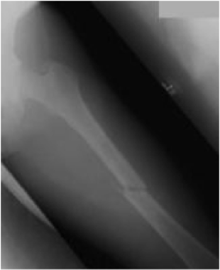 An example of an isolated, short, oblique midshaft femur fracture. Although not seen in this x-ray film, radiographic visualization of both the proximal and distal joints should be performed for all diaphyseal fractures.
Traumatic femur fractures in the young individual are generally caused by high-energy forces and are often associated with multisystem trauma. In the elderly population, femur fractures are typically caused by a low energy mechanism such as a fall from standing height. Isolated injuries can occur with repetitive stress and in the presence of metabolic bone diseases, metastatic disease or primary bone tumors. [1, 2, 3]
The femur is very vascular, and fractures can result in significant blood loss into the thigh. Up to 40% of isolated fractures may require transfusion as such injuries can result in loss of up to three units of blood. [4] This factor is significant, especially in elderly patients who have less cardiac reserve.
Femur fracture patterns vary according to the direction of the force applied and the quantity of force absorbed. A perpendicular force results in a transverse fracture pattern, an axial force may injure the hip or knee, and rotational forces may cause spiral or oblique fracture patterns. The amount of comminution present increases with increasing amounts of force. [1, 2, 5, 6]
Most femur fractures are treated surgically. The goal of early surgical treatment is stable, anatomic fixation, allowing mobilization as soon as possible. Surgical stabilization is also important for early extremity function, allowing both hip and knee motion and strengthening. Injuries and fractures of the femur may have significant short and long-term effects on gait kinematics and function if alignment is not restored.
Related Medscape resources
Resource Center Exercise and Sports Medicine
Specialty Site Emergency Medicine
Specialty Site Orthopaedics
Etiology
Traumatic causes of femoral fractures include the following:
-
Motor vehicle trauma (eg, motor vehicle collision, motorcycle collision, auto/pedestrian collision)
-
Sports (eg, high-speed and contact/collision sports with direct trauma, skiing, football, hockey)
-
Falls (eg, from height, mountain climbing, pole vaulting)
-
Gunshot wounds
-
An update to the American Academy of Pediatrics’ 2006 guidelines for differential diagnosis of fractures caused by child abuse reported that abuse causes between 12% and 20% of all fractures in infants and children; however, in children younger than 3 years, physicians misdiagnose as many as 20% of fractures caused by abuse. The femur, humerus, and tibia are the most common long bones fractured in child abuse. The report further added that in the nonambulatory child, femoral fractures are more likely to be caused by child abuse, whereas these fractures in ambulatory children are rarely inflicted by abuse. [7, 8]
Pathologic causes include the following:
-
Metabolic bone disease
-
Primary bone tumor
-
Metastatic tumor
-
Infection
-
Prolonged bisphosphonate use
Causes of stress fracture include the following:
-
Repetitive impact activities such as running (jogging) and jumping
-
Metabolic bone disease
-
Amenorrheic or oligomenorrheic female runners
-
Abnormal bone mineral density
-
Improper training
-
Improper footwear
Epidemiology
United States statistics
The incidence of femoral shaft fractures ranges from 9.5 to 18.9 per 100,000 annually. [9] From 0.7% to 1.7% of all fractures in children involve the femoral shaft. [10]
Approximately 250,000 proximal femur fractures occur in the United States annually. This number is anticipated to double by the year 2050. [11]
High-energy injuries are most common in younger males. See the images below.
The incidence of femur fractures increases in elderly patients.
Fatigue fractures of the femur occur at a rate of 19.9 per 10,000 persons per year. [12]
Stress fractures as a whole occur in up to 37% of runners. [9] The femur accounts for 11% of stress fractures. Approximately 53% of these fractures occur about femoral shaft. [9]
Functional Anatomy
The femur is the strongest, longest, and heaviest bone in the body and is essential for normal ambulation. The femoral shaft is tubular with a slight anterior bow, extending from the lesser trochanter to the flare of the femoral condyles. The femur is subject to many forces during ambulation including axial loading, bending, and torsional forces. During weight bearing, the medial cortex is subject to compressive forces while tensile forces are placed on the lateral cortex during contraction, the large muscles surrounding the femur account for a large portion of the applied forces. [1, 2, 5, 6, 13]
Several large muscles attach to the femur which can affect displacement of certain fracture patterns. Proximally, the gluteus medius and minimus attach to the greater trochanter. The forces from these muscles may result in an abduction deformity to the proximal fragment of proximal femoral shaft and subtrochanteric femur fractures. The iliopsoas attaches to the lesser trochanter, resulting in a flexion deformity of this same fragment, in fractures occurring below the level of the lesser trochanter. The linea aspera (rough line on the posterior shaft of the femur) reinforces the strength of the femur and is an attachment for the gluteus maximus, adductor magnus, adductor brevis, vastus lateralis, vastus medialis, vastus intermedius, and short head of the biceps. Distally, the large adductor muscle mass attaches medially, resulting in an apex lateral deformity seen in certain distal femur fractures. The medial and lateral heads of the gastrocnemius attach over the posterior femoral condyles, resulting inflexion deformity in distal-third fractures.
The blood supply enters the femur through metaphyseal arteries and branches of the profunda femoris artery, penetrating the diaphysis and forming medullary arteries extending proximally and distally. Healing of femur fracture is enhanced by the surrounding muscles and soft tissues contributing to local recruitment of blood supply around the callus. The femoral artery courses down the medial aspect of the thigh to the adductor hiatus, at which time it becomes the popliteal artery. Injuries to the artery occur at the level of the adductor hiatus, where soft-tissue attachments may cause tethering.
See also Medscape Drugs & Diseases articles Nerve Entrapment Syndromes and Nerve Entrapment Syndromes of the Lower Extremity.
Sport-Specific Biomechanics
Trauma-induced fractures of the femur occur with contact and during high-speed sports. A significant amount of energy is transferred to the limb in a femur fracture, such as might be generated in skiing, football, hockey, rodeo, and motor sports.
Stress fracture
A femoral stress fracture is the result of cyclic overloading of the bone. With prolonged activity, the muscles that dissipate force to bone begin to fatigue placing increased force on the bone itself. Muscle fatigue may also contribute by altering gait kinematics. Stress becomes concentrated at susceptible areas with eventual weakening and subsequent microfracture, as these repetitive stresses overcome the ability of the bone to remodel. The area most susceptible to stress fractures is the medial junction of the proximal and middle third of the femur. Fractures in this location occur as a result of the compression forces on the medial femur. [2]
A study suggested that the lateral cortex of the femoral shaft may also be susceptible to stress fracture due to tensile forces. [14]
Stress fractures can also occur on the lateral aspect of the femoral neck in areas of distraction and are less likely to heal non-operatively than compression-side stress fractures on the medial side. Stress fractures occur most often in repetitive overload sports such as in runners and basketball players. For more information, refer to the Medscape Drugs & Diseases article Femoral Neck Stress Fracture.
Prognosis
Long-term symptoms after fracture include muscular weakness, limited standing and walking, gait abnormalities, some intermittent pain, and inability to return to preinjury work.
Surgical management is rarely needed to treat femoral stress fractures; however, surgical stabilization is recommended for recalcitrant cases.
Complications
Complications associated with traumatic femoral fractures include the following:
-
Nonunion
-
Delayed union
-
Malunion
-
Infection
-
Venous thromboembolic event (ie, deep venous thrombosis or pulmonary embolism)
-
Pain
-
Ambulatory dysfunction
-
Refracture
-
Hardware failure
-
Prominent hardware
-
Neurologic injury
-
Peroneal nerve palsy - Most commonly due to traction
-
Pudendal nerve injury - Due to compression at the perineal post
-
Sciatic nerve injury
-
Vascular injury
-
False aneurysm
-
Atrioventricular fistula
-
Compartment syndrome
Complications associated with femoral stress fractures include the following:
-
Progression to a complete fracture
-
Refracture
-
Nonunion
-
Osteonecrosis
-
Arthritis
-
Continued pain
-
An example of an isolated, short, oblique midshaft femur fracture. Although not seen in this x-ray film, radiographic visualization of both the proximal and distal joints should be performed for all diaphyseal fractures.
-
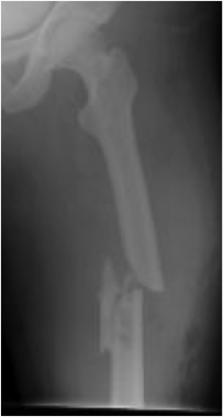
Radiograph of a high-energy femoral shaft fracture.
-
Radiograph of a high-energy femoral shaft fracture.
-
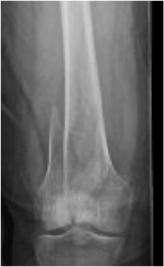
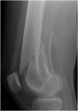
Radiograph of an intra-articular distal femur fracture.
-
Radiograph of an intra-articular distal femur fracture.
-
Full length AP radiograph of an intertrochanteric fracture.
-
MRI of a patient with a stress fracture at the base of the femoral neck.
-
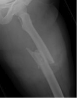
AP radiograph of a healing femoral shaft fracture after intramedullary nailing.
-
Lateral radiograph of a healing femoral shaft fracture after intramedullary nailing.
-
An intra-articular distal femur fracture treated with intramedullary nailing as well as independent screw fixation.
-
An intra-articular distal femur fracture treated with intramedullary nailing as well as independent screw fixation.


