Continually Updated Clinical Reference
 
 
  All Sources     eMedicine     Medscape     Drug Reference     MEDLINE
 
eMedicine - Elbow Dislocation : Article by

Quick Find
Authors & Editors
Introduction
Clinical
Differentials
Workup
Treatment
Medication
Follow-up
Miscellaneous
Multimedia
References

Related Articles
Compartment Syndrome, Upper Extremity


Patient Education
Breaks, Fractures, and Dislocations Center

Elbow Dislocation Overview

Elbow Dislocation Causes

Elbow Dislocation Symptoms

Elbow Dislocation Treatment

Broken Elbow Overview


Author: Mark E Halstead, MD, Clinical Instructor, Departments of Pediatrics and Orthopedics, Washington University School of Medicine; Team Physician, St Louis Rams, Washington University Athletics

Mark E Halstead is a member of the following medical societies: American Academy of Pediatrics, American College of Sports Medicine, and American Medical Society for Sports Medicine

Coauthor(s): David T Bernhardt, MD, Director of Adolescent and Sports Medicine Fellowship, Associate Professor, Department of Pediatrics, University of Wisconsin

Editors: Joseph P Garry, MD, Director of Sports Medicine and Sports Medicine Fellowship, Associate Professor of Family Medicine and Exercise and Sport Science, Department of Family Medicine, East Carolina University Brody School of Medicine; Francisco Talavera, PharmD, PhD, Senior Pharmacy Editor, eMedicine; Henry T Goitz, MD, Chief, Sports Medicine, Associate Professor, Department of Orthopaedic Surgery, Medical College of Ohio; Jon B Whitehurst, MD, Clinical Instructor of Surgery, University of Illinois College of Medicine; Partner and Executive Board Member, Rockford Orthopedic Associates; Orthopedic Chairman, Rockford Memorial Hospital; Sherwin SW Ho, MD, Associate Professor, Department of Surgery, Section of Orthopedic Surgery and Rehabilitation Medicine, University of Chicago

Author and Editor Disclosure

Synonyms and related keywords: elbow dislocation, dislocation of elbow, dislocated elbow, radial head dislocation, ulnar dislocation, radial head subluxation, FOOSH injury, falling on an outstretched hand, nursemaid's elbow, elbow injury, elbow trauma

Background

Elbow dislocation is the most common dislocation in children; in adults, it is the second most common dislocation after that of the shoulder.1, 2, 3, 4 The elbow is amazingly stable, relying more on bony anatomy configuration for stability rather than ligaments. Considerable force is necessary to dislocate the elbow; sports activities account for up to 50% of elbow dislocations, and this type of injury is more commonly seen in adolescent and young adult populations.

Posterior elbow dislocations comprise over 90% of elbow injuries. Early recognition of this injury is required due to the need for early reduction, given a higher likelihood for poor function and possible neurovascular compromise with delays in reduction.1, 2, 3, 4, 5 Associated fractures are not infrequent with elbow dislocations, given the force that is required to dislocate the elbow.

Anterior dislocations are seen much less commonly than posterior dislocations. Divergent dislocations, which result in the ulna and radius dislocating in opposite directions, are even more rare. In the pediatric population, radial head subluxation is the main cause of elbow dislocations.

For excellent patient education resources, visit eMedicine's Breaks, Fractures, and Dislocations Center. Also, see eMedicine's patient education articles Elbow Dislocation and Broken Elbow.

Related eMedicine topics:
Joint Reduction, Elbow Dislocation, Posterior
Joint Reduction, Radial Head Dislocation
Posttraumatic Heterotopic Ossification

Related Medscape topics:
Resource Center Adolescent Medicine
Resource Center Exercise and Sports Medicine
Resource Center Joint Disorders
Specialty Site Neurology & Neurosurgery
Specialty Site Orthopaedics
Specialty Site Pediatrics

Frequency

United States

The rate of elbow dislocation is 6-13 cases per 100,000 people, and this injury occurs more frequently in males than in females. Of all elbow dislocations, 10-50% are sports related. More than 90% of elbow dislocations are posterior dislocations.

Functional Anatomy

The elbow is primarily a flexion-extension hinge joint, which also allows for pronation and supination. Normal range of motion (ROM) at the elbow should be extension to 0° and flexion to 150°.1, 2

The humerus and ulna form a very stable unit, which is generally resistant to disruption unless considerable force is applied. This inherent stability also reduces the likelihood of redislocation. The primary bony stabilizers are the coronoid and radial head.

The medial collateral ligament (MCL) and lateral collateral ligament (LCL) comprise the ligamentous stability of the elbow and act as a back-up system to the elbow's natural bony stability. The MCL consists of 3 bands, the anterior oblique, posterior oblique, and the transverse. The anterior band provides most of the resistance to valgus stress. The LCL has 2 bands, the ulnar collateral and radial collateral.

The 2 main compartments of the elbow are the anterior and posterior compartments. The anterior compartment contains the brachial artery and the ulnar and median nerves. This compartment is more commonly affected by dislocations and is the reason for clinical concern regarding brachial artery disruption and median or ulnar nerve entrapment.1, 2, 4, 6

The ulnar nerve passes posteriorly to the medial epicondyle of the humerus, and then it travels deep in the forearm before becoming more superficial again at the wrist. The close proximity of the ulnar nerve to the medial epicondyle allows for the increased likelihood of entrapment when a dislocation occurs. The median nerve is also frequently affected and travels intimately with the brachial artery, which predisposes to simultaneous injury for both the artery and nerve. The posterior compartment contains the radial nerve and triceps brachii muscle.

Anatomically, the mechanism for elbow dislocations is believed to occur as a continuum of damaged/torn structures, beginning laterally with the ulnar portion of the LCL, followed by complete LCL disruption, then damage to the anterior and posterior compartments. The posterior MCL can then become damaged, leaving the anterior portion intact. Further force can allow the elbow to pivot about the anterior bundle of the MCL, potentially damaging it. The LCL, therefore, is considered to be the initial weak link in elbow dislocations.

In the pediatric population, the clinician should be aware of the 6 ossification centers of the elbow joint as well as the annular ligament. The capitellum, radial head, internal (medial) epicondyle, trochlea, olecranon, and external (lateral) epicondyle (CRITOE) is the order in which the ossification centers appear. These centers may often be mistaken for fractures on x-rays. NOTE: A general rule of thumb for the time of appearance of the ossification centers is "1-3-5-7-9-11," which are the ages in years, corresponding to the CRITOE pneumonic.

In cases in which there is radial head subluxation, the radial head slips under the annular ligament and becomes trapped.

Sport-Specific Biomechanics

Biomechanically, no single sport definitively increases the risk of elbow dislocations; however, sports that increase the likelihood of a person falling onto an outstretched hand (ie, FOOSH injury) (eg, gymnastics, rollerblading, cycling) may theoretically increase the risk of elbow dislocation.


History

  • In posterior elbow dislocations, the patient often describes falling on an outstretched hand (ie, the FOOSH injury) as the mechanism of injury. Some clinicians speculate that the elbow is more likely to dislocate when it is slightly abducted and flexed. When compressive forces are directed on to the outstretched hand, the radius and ulna, along with the valgus force at the elbow, suffer the common posterolateral dislocation. These forces also contribute to associated fractures. In addition, hyperextension at the elbow has been seen with elbow dislocations.
  • Anterior dislocations are usually the result of a direct posterior blow to a flexed elbow. Associated fractures of the olecranon are commonly seen.
  • Divergent dislocations are very rare injury and are associated with significant high-energy trauma to the elbow.
  • In children, radial head subluxations often occur when the arm is pulled. The child commonly holds the arm pronated, mildly flexed, and abducted against the body and refuses or fights any manipulation of the affected arm.
  • Essential elements of the dislocation history include the mechanism of the injury, the time between the injury and presentation, functioning, previous attempts at reduction/manipulation, swelling, location, and the type of pain.

Related Medscape topics:
Resource Center Fracture
Resource Center Joint Disorders
Resource Center Trauma

Physical

Neurovascular assessment and documentation of the clinical evaluation are essential in any elbow dislocation because associated brachial artery and ulnar nerve injuries are frequent. Median nerve injuries are also common.

  • Evaluate the injury for swelling.
  • Note any deformities that are present.
  • Posterior elbow dislocations often have a very prominent olecranon and a forearm that appears foreshortened.
  • Anterior elbow dislocations have the appearance of an elongated forearm, and the arm is held in extension.
  • Touch sensation of the median and ulnar nerves can be quickly assessed by testing the distal palmar aspect of the first through fifth digits. (The ulnar nerve innervates the medial one half of the fourth digit and the fifth digit, as well as the dorsal side of the same digits.)
  • Motor function of the median and ulnar nerve can be quickly assessed by evaluating the abduction and adduction strength of the digits (ulnar nerve) and the opposability of the thumb (median nerve).

Related Medscape topics:
Resource Center Vascular Surgery
Specialty Site Neurology & Neurosurgery
Specialty Site Orthopaedics

Causes

Unlike the shoulder, a previous elbow dislocation does not predispose a patient to future dislocations. Elbow dislocations are commonly caused by a fall on an outstretched hand or by a traumatic event. Radial head subluxations in children are usually caused by pulling or yanking on the child's arm when the child's elbow is extended.


Compartment Syndrome, Upper Extremity

Other Problems to Be Considered

Elbow fractures

Monteggia fracture

Pediatric radial head subluxation (nursemaid's elbow)


Imaging Studies

  • Before reduction of the injury, obtain anteroposterior and lateral radiographs of the elbow. If clinicians feel comfortable trying a reduction in the field, they may do so before obtaining radiographs; however, postreduction films should be obtained and the affected limb's neurovascular status should be documented pre- and postreduction.
  • Postreduction films are also necessary to ensure adequate reduction and to evaluate for fractures. The recreation of a normal radiocapitellar line (the line drawn through the shaft of the radius through the center of the capitellum) should be evident on all radiographic views.
  • Computed tomography (CT) scanning may be useful in the evaluation for the full extent and location of fractures that may occur with a complex elbow dislocation.
  • Magnetic resonance imaging (MRI) has little utility in an acute elbow dislocation.

Related Medscape topic:
Specialty Site Radiology

Other Tests

  • A role may exist for angiography in the evaluation of a suspected associated arterial injury; however, if angiography would significantly delay vascular repair, the operating room may be a more appropriate setting for its use.

Procedures

  • Several methods for reducing a posterior elbow dislocation have been suggested.1, 2, 3, 4, 7, 8, 9, 10, 11 Before reduction, adequate patient analgesia and sedation are necessary not only for patient comfort but also for the reduction. Radial head subluxations in children (ie, nursemaid's elbow) can generally be reduced without the need for sedation or analgesia. The 2 most commonly used techniques for the posterior dislocation are as follows:
    • The author's preferred method is to place the patient in a prone position with the affected elbow flexed at 90° and the humerus supported by the table (see Image 1). The hand of the affected arm should be pointing toward the ground. Apply downward traction to the forearm, which is held in slight pronation, while using the other hand to grasp the humerus, and apply pressure to the olecranon in a downward motion to facilitate reduction.
    • A second method involves the patient lying supine with the affected arm extended to the side in slight flexion. An assistant applies traction to the humerus toward the patient's body, while a second individual applies in-line traction to the forearm, the latter of which is held slightly flexed and supinated to facilitate reduction.
  • Perform anterior elbow dislocation reductions by grasping the humerus with 2 hands to apply countertraction, while an assistant provides in-line traction to the forearm. Orthopedic assistance may be beneficial given the infrequency of anterior dislocations.
  • Treat radial head subluxations in children by supporting the child's affected arm with your nondominant hand, with moderate pressure placed on the radial head. With your dominant hand on the child's wrist, apply gentle traction to the arm, and in 1 motion, supinate and fully flex the affected elbow. You can palpate for a click over the radial head with your nondominant hand.

    Some studies have suggested that hyperpronation may be a more effective way to reduce the subluxation.12 The hyperpronation method also may be attempted if a subluxation cannot be reduced by the more commonly used supination and flexion method.

  • Postreduction: Place the elbow through gentle ROM testing. (NOTE: Extending the elbow beyond 20° from full extension may cause the elbow to redislocate and is not recommended.) Inability to move the elbow smoothly through ROM following reduction should raise the suspicion of a trapped medial epicondyle fracture. Reexamination for neurovascular integrity should take place postreduction, followed by follow-up elbow radiographs. The elbow joint is expected to be unstable following a dislocation. (NOTE: Assessment for stability following reduction is likely to be painful for the patient and does not provide further information for the management of this condition.)
  • If near-full ROM is present in the affected elbow without associated fractures, the elbow may be splinted using a posterior splint, with the forearm in slight pronation and the elbow flexed at 90°.
  • If there are any concerns regarding potential neurovascular compromise or excessive swelling, or the risk of compartment syndrome exists, it would be beneficial to admit the patient to a hospital for a 24-hour period of observation following the injury.
  • For on-site reductions, such as at a game or in a training room where radiographs may not be readily available, a physician who feels comfortable reducing an elbow may do so, provided the physician performs and documents a complete neurovascular examination before and after the reduction.4 Because of the high association of fractures with elbow dislocations, the patient should be advised to obtain radiographs of the elbow soon after the reduction.


Acute Phase

Rehabilitation Program

Physical Therapy

Early ROM exercises in stable, reduced elbow dislocations has been shown to be associated with an improved outcome. However, immobilization of the affected elbow for longer than 3 weeks in patients following an elbow dislocation has been associated with loss of ROM compared with patients who start early ROM exercises.10

Medical Issues/Complications

  • Brachial artery disruption may be seen in any elbow dislocation and special attention should be made in open dislocations.
  • Ulnar nerve injury may occur in up to 15% of elbow dislocations. Perform an initial neurovascular assessment and frequent reassessments.
  • Median nerve injury is frequently seen in conjunction with brachial artery injuries because both of these structures are in close anatomic proximity. The median nerve function should always be assessed with a higher suspicion of injury if a brachial artery injury is known to exist. The median nerve may also be injured or entrapped during a reduction.
  • Associated fractures for elbow dislocation include those of the radial head or neck (5-10%), medial/lateral epicondyle avulsions (10%), those of the coronoid process (10%), and fractures of the distal radius, ulna, and proximal humerus (10%).
  • Compartment syndrome may develop in the forearm fascia or biceps tendon due to massive swelling, which may occur in an acute elbow dislocation. Compartment syndrome must be considered in the differential diagnosis in the presence of persistent patient pain, particularly when exacerbations of pain occur with passive finger and wrist extension of the dislocated arm.
  • Ectopic calcification, primarily around the collateral ligaments, is common after an elbow dislocation, provides no limitations and requires no intervention.
  • Myositis ossificans may also be seen if significant hemarthrosis developed with the elbow dislocation.

Surgical Intervention

Seek surgical intervention by an orthopedist if any signs of neurovascular compromise, associated fractures, or nonreducible dislocations are present.

Related Medscape topics:
Resource Center Fracture
Resource Center Joint Disorders
Resource Center Vascular Surgery
Specialty Site Neurology & Neurosurgery
Specialty Site Orthopaedics
Specialty Site Surgery

Consultations

Obtain orthopedic consultation if any signs of neurovascular compromise, associated fractures, or nonreducible dislocations are present.

Recovery Phase

Rehabilitation Program

Physical Therapy

Patients with limitations in ROM on follow-up evaluation may benefit from more aggressive physical therapy to regain loss of mobility.

Maintenance Phase

Rehabilitation Program

Physical Therapy

Depending on the severity of the elbow dislocation, it may take several months for the elbow to fully heal. Muscle-strengthening activities, in addition to the ROM program, are important to improve endurance of the elbow. Incorporate sport-specific training as the athlete progresses through rehabilitation to ensure a safe return to his/her sport.13

Related Medscape topic:
Resource Center Exercise and Sports Medicine

Surgical Intervention

Surgical intervention may be needed for a functional flexion contracture or for chronic residual instability.14, 15, 16


Use of medication for elbow dislocations is beneficial in the acute setting when reduction of the dislocation is to take place. Choosing both an anxiolytic and a pain medication is ideal for a conscious sedation to facilitate reduction. Once reduction has occurred, pain may still be an issue, and it would be reasonable to provide the patient with oral pain medication to use in the outpatient setting.

Drug Category: Anxiolytics

Anxiolytics allow for relaxation and mild sedation when reduction of a dislocated elbow is attempted. These agents also allow for a lower dose of analgesics to be used.

Drug NameMidazolam (Versed)
DescriptionDOC for anxiolytics. Shorter-acting benzodiazepine sedative-hypnotic that is useful in patients requiring acute and/or short-term sedation. Midazolam is also useful for its amnestic effects.
Adult Dose2.5-5 mg IV; not to exceed 2.5 mg IV over 2 min; not to exceed a total of 10 mg

Allow 2-3 min between doses to assess effect.

May start with 1 mg IV and slowly titrate to effect.
Pediatric Dose0.05-0.1 mg/kg IV; not to exceed the total dose of 10 mg

0.05-0.15 mg/kg IM 30-60 min before the procedure; not to exceed the total dose of 10 mg

0.25-0.5 mg/kg PO; not to exceed the total dose of 10 mg

Intranasal: 0.2 mg/kg; may repeat in 5-15 min
ContraindicationsDocumented hypersensitivity to midazolam, any components, and cherries (syrup only); acute narrow-angle glaucoma; existing CNS depression
InteractionsCNS depressants; alcohol may increase sedation and respiratory depression; narcotic agents may increase hypotension; may increase midazolam serum concentrations with cimetidine, ranitidine, erythromycin, diltiazem, verapamil, fluconazole, ketoconazole, itraconazole; protease inhibitors may decrease the midazolam metabolism
PregnancyD - Fetal risk shown in humans; use only if benefits outweigh risk to fetus
PrecautionsCaution in patients with renal and hepatic impairment, CHF, and pulmonary disease; respiratory/cardiac monitoring is essential during sedation in children

Drug NameLorazepam (Ativan)
DescriptionSedative hypnotic with short onset of effects and a relatively long half-life. By increasing the action of gamma-aminobutyric acid (GABA), which is a major inhibitory neurotransmitter in the brain, this agent may depress all levels of CNS, including the limbic and reticular formation. When the patient needs to be sedated for longer than a 24-hour period, this medication is excellent.
Adult Dose1-4 mg/dose IV given slowly over 2-5 min; may repeat in 10-15 min; not to exceed 8 mg/12 h
Pediatric Dose0.05 mg/kg PO/IM (range 0.02-0.09 mg/kg)

0.05 mg/kg IV (range 0.02-0.09 mg/kg; may consider smaller doses 0.01-0.03 mg/kg) and repeat q20 min to achieve effect; give slow IV
ContraindicationsDocumented hypersensitivity; narrow-angle glaucoma; severe hypotension
InteractionsOther CNS/respiratory depressants may increase effects
PregnancyD - Fetal risk shown in humans; use only if benefits outweigh risk to fetus
PrecautionsUse with caution in patients with impaired renal or hepatic function or who have compromised pulmonary function.

Drug NameDiazepam (Valium)
DescriptionDepresses all levels of the CNS (eg, limbic and reticular formation), possibly by increasing the activity of GABA. Individualize the dosage and increase cautiously to avoid adverse effects.
Adult Dose10 mg PO

5 mg IV; may repeat with 2.5 mg if needed
Pediatric Dose0.2-0.3 mg/kg PO; not to exceed 10 mg; 45-60 min before the procedure

0.04-0.3 mg/kg/dose IV/IM; not to exceed 0.6 mg/kg in 8h period
ContraindicationsDocumented hypersensitivity to diazepam or any component (emulsified diazepam injection contains soybean oil, egg yolk, phospholipids); comatose patients; narrow-angle glaucoma
InteractionsCNS depressants, cimetidine, and erythromycin may decrease metabolism; valproic acid may displace diazepam from the binding sites, resulting in increased sedation; use with ritonavir is not recommended
PregnancyD - Fetal risk shown in humans; use only if benefits outweigh risk to fetus
PrecautionsCaution in patients with other CNS depressants; hypoalbuminemia, and liver or hepatic dysfunction

Drug Category: Analgesics

Pain control is essential to quality patient care. Analgesics ensure patient comfort, promote pulmonary toilet, and have sedating properties, which are beneficial for patients who have sustained trauma or who have sustained injuries.

Related eMedicine topics:
Toxicity, Acetaminophen
Toxicity, Narcotics

Related Medscape topics:
Resource Center Adverse Drug Events Reporting
Resource Center Opioids: A Guide to State Opioid Prescribing Policies
Resource Center Pain Management: Advanced Approaches to Chronic Pain Management
Resource Center Pain Management: Pharmacologic Approaches

Drug NameMorphine (Duramorph, Astramorph, MS Contin)
DescriptionIndicated for moderate to severe acute and chronic pain.
Adult Dose2.5-20 mg/dose IV/IM/SC; usual dose of 10 mg; dosing q2-6h
Pediatric Dose<12 years: 0.05-0.1 mg/kg IV 5 min before the procedure

>12 years: 3-4 mg IV; may repeat in 5 min prn
ContraindicationsDocumented hypersensitivity to morphine sulfate; increased intracranial pressure; severe respiratory depression; severe liver or renal insufficiency
InteractionsPhenothiazines may antagonize the analgesic effects of opiate agonists; tricyclic antidepressants, MAO inhibitors, and other CNS depressants may potentiate the adverse effects of morphine.
PregnancyB - Fetal risk not confirmed in studies in humans but has been shown in some studies in animals
PrecautionsAvoid in the presence of hypotension, respiratory depression, nausea, emesis, constipation, and urinary retention; caution in patients with atrial flutter and other supraventricular tachycardias; has vagolytic action and may increase the ventricular response rate

Drug NameFentanyl (Duragesic, Sublimaze)
DescriptionPotent narcotic analgesic with a much shorter half-life than morphine sulfate. DOC for conscious sedation analgesia. Ideal for analgesic action of short duration during anesthesia and immediate postoperative period.

Excellent choice for pain management and sedation with short duration (30-60 min) and easy to titrate. Easily and quickly reversed by naloxone. After initial dose, the subsequent doses should not be titrated more frequently than q3h or q6h thereafter.

When using the transdermal dosage form, most patients are controlled with 72 h dosing intervals. However, some patients require dosing intervals of 48 h.
Adult Dose25-50 mcg IV; repeat doses of 25 mcg up to 4-5 times q5 min if needed
Pediatric Dose1-2 mcg/kg/dose IV/IM with repeated doses at 30-min intervals prn
ContraindicationsDocumented hypersensitivity; increased intracranial pressure; severe hepatic or renal insufficiency; severe respiratory depression
InteractionsPhenothiazines may antagonize the analgesic effects of opiate agonists; tricyclic antidepressants may potentiate the adverse effects of fentanyl when both drugs are used concurrently.
PregnancyC - Fetal risk revealed in studies in animals but not established or not studied in humans; may use if benefits outweigh risk to fetus
PrecautionsCaution in the presence of hypotension, respiratory depression, constipation, nausea, emesis, and urinary retention; idiosyncratic reaction, known as chest wall rigidity syndrome, may require neuromuscular blockade in order to increase ventilation.

Drug NameOxycodone and acetaminophen (Percocet, Roxicet)
DescriptionDrug combination indicated for the relief of moderate to severe pain.
Adult Dose1-2 tab PO q4-6h prn
Pediatric Dose0.05-0.15 mg/kg/dose up to 5 mg/dose PO q4-6h based on oxycodone component
ContraindicationsDocumented hypersensitivity to oxycodone, acetaminophen; severe liver or renal insufficiency
InteractionsPhenothiazines may decrease the analgesic effects of this medication; the toxicity increases with the coadministration of either CNS depressants or tricyclic antidepressants.
PregnancyC - Fetal risk revealed in studies in animals but not established or not studied in humans; may use if benefits outweigh risk to fetus
PrecautionsThe duration of action may increase in the elderly; be aware of the total daily dose of acetaminophen the patient is receiving; do not exceed 4,000 mg/24h of acetaminophen; higher doses may cause liver toxicity.

Drug NameAcetaminophen and codeine (Tylenol with codeine, Tylenol #3)
DescriptionIndicated for the treatment of mild to moderate pain.
Adult Dose1-2 tab PO q4h; not to exceed 12 tab/24 h
Pediatric Dose<3 years: 0.5-1 mg codeine/kg/dose PO q4-6h

3-6 years: 5 mL PO tid/qid prn

7-12 years: 10 mL PO tid/qid prn

>12 years: 15 mL PO q4h prn
ContraindicationsDocumented hypersensitivity to acetaminophen, codeine, or any component
InteractionsCNS depressants, alcohol, phenothiazines, and tricyclic antidepressants can increase the adverse effects of codeine or increase the hepatotoxicity of acetaminophen
PregnancyC - Fetal risk revealed in studies in animals but not established or not studied in humans; may use if benefits outweigh risk to fetus
PrecautionsCaution in patients with hypersensitivity to morphine, hydromorphone, levorphanol, oxycodone, oxymorphone, and hydrocodone

Drug NameAcetaminophen and hydrocodone (Vicodin, Hydrocet, Lorcet)
DescriptionDrug combination indicated for moderate to severe pain.
Adult Dose1-2 tab or cap PO q4-6h prn
Pediatric DoseNot established
ContraindicationsDocumented hypersensitivity to hydrocodone, acetaminophen; CNS depression; severe respiratory depression
InteractionsCoadministration with phenothiazines may decrease analgesic effects; the toxicity increases with CNS depressants or tricyclic antidepressants
PregnancyC - Fetal risk revealed in studies in animals but not established or not studied in humans; may use if benefits outweigh risk to fetus
PrecautionsThe tablets contain metabisulfite, which may cause hypersensitivity; caution in patients who are dependent on opiates because this substitution may result in acute opiate-withdrawal symptoms; caution in patients with severe renal or hepatic dysfunction


Return to Play

A follow-up examination before the patient's return to play is necessary to reassess motion of the formerly dislocated elbow following the immobilization and early ROM period. Most individuals can return to play 3-6 weeks following an elbow dislocation.

In sports in which an elbow dislocation occurs in a player's nondominant arm, return to play may occur at the earlier end of the rehabilitation spectrumas long as motion is back to a level that is suitable to the physician and athlete. For elbow dislocations that occur in a player's dominant arm, return to play may take a longer time period. Throwing sports, such as baseball, may require the patient to undergo periods of rest up to 3 months following a dislocation, followed by a strengthening rehabilitative program once full motion is achieved.

Complications

Complications of elbow dislocation primarily include neurovascular compromise, compartment syndrome, and loss of ROM. Chronic regional pain syndrome may occur. Close attention to the neurologic examination pre- and postreduction as well as at the follow-up visit may alert the physician to potential neurologic problems.

Prevention

Elbow dislocations in children due to radial head subluxation (nursemaid's elbow) are often preventable. A child should not be forcibly pulled, lifted, or swung in the air by the hand or wrist. Always lift a small child from under the arms, rather than by the hand or wrist. Athletes who participate in high-risk sports, where falling on an outstretched arm is common, may be advised to wear protective gear to prevent elbow injuries.

Prognosis

Approximately 50% of patients with dislocated elbows achieve a full recovery, including full ROM. One third of patients experience some limitation of motion at the elbow, usually less than 10° of compromised motion. The remaining 10-15% of patients have more significant losses in function, primarily related to limited ROM. Some correlation exists between the severity of the initial injury and the likelihood of having significant motion limitations further in time from the injury occurrence.


Medical/Legal Pitfalls

  • Neurovascular compromise is the most crucial potential complication of elbow dislocations and can be quickly and easily assessed. Perform frequent neurovascular assessments and document the patient's neurovascular function, both pre- and postreduction. The higher occurrence of associated injuries to the median and ulnar nerves and brachial artery necessitate these evaluations.
  • Elbows may spontaneously reduce before clinical evaluation or any treatment. The clinician should be aware of this and suspect a reduced dislocation if swelling is noted and the mechanism of injury corresponds with a dislocation.
  • In-field reduction without a radiograph might invite legal inquiry, particularly if a postreduction radiograph demonstrates evidence of a fracture. Despite this, clinician comfort and judgment (ie, time delay to reducing the injury if awaiting a prereduction radiograph) must be exercised for each suspected injury.

Related Medscape topics:
Resource Center Exercise and Sports Medicine
Resource Center Joint Disorders
Resource Center Medical Malpractice and Legal Issues
Resource Center Trauma
Specialty Site Orthopaedics


Media file 1:  The preferred method for posterior elbow dislocation reduction is to lay the patient prone with the humerus supported by the exam table. Place one hand around the wrist of the affected arm and apply downward traction, while the other hand stabilizes the humerus and the thumb is placed over the olecranon, with gentle pressure applied to facilitate reduction.
Click to see larger pictureClick to see detailView Full Size Image
Media type:  Photo

Media file 2:  
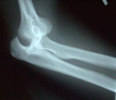
Posterior and lateral dislocation of the left elbow in a soccer goalie. A small avulsion fracture of the olecranon is present.
Click to see larger pictureClick to see detailView Full Size Image
Media type:  X-RAY


  1. O'Driscoll SW. Elbow dislocations. In: Morrey BF, ed. The Elbow and Its Disorders. 3rd ed. Philadelphia, Pa: WB Saunders; 2000:409-17.
  2. Rockwood CA Jr, Green DP, Bucholz RW, eds. Rockwood and Green's Fractures in Adults. 4th ed. Philadelphia, Pa: Lippincott Williams & Wilkins; 1996:971-85.
  3. Kuhn MA, Ross G. Acute elbow dislocations. Orthop Clin North Am. Apr 2008;39(2):155-61, v. [Medline].
  4. Ross G. Acute elbow dislocation: on-site treatment. Phys Sportsmed. Feb 1999;27(2):121-2. [Full Text].
  5. Sheps DM, Hildebrand KA, Boorman RS. Simple dislocations of the elbow: evaluation and treatment. Hand Clin. Nov 2004;20(4):389-404. [Medline].
  6. Nelson AJ, Izzi JA, Green A, Weiss AP, Akelman E. Traumatic nerve injuries about the elbow. Orthop Clin North Am. Jan 1999;30(1):91-4. [Medline].
  7. Mehta JA, Bain GI. Elbow dislocations in adults and children. Clin Sports Med. Oct 2004;23(4):609-27, ix. [Medline].
  8. Cohen MS, Hastings H 2nd. Acute elbow dislocation: evaluation and management. J Am Acad Orthop Surg. Jan-Feb 1998;6(1):15-23. [Medline].
  9. Ross G, McDevitt ER, Chronister R, Ove PN. Treatment of simple elbow dislocation using an immediate motion protocol. Am J Sports Med. May-Jun 1999;27(3):308-11. [Medline].
  10. Schippinger G, Seibert FJ, Steinböck J, Kucharczyk M. Management of simple elbow dislocations. Does the period of immobilization affect the eventual results?. Langenbecks Arch Surg. Jun 1999;384(3):294-7. [Medline].
  11. Villarin LA Jr, Belk KE, Freid R. Emergency department evaluation and treatment of elbow and forearm injuries. Emerg Med Clin North Am. Nov 1999;17(4):843-58, vi. [Medline].
  12. Macias CG, Bothner J, Wiebe R. A comparison of supination/flexion to hyperpronation in the reduction of radial head subluxations. Pediatrics. Jul 1998;102(1):e10. [Medline][Full Text].
  13. Duckworth AD, Kulijdian A, McKee MD, Ring D. Residual subluxation of the elbow after dislocation or fracture-dislocation: treatment with active elbow exercises and avoidance of varus stress. J Shoulder Elbow Surg. Mar-Apr 2008;17(2):276-80. [Medline].
  14. Micic I, Kim SY, Park IH, Kim PT, Jeon IH. Surgical management of unstable elbow dislocation without intra-articular fracture. Int Orthop. Aug 2 2008;epub ahead of print. [Medline].
  15. Jeon IH, Kim SY, Kim PT. Primary ligament repair for elbow dislocation. Keio J Med. Jun 2008;57(2):99-104. [Medline][Full Text].
  16. Duckworth AD, Ring D, Kulijdian A, McKee MD. Unstable elbow dislocations. J Shoulder Elbow Surg. Mar-Apr 2008;17(2):281-6. [Medline].
  17. Burra G, Andrews JR. Acute shoulder and elbow dislocations in the athlete. Orthop Clin North Am. Jul 2002;33(3):479-95. [Medline].
  18. Sano S, Rokkaku T, Imai K, et al. Radial head dislocation with ulnar plastic deformation in children: An osteotomy within the middle third of the ulna. J Shoulder Elbow Surg. Jul 19 2008;epub ahead of print. [Medline].

Elbow Dislocation excerpt

Article Last Updated: Aug 12, 2008