Practice Essentials
Cervical spine fractures lead to substantial morbidity and mortality. Neck injury in athletes can quickly end or change the future of an athlete. Failure to properly recognize and provide early care in cervical spine fracture cases may lead to devastating complications. [1, 2, 3, 4]
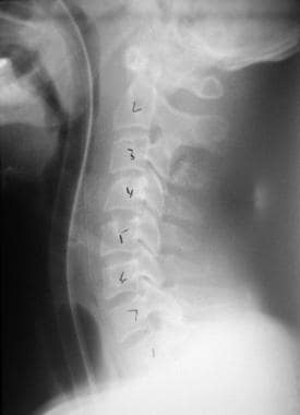
A C3 spinous fracture is depicted in the image below.
Signs and symptoms
Ask the athlete if neck pain is present. Determine location and quality of any pain. Ask if the pain radiates distally or to the extremities.
Determine if the patient is experiencing paresthesias or weakness.
Palpate the neck, and specifically feel for midline bony pain, muscle spasm, step-off, and crepitus.
Determine if extremity sensation is intact. Determine if the athlete can move all extremities without deficits.
Determine if the athlete can perform range of motion (ROM) in all directions without pain or symptoms. NOTE: Do not perform passive ROM of the neck.
Determine if head compression elicits pain or symptoms.
See Presentation for more detail.
Diagnosis
Imaging studies
Plain radiography is useful but is being replaced by the availability and performance of computed tomography (CT).
CT scanning provides much greater visualization of fractures and the capability to form 3-dimensional reconstructions of the vertebrae, which may be helpful preoperatively.
Magnetic resonance imaging serves best in evaluating the soft structures of the spinal column.
See Workup for more detail.
Management
Treat the site of injury with spinal precautions, and address airway, breathing, and circulation. Immobilize the athlete's neck in neutral position. Immobilize the spinal column on a backboard.
Transport the athlete to a facility with the ability to stabilize the athlete and to radiographically evaluate the neck.
If a fracture is detected, immediately consult a spinal orthopedic surgeon or neurosurgeon.
As with all fractures, pain management should be a primary concern.
See Treatment and Medication for more detail.
Epidemiology
United States statistics
The incidence of all spinal injuries in the United States has been reported at approximately 10,000 cases per year. Nearly 200,000 people in the United States have a history of spinal injuries. These statistics do not differentiate between injuries with fracture and injuries without fracture. [5, 6, 7]
Sports-related activities represent 10-15% of these injuries, and spinal injuries represent 2-3% of all sports-related injuries. Certain sports (eg, American football, diving, gymnastics, skiing, wrestling, rugby, hang gliding, paragliding, surfing, equestrian events, ice hockey) are more frequently associated with the risk of spinal trauma. [2, 3, 4, 6, 7, 8, 9, 10, 11, 12, 13, 14]
The most common spinal injuries cited in the literature are injuries secondary to contact sports such as football. Nearly 1.2 million high school athletes and 200,000 college and professional athletes participate in football. The National Football Head and Neck Injury Registry contains data on cervical spine injuries as a result of participation in football. A trend can be seen over time, as equipment and helmets improved. The incidence of cervical spine injuries increased until 1976. In that year, antispearing rules were established to prevent the athlete from using the helmet as driving force in tackles. Direct collision created higher axial loads than the neck could withstand, leading to high injury rates. This rule, along with better coaching of blocking and tackling techniques, has resulted in a significant decrease in the number of spinal injuries. [10]
Diving is often cited as another significant cause of cervical spine injuries. Injuries resulting from diving are often associated with devastating outcomes. Diving rules (eg, depth of starting areas) and proper technique have lowered the probability of injury during supervised athletic events. However, unsupervised swimming and diving into shallow water present significant risks. Public awareness of this problem has led to the development of special awareness programs, but the risk of injury remains high.
Leonard et al studied 540 children with cervical spine injuries and found that while motor vehicle crashes accounted for the most common injury in the axial region for children 2 and 2 to 7 years old, sports accounted for as many injuries as motor vehicle crashes (53% being subaxial) in children 8 to 15 years old. [15]
Functional Anatomy
The human spine serves to provide structural support and bony protection of the spinal cord. The cervical spine consists of 7 bony vertebrae separated by flexible intervertebral discs. They are joined together by an intricate network of ligaments, which helps form the normal lordotic curve of the cervical neck. [16]
The spinal column can be divided into 2 separate columns based on function and injury patterns. The anterior column consists of the bodies of the vertebrae, intervertebral discs, and the anterior and posterior longitudinal ligaments. The function of the vertebral body is to support weight. The posterior column contains the spinal canal and consists of the pedicles, laminae, articulating facets, and transverse and spinous processes. These structures form the vertebral arch, which encloses the vertebral foramen and protects the neural tissues.
The arch is formed by bilateral pedicles that are oriented posteriorly and join 2 laminae. The spinous process arises posteriorly from the vertebral arch. The cervical transverse processes and 4 articular processes also arise from the arch. The cervical transverse processes are unique to the vertebral column with an oval foramen transversarium. The vertebral arteries pass through these foramina. The posterior column also includes a group of ligaments including the supraspinous, infraspinous, interspinous, and nuchal ligaments.
The first 2 cervical vertebrae are atypical in form and function. The next 5 vertebrae are all similar in structure and function. The atlas, C1, is a ring-shaped bone that supports the skull. Two concave, superior articular facets articulate with the occipital condyles. The atlas does not have a body or spinous process. The atlas has an anterior and posterior arch, each with a tubercle and lateral mass. The axis, C2, is the strongest cervical vertebrae. The atlas rotates on 2 large articulating surfaces. The odontoid process (dens) projects superiorly from the C2 body and is the bony structure that the atlas rotates on. The odontoid process is held in place by the transverse ligament of the atlas.
Sport-Specific Biomechanics
Contact sports, falls, and diving in sports may lead to vertebral stress and fractures. Sports that involving tackling can increase exposure to mechanisms causing fractures.
Prognosis
The prognosis for the athlete is completely dependent on the type and extent of his or her injuries as well as associated problems.
Complications
An extensive list of complications of cervical fractures exists. Neurologic impairment is the most obvious and severe. The neurologic complications may range from paresthesias to complete loss of function. Cervical spinal cord injuries can be devastating because they may involve respiratory function and death. Spinal shock is also challenging to care for in the initial phase of injury. Long-term complications are related to immobilization and loss of function. These complications include skin breakdown, infections, loss of muscle mass, depression, and increased risk of suicide. Halo immobilization is associated with pin-site infections and osteomyelitis. Long-term collar immobilization is associated with skin breakdown.
Patient Education
A strong educational program should be included in all sports, especially contact and high-risk sports. Proper tackling must be taught from the beginning. A community education program should also be encouraged to prevent unsupervised sports injuries. Water and pool safety must be widely encouraged, including emphasis on feet-first water entry and avoidance of chemical impairment while engaging in water sports.
Everyone exposed to athletes (eg, physicians, coaches, trainers, referees, parents) should aid in providing this education. The rules of play for sports should reflect an effort to prevent injury and promote safe play. Paramedics and hospital personnel should be educated in proper care of a patient wearing equipment such as helmets and pads.
For patient education resources, see the Neck Strain, Neck Pain, and Whiplash.
-
Anteroposterior view of atlantooccipital dislocation.
-
Odontoid view of a Jefferson fracture.
-
Lateral view of a C2 fracture dislocation.
-
Odontoid type 2 fracture.
-
Lateral view of type 3 odontoid fracture.
-
Computed tomography scans of odontoid type 3 fracture.
-
Lateral view of a C3 spinous fracture.
-
Lateral view of hangman's fracture.
-
C3 flexion fracture.
-
C4 burst fracture.
-
Clay shoveler's fracture.
-
Unilateral locked facets on C5 and C6.
-
Bilateral facet fracture/dislocation at C6/C7.
-
Child with C6 flexion wedge fracture.
-
C7 lamina fracture.


