| Patient Education |
|
Click here for patient education.
|
|
You are in: eMedicine Specialties >
Sports Medicine > Spine
Thoracic Outlet Syndrome
Article Last Updated: Aug 30, 2006
AUTHOR AND EDITOR INFORMATION
Section 1 of 11
Author: Daryl A Rosenbaum, MD, Assistant Professor, Director of Sports Medicine Fellowship, Department of Family and Community Medicine, Wake Forest University School of Medicine; Team Physician, United States Soccer Federation
Daryl A Rosenbaum is a member of the following medical societies: American Academy of Family Physicians, American Medical Society for Sports Medicine, North Carolina Medical Society, and Society of Teachers of Family Medicine
Coauthor(s):
Matthew L Silvis, MD, Sports Medicine Fellow, Department of Family Medicine, Wake Forest University Baptist Medical Center;
Ajay Kalra, MD, Consulting Surgeon, Surgical Specialists;
Matt Thornburg, MD, Staff Physician, Department of Family and Community Medicine, University of Missouri Health Care at Columbia;
Donald Spadone, MD, Department of Surgery, Division of Vascular Surgery, Assistant Professor, University of Missouri Health Sciences Center
Editors: Craig C Young, MD, Medical Director of Sports Medicine, Departments of Orthopedic Surgery and Community and Family Medicine, Sports Medicine Fellowship Director, Associate Professor, Medical College of Wisconsin; Francisco Talavera, PharmD, PhD, Senior Pharmacy Editor, eMedicine; Russell D White, MD, Professor of Medicine, Department of Community and Family Medicine, University of Missouri-Kansas City School of Medicine, Truman Medical Center Lakewood; Jon Whitehurst, MD, Consulting Staff, Rockford Orthopedic Associates; William Jay Bryan, MD, Clinical Professor, Department of Orthopedic Surgery, Baylor University College of Medicine
Author and Editor Disclosure
Synonyms and related keywords:
TOS, vascular TOS, neurogenic TOS, disputed TOS, vascular thoracic outlet syndrome, neurogenic thoracic outlet syndrome, disputed thoracic outlet syndrome, nonspecific-type TOS, nonspecific TOS, nonspecific thoracic outlet syndrome, effort thrombosis, Paget-von Schrötter variant, Paget-Schroetter syndrome, Paget-von Schroetter syndrome, Paget-Schrötter syndrome, cervical rib syndrome, costoclavicular syndrome, scalenus syndrome, thoracic outlet compression syndrome, scalene triangle, costoclavicular space, pectoralis minor space, shoulder pain, arm pain, hand pain
Background
The thoracic outlet is bordered by the scalene muscles, first rib, and clavicle. Neurovascular structures pass from the neck and thorax into the axilla through this space. Thoracic outlet syndrome (TOS) is not the name of a single entity, but rather a collective title for a variety of conditions attributed to compression of these neurovascular structures as they traverse the thoracic outlet. It remains one of the most controversial clinical entities in medicine (Cuetter, 1989; Roos, 1999; Wilbourn, 1999).
Frequency
United States
The wide variability of patient symptoms and signs and the lack of an objective confirmatory test for the diagnosis of TOS makes correctly identifying patients with TOS difficult. Therefore, determining its exact incidence remains elusive; estimates range from 3-80 cases per 1000 population (Huang, 2004). TOS is more common in women, particularly those with poor muscular development, poor posture, or both (Safran, 2004).
Functional Anatomy
The neurovascular bundle courses through 3 spaces, or triangles, as it exits the neck to reach the axilla and proximal arm. All 3 spaces can be the source of compression of the various components of the neurovascular bundle, including the brachial plexus and the subclavian vessels. These spaces are small at rest and become even smaller with certain arm maneuvers such as abduction and external rotation. This can aid in diagnosis and forms the basis for provocative testing discussed later. The first space is the interscalene triangle. It is bordered by the anterior scalene muscle, the middle scalene muscle, and the upper border of the first rib. This space contains the trunks of the brachial plexus and subclavian artery. This is the most common site for neural compression, vascular compression, or both (Huang, 2004).
The second space is the costoclavicular triangle. It is bordered by the clavicle, first rib, and scapula and contains the subclavian artery and vein and the brachial nerves. The third and final space is beneath the coracoid process just deep to the pectoralis minor tendon and is referred to as the subcoracoid space.
Sport Specific Biomechanics
TOS is most often seen in patients who engage in repetitive motions that place the shoulder at the extreme of abduction and external rotation. An example of such activity is swimming, especially with the freestyle stroke, butterfly stroke, and backstroke. When a swimmer reports tightness and pain around the shoulder, neck, and clavicle as his or her hand enters the water, TOS should be suspected. In addition to swimmers, other athletes affected by this condition include water polo, baseball, and tennis players and athletes in any other activity that places repetitive stress on the shoulder at the extremes of abduction and external rotation. These individuals may present with neurologic and arterial or venous symptoms. Venous TOS most commonly develops in young male athletes in whom upper extremity musculature is overdeveloped as a result of work or physical conditioning. Baseball players, whose sport requires repetitive throwing motions, are at increased risk for arterial TOS in their dominant arm.
History
The initial presentation is dependent on whether the compression is primarily vascular, neurogenic, or a combination of both. It is also dependent on the underlying continuum of histopathologic changes noted with chronic nerve compression, ranging from intermittent to constant debilitating symptoms (Mackinnon, 2002). Three symptomatic patterns emerge; these are vascular TOS, true neurogenic TOS, and disputed or nonspecific-type TOS. Vascular TOS is rare and can involve the subclavian artery or vein. Both forms of vascular TOS tend to occur in young patients who perform vigorous overhead arm activity such as throwing. With venous obstruction (if secondary to thrombosis, Paget-von Schrötter syndrome), patients may present with upper extremity swelling, venous distension, or diffuse arm or hand pain (including the forearm). With arterial obstruction, patients may report color changes of their affected upper extremity, claudication, or diffuse arm or hand pain (including the forearm). Because of arterial collateral blood flow, initial symptoms tend to be mild, with arm ache and fatigue particularly after overhead activity. Patients typically seek medical evaluation after ischemic events (eg, ulceration, gangrene, absent pulses) occur (Mackinnon, 2002). Neurogenic TOS involves compression of the brachial plexus. Similar to vascular TOS, a pure neurogenic presentation is also rare. Patients present with painless atrophy of the intrinsic muscles of the hand, and athletes may report difficulty grasping a racket or ball as a result of intrinsic muscle weakness. They also may report sensory loss or paresthesias. Pain is often reported but is not as dramatic as in the nonspecific-type TOS (Huang, 2004). Again, neurogenic TOS tends to affect individuals who perform overhead arm activities. The disputed or nonspecific-type TOS refers to a large group of patients with unexplained pain in the arm, scapular region, and cervical region. Typically, their symptoms begin after a traumatic event (eg, motor vehicle accident). Much debate surrounds this diagnosis, with certain providers believing the disorder is underdiagnosed (Roos, 1999) and others believing it is overdiagnosed (Wilbourn, 1999).
Physical
The examination should begin with an assessment of the patient’s posture. A slumped posture of the shoulders and upper back and a “poked-forward” position of the head and neck are comfortable but potentially damaging for the scapular and neck muscles and are thought to contribute to the susceptibility for TOS (Mackinnon, 2002). Symmetry of both arms should be evaluated. Cervical active range of motion assessment and the Spurling test (ie, patient’s head placed in extension and lateral flexion with axial compression applied by the examiner to the patient’s head in an effort to recreate radicular pain) should be performed. Active and passive range of motion of both shoulders should be examined. A careful neurovascular examination of both upper extremities is needed, taking care to remember the muscles and nerves supplied by the lower brachial plexus are most commonly affected. Vascular TOS has different examination signs depending on whether the venous or arterial vessels are affected. With venous compression, patients often present with edema and cyanosis of the upper extremity. They may also have distended veins in the shoulder or chest. With arterial compression, patients often present with pallor, a weak or absent pulse, and coolness of the upper extremity. Decreased blood pressure greater than 20 mm Hg in the affected arm compared with the contralateral arm is sometimes noted and is a reliable indicator of arterial involvement (Huang, 2004). Rarely, small infarcts are noted in the hands and fingers and are due to embolization. The classic finding in a person with neurogenic TOS is the Gilliatt-Sumner hand. This physical examination finding includes atrophy of the abductor pollicis brevis with lesser involvement of the interossei and hypothenar muscles (Huang, 2004). Patients may also have decreased sensation that follows the ulnar nerve distribution because the lower trunks of the brachial plexus are usually more involved than the upper trunks. Patients with disputed or nonspecific-type TOS tend to have diffuse upper extremity pain with guarding. Examination tends to be difficult and findings nonfocal. Weakness and decreased sensation tend to be unreliable signs that are difficult to quantify.
Because of the variability of the structures involved in TOS, many provocative maneuvers have been described to aid in diagnosis. They include the Adson maneuver, Wright test, and Roos stress test (Mackinnon, 2002; Safran, 2004). Note, however, that these tests have high rates of false-positive and false-negative results (Mackinnon, 2002). The Adson maneuver is performed by positioning the tested shoulder in slight abduction and extension. Then, the patient extends his or her neck and turns the head toward this shoulder. The patient inhales while the examiner simultaneously palpates the ipsilateral radial pulse. If the pulse diminishes or the patient has paresthesias, the test result is considered positive as long as this maneuver does not cause symptoms on the asymptomatic contralateral side.
The Wright test is performed by progressively hyperabducting and externally rotating the patient’s arm while assessing the ipsilateral radial pulse. Again, the test result is considered positive if the pulse diminishes or paresthesias develop (Safran, 2004).
The Roos stress test is performed with the patient positioning his or her shoulders in abduction and external rotation of 90° with elbow flexion at 90°. The patient then opens and closes his or her hands for several minutes. Reproduction of symptoms or a sensation of heaviness or fatigue is considered a positive test result (Safran, 2004).
Causes
Causes of TOS can be divided into bony and soft tissue factors. Bony factors include abnormalities such as anomalous cervical ribs, hypoplastic first thoracic ribs, and exostoses of the first rib or clavicle (Roos, 1976; Rayan, 1988). The rate of anomalous cervical ribs is considered to be 0.17-0.74% in the general population and the rate of rudimentary first ribs is 0.29-0.76% (Mackinnon, 2002).
Soft tissue factors include congenital anomalies such as anomalous fibrous muscular bands near the brachial plexus and hypertrophic muscles in athletes and weight lifters (Roos, 1976; Esposito, 1997). Space-occupying lesions (eg, tumors, cysts) and inflammatory processes also occur in the soft tissues and can cause TOS. Trauma or mechanical stress to the neck, shoulders, or upper extremities can lead to TOS. In fact, a combination of neck trauma and anatomic predisposition (ie, cervical rib) is considered the main etiology of TOS. Posttraumatic conditions such as hematoma, myositis ossificans, and scar formation can be important variables, as can a droopy shoulder secondary to trapezius muscle weakness (Al-Shekhlee, 2003). TOS can be secondary to malunion of a clavicle fracture (Fujita, 2001).
Interestingly, multiple points of compression may be present as the peripheral nerves descend from the thoracic outlet to the hand (simultaneous TOS and ulnar nerve compression at the elbow or carpal tunnel syndrome in the wrist). This has been referred to as double- (Upton, 1973) or multiple-crush syndrome (Urschel, 1998).
Acromioclavicular Joint Injury
Brachial Plexus Injury
Cervical Disc Injuries
Cervical Discogenic Pain Syndrome
Cervical Radiculopathy
Clavicular Injuries
Elbow and Forearm Overuse Injuries
Shoulder Impingement Syndrome
Thoracic Disc Injuries
Thoracic Discogenic Pain Syndrome
Other Problems to be Considered
Acute coronary syndrome Fibromyalgia Carpal tunnel syndrome Cervical cord tumor Dead arm syndrome (ie, multidirectional shoulder instability) Epicondylitis Hypothyroidism Multiple sclerosis Pectoralis minor compression Reflex sympathetic dystrophy (complex regional pain syndrome) Syringomyelia Superior pulmonary sulcus tumors and Pancoast syndrome (Arcasoy, 1997) Tendinitis Vasculitis
In TOS with vascular compromise or nerve compression, with resultant atrophy of the intrinsic hand muscles, the diagnosis is not controversial and specific tests can confirm the diagnosis. However, no infallible clinical tests, laboratory tests, radiographic tests, or electrical studies establish the diagnosis of TOS syndrome in patients with disputed or nonspecific-type TOS (Wood, 1988). Many tests are available to refine the differential diagnosis and confirm or exclude other potential conditions.
Lab Studies
To exclude systemic disease and inflammation, a few simple blood tests may refine the differential diagnosis, including a blood glucose level, complete blood cell count, erythrocyte sedimentation rate, basic metabolic panel, thyrotropin level, and rheumatologic workup, if indicated.
Imaging Studies
Radiography
Cervical spine and upper thoracic spine radiographs may demonstrate bony abnormalities. Chest, shoulder, and clavicle radiographs may also identify bony abnormalities. CT scanning and MRI CT scanning and MRI are more useful for identifying other conditions that might cause similar symptoms, rather than for establishing the diagnosis of TOS (Novak, 1993). Magnetic resonance angiography
This can be useful for the diagnosis of arterial vascular TOS.
Venography and duplex scanning
Venography and duplex scanning (ie, ultrasonography combined with Doppler velocity waveforms) are used to assist in the diagnosis of subclavian vein compression (thrombosis). These tests can be performed dynamically with positions that recreate the tension placed on the thoracic outlet during certain motions such as abduction and external rotation.
Other Tests
Electrodiagnostic studies can be helpful for classic cases of neurogenic TOS and therefore can be useful when results are positive. However, many symptoms are intermittent in neurogenic TOS; therefore, negative test results do not rule out this diagnosis. Electrodiagnostic testing can also be helpful in diagnosing other neuromuscular disorders. Nerve conduction velocity has been used for the diagnosis of TOS as defined by a reduction to less than 85 m/s of either the ulnar or median nerves across the thoracic outlet and was found to corroborate the clinical diagnosis. A nerve conduction velocity of less than 60 m/s was considered an indication for surgery (Urschel, 1998). However, as with many aspects of TOS, this remains controversial and has not been universally accepted. Somatosensory evoked potentials are equally controversial, with some studies favoring their use (Machleder, 1987) and others not (Komanetsky, 1996). Electromyography may be helpful in confirming the presence or absence of a specific alternative diagnosis.
Acute Phase
Surgery is indicated for acute vascular insufficiency and progressive neurologic dysfunction. For subclavian venous thrombosis, treatment addresses 3 problems: the clot, the extrinsic compression, and the intrinsic damage to the vein (Sanders, 2004). Thrombolysis with urokinase is the most commonly recommended treatment, with continued anticoagulation for several months. The timing of surgical decompression is debated, but surgical decompression is needed for long-term improvement (Machleder, 1993; Urschel, 1998; Urschel, 2000). Patients with acute ischemia of the upper extremity require prompt diagnosis and surgical treatment (Patton, 2004). All other patients should receive nonoperative treatment that includes relative rest, nonsteroidal anti-inflammatory medications, cervicoscapular strengthening exercises, and modalities such as ultrasound, transcutaneous nerve stimulation, and biofeedback. Conservative care has been shown to be successful in most patients (Dale, 1975). In those patients in whom pain is refractory to conservative care, surgery should be considered.
Rehabilitation Program
Physical Therapy
Physical therapy that addresses postural abnormalities and muscle imbalance relieves symptoms in most patients with TOS by relieving pressure on the thoracic outlet. This is based on 3 potential effects of abnormal static or repetitive postures and positions. First, increased pressure directly around nerves at various entrapment points or increased tension on nerves creates chronic nerve compression. Second, certain postures maintain muscles in abnormally shortened positions, resulting in a new length. When these adapted muscles are stretched, pain occurs. Third, abnormal posture results in some muscles being stretched and others being shortened to new lengths, resulting in both being placed at a mechanical disadvantage and leading to muscle imbalance (Mackinnon, 2002). This is the basis for physical therapy. Although, many conservative protocols for physical therapy are described, few outcome studies have been published. The few studies available demonstrate positive outcomes for most patients (Kenny, 1993; Lindgren, 1997; Novak, 1995). Patient treatment includes several components that address the brachial plexus nerve compression and muscle imbalance in the cervicoscapular region. Key points emphasized in treatment begin with education. Postural correction focuses on positions of most risk and least risk for compression, with integration into activities of daily living at work, home, and sleep. For example, patients should avoid overhead arm positions while sleeping. Postural and position correction can be aided by wrist splints, elbow pads, soft neck rolls for nighttime use, and lumbar supports for sitting. In addition, the impact of body habitus and general physical conditioning should be evaluated and discussed (ie, obesity, breast hypertrophy). Physiotherapy focuses on pain control and range of motion with specific stretching exercises. Stretching should begin with short, tight muscles (ie, upper trapezius, levator scapulae, scalenes, sternocleidomastoid, pectoralis major/minor, suboccipitalis) and should not be aggressive. Once pain control and cervical motion are regained, strengthening exercises of the lower scapular stabilizers are begun, as is an aerobic conditioning program (Novak, 1995; Novak, 1996). The importance of patient compliance should not be overlooked.
Surgical Intervention
Little argument exists for the surgical treatment of a patient with severe compression or compromise of the subclavian vein or artery. However, less severe cases are more controversial. Likewise, patients with atrophy of the intrinsic muscles of the hand secondary to TOS with no distal sites of compression need surgical intervention (Mackinnon, 2002). Because of the high prevalence of surgical complications and variable reports of success, many surgeons offer surgery to patients with disputed or nonspecific-type TOS only as a last resort after prolonged conservative management and a detailed discussion regarding the risks and complications of surgery. Potential complications from surgery can include pneumothorax, injury to the subclavian artery or vein, injury to the brachial plexus and long thoracic nerve, apical hematoma, intercostobrachial nerve injury, and injury to the thoracic duct (Leffert, 2004) The surgical approach used varies and may be specialty dependent, with the transaxillary approach preferred by many thoracic and vascular surgeons and the anterior supraclavicular approach favored by most neurosurgeons (Huang, 2004). Both approaches allow for first rib removal and part or total scalene muscle removal.
Success rates for surgery vary dramatically in the literature. One review of 47 patients with TOS revealed 75% lower plexus and 50% upper plexus compressions remained asymptomatic at 4.6 years (Balci, 2003). Morbidity in this study involved 17% of patients and most frequently was the result of incisional pain. However, not all studies have been so impressive. One retrospective analysis of patients with nonspecific neurogenic TOS demonstrated work disability at 1 year after surgery in 60% of patients. At 4.8 years of follow-up, 72.5% patients were limited in activities (Franklin, 2000).
This has led many surgeons to agree with Wood et al who empathically stated in 1988 that some errors always occur in diagnosis, and, therefore, surgery should be advised "on a basis of exclusion and with great reservation" (Wood, 1988). This is especially true for disputed or nonspecific-type TOS (Huang, 2004).
Consultations
Consultation with a sports medicine specialist and surgeon is recommended.
Recovery Phase
Rehabilitation Program
Physical Therapy
Postoperative physical therapy is essential for strengthening and range of motion.
Maintenance Phase
Rehabilitation Program
Physical Therapy
Continued regular stretching of the muscles around the cervical girdle (eg, scalene, pectoralis major and minor, trapezius, levator scapulae, and sternocleidomastoid muscles) is essential.
Medical Issues/Complications
- Patients may require continued postoperative anticoagulation with warfarin.
- To help prevent recurrence, the patient should avoid sleeping with his or her arms overhead.
Acute findings of ischemia or thrombosis require immediate evaluation and anticoagulation.
Drug Category: Anticoagulants
Anticoagulants are used to treat acute arterial or venous occlusion.
| Drug Name | Heparin (HEP-LOCK U/P, Liquaemin) |
| Description | Potentiates antithrombin III and prevents conversion of fibrinogen to fibrin. Inhibits thrombogenesis. |
| Adult Dose | Loading dose: 80 U/kg IV Maintenance infusion: 15-25 U/kg/h IV; check aPTT q6h to maintain a range of 40-60 seconds |
| Pediatric Dose | Loading dose: 50 U/kg/h IV Maintenance infusion: 15-25 U/kg/h IV Increase dose by 2-4 U/kg/h IV q6-8h prn according to aPTT results |
| Contraindications | Documented hypersensitivity; subacute bacterial endocarditis, active bleeding; history of heparin-induced thrombocytopenia |
| Interactions | Digoxin, nicotine, tetracycline, and antihistamines may decrease effects; NSAIDs, acetylsalicylic acid, dextran, dipyridamole, and hydroxychloroquine may increase toxicity |
| Pregnancy | C - Safety for use during pregnancy has not been established.
|
| Precautions | In neonates, preservative-free heparin is recommended to prevent possible toxicity (gasping syndrome) caused by benzyl alcohol (a preservative); caution in severe hypotension and shock; monitor for bleeding with peptic ulcer disease, menstruation, increased capillary permeability, and IM injections |
| Drug Name | Warfarin (Coumadin) |
| Description | Interferes with hepatic synthesis of vitamin K–dependent coagulation factors. Tailor dose to maintain an INR in the range of 2-3. |
| Adult Dose | 5 mg PO qd for 2-4 d initially; adjust dose to desired PT and/or INR |
| Pediatric Dose | 0.05-0.34 mg/kg/d PO; adjust for desired INR |
| Contraindications | Documented hypersensitivity; severe liver or kidney disease; open wounds; GI ulcers |
| Interactions | Griseofulvin, carbamazepine, glutethimide, estrogens, nafcillin, phenytoin, rifampin, barbiturates, cholestyramine, colestipol, vitamin K, spironolactone, oral contraceptives, and sucralfate may decrease anticoagulant effects Oral antibiotics, phenylbutazone, salicylates, sulfonamides, chloral hydrate, clofibrate, diazoxide, anabolic steroids, ketoconazole, ethacrynic acid, miconazole, nalidixic acid, sulfonylureas, allopurinol, chloramphenicol, cimetidine, disulfiram, metronidazole, phenylbutazone, phenytoin, propoxyphene, sulfonamides, gemfibrozil, acetaminophen, and sulindac may increase anticoagulant effects |
| Pregnancy | D - Unsafe in pregnancy
|
| Precautions | Do not switch brands after achieving therapeutic response; caution in active tuberculosis or diabetes; patients with protein C or protein S deficiency are at risk of skin necrosis |
Drug Category: Analgesics
The use of analgesics may aid in relieving the discomfort of an acute occlusion of the vascular structures or nervous impingement.
| Drug Name | Acetaminophen (Tylenol, Feverall, Aspirin Free Anacin) |
| Description | DOC for pain in patients with documented hypersensitivity to aspirin or NSAIDs, those with upper GI disease, and those who are taking oral anticoagulants. |
| Adult Dose | 325-650 mg PO q4-6h or 1000 mg PO tid/qid; not to exceed 4 g/d |
| Pediatric Dose | <12 years: 10-15 mg/kg/dose PO q4-6h prn; not to exceed 2.6 g/d >12 years: Administer as in adults; not to exceed 5 doses in 24 h |
| Contraindications | Documented hypersensitivity; G-6-PD deficiency |
| Interactions | Rifampin can reduce analgesic effects; coadministration with barbiturates, carbamazepine, hydantoins, and isoniazid may increase hepatotoxicity |
| Pregnancy | B - Usually safe but benefits must outweigh the risks.
|
| Precautions | Hepatotoxicity possible in patients with chronic alcoholism at various dose levels; severe or recurrent pain or severe or continued fever may indicate serious illness; many OTC products contain acetaminophen, and combined use may result in cumulative doses exceeding recommended maximum dose |
| Drug Name | Ibuprofen (Motrin, Ibuprin) |
| Description | DOC for patients with mild to moderate pain. Inhibits inflammatory reactions and pain by decreasing prostaglandin synthesis. |
| Adult Dose | 200-400 mg PO q4-6h prn; not to exceed 3.2 g/d; may administer 600-800 mg PO tid when used as anti-inflammatory medication |
| Pediatric Dose | 6 months to 12 years: 30-70 mg/kg/d PO divided tid/qid; start at lower end of dosing range and titrate to maximum of 2.4 g/d >12 years: Administer as in adults |
| Contraindications | Documented hypersensitivity to other NSAIDs (high cross-reactivity exists); avoid in patients with recent GI bleeding, peptic ulcer disease, or renal insufficiency and in those at high risk for bleeding |
| Interactions | Coadministration with aspirin increases risk of inducing serious NSAID-related adverse effects; probenecid may increase concentrations and, possibly, toxicity of NSAIDs; may decrease effect of hydralazine, captopril, and beta-blockers; may decrease diuretic effects of furosemide and thiazides; coadministration of anticoagulants may increase PT (instruct patients to watch for signs of bleeding); may increase risk of methotrexate toxicity; phenytoin levels may be increased when administered concurrently |
| Pregnancy | B - Usually safe but benefits must outweigh the risks. D - Unsafe in pregnancy
|
| Precautions | Caution in congestive heart failure, hypertension, and decreased renal and hepatic function; caution in anticoagulation abnormalities or during anticoagulant therapy |
| Drug Name | Acetaminophen and codeine (Tylenol #3) |
| Description | Oral analgesic indicated for treatment of moderate pain. |
| Adult Dose | 30-60 mg/dose (based on codeine content) PO q4-6h or 1-2 tabs PO q4-6h; not to exceed 12 tab/24h |
| Pediatric Dose | 0.5-1 mg/kg/dose (based on codeine content) PO q4-6h; 10-15 mg/kg/dose (based on acetaminophen content) PO; not to exceed 2.6 g acetaminophen per 24h |
| Contraindications | Documented hypersensitivity |
| Interactions | Toxicity of codeine increases with CNS depressants, TCAs, MAOIs, neuromuscular blockers, CNS depressants, phenothiazines, and narcotic analgesics; rifampin can reduce analgesic effects; coadministration with barbiturates, carbamazepine, hydantoins, and isoniazid may increase hepatotoxicity |
| Pregnancy | C - Safety for use during pregnancy has not been established.
|
| Precautions | Caution in patients dependent on opiates (substitution may result in acute opiate-withdrawal symptoms); caution in severe renal or hepatic dysfunction; hepatotoxicity possible in chronic alcoholism at various dose levels; severe or recurrent pain or severe or continued fever may indicate serious illness; many OTC products contain acetaminophen, and combined use with these products may result in cumulative doses exceeding recommended maximum dose |
Drug Category: Fibrinolytic Agent
Thrombolytics are used to promote fibrinolysis of intraluminal thrombus or embolus in occluded vessels.
| Drug Name | Urokinase (Activase) |
| Description | Direct plasminogen activator that acts on endogenous fibrinolytic system and converts plasminogen to the enzyme plasmin, which, in turn, degrades fibrin clots, fibrinogen, and other plasma proteins. Advantage is that agent is nonantigenic. Most often used for local fibrinolysis of thrombosed catheters and superficial vessels. When used for local fibrinolysis, urokinase is given as local infusion directly into area of thrombus and with no bolus given. Dose should be adjusted to achieve clot lysis or patency of affected vessel. |
| Adult Dose | Loading dose: 4400 U/kg IV over 10 min and then increase to 6000 U/kg/h Maintenance dose: 4400-6000 U/kg/h IV |
| Pediatric Dose | Administer as in adults |
| Contraindications | Documented hypersensitivity; internal bleeding, recent trauma, history of intracranial or intraspinal surgery or trauma, cerebrovascular accident, intracranial neoplasm |
| Interactions | Thrombolytic enzymes, alone or in combination with anticoagulants and antiplatelets, may increase risk of bleeding complications |
| Pregnancy | B - Usually safe but benefits must outweigh the risks
|
| Precautions | Caution with IM administration of medications, severe hypertension, or trauma or surgery in previous 10 d; to avoid dislodging a possible deep vein thrombi, do not measure blood pressure in lower extremities; monitor therapy by assessing PT, aPTT, TT, or fibrinogen level approximately 4 h after initiation of therapy |
Return to Play
Return to play is difficult to generalize and depends on multiple variables, including the type of TOS, presence of contributing factors, treatment plan, response to treatment, and sport played.
Complications
Ischemic changes, including gangrene, are potential complications of arterial TOS. Pulmonary embolism is reported in 0-28.5% of patients with subclavian-axillary venous thrombosis. Venous gangrene and upper extremity phlegmasia cerulea dolens account for 2-5% of all cases of phlegmasia. Nerve injury (eg, brachial plexus neurapraxia) is the most serious postoperative complication after thoracic outlet decompression. Bleeding problems from the subclavian vessels and lymph leakage from the thoracic duct occur infrequently.
Prevention
The patient should avoid repetitive motions, stressful lifting, and overhead work. Performing a regular exercise program for improving flexibility and strength is beneficial. Shoulder-elevating movements (eg, shrugs, hand circles) increase range of motion and aide in prevention, but they are not a treatment modality.
Prognosis
Symptoms resolve with conservative therapy in approximately 90% of individuals. Postsurgical success rates over 1 year vary from 43-78%. A good surgical result means improvement, not total cure. Most patients are able to return to their previous lifestyle without difficulty. Job modification is required in individuals who perform repetitive activities, work on assembly lines, perform heavy laboring, or work with their arms elevated.
Education
Inform patients that symptoms recur in 15-20% of patients. Initial treatment is conservative in nature and includes a search for other diagnoses. Chronic pain may improve with the continued use of analgesics and a routine exercise and strengthening program.
Medical/Legal Pitfalls
- Advise patients that surgical decompression may not completely alleviate all symptoms.
- Recurrence is likely if repetitive and overhead activities are continued.
- All possible causes of symptoms should be excluded prior to surgical decompression of the thoracic outlet.
| Media file 1:
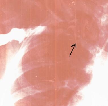
Thoracic outlet syndrome in a 16-year-old volleyball player with stenotic right subclavian vein (arrow) secondary to fibrosis. |
 | View Full Size Image | |
Media type: X-RAY
|
- Al-Shekhlee A, Katirji B. Spinal accessory neuropathy, droopy shoulder, and thoracic outlet syndrome. Muscle Nerve. Sep 2003;28(3):383-5. [Medline].
- Arcasoy SM, Jett JR. Superior pulmonary sulcus tumors and Pancoast's syndrome. N Engl J Med. Nov 6 1997;337(19):1370-6. [Medline].
- Atasoy E. Recurrent thoracic outlet syndrome. Hand Clin. Feb 2004;20(1):99-105. [Medline].
- Balci AE, Balci TA, Cakir O, Eren S, Eren MN. Surgical treatment of thoracic outlet syndrome: effect and results of surgery. Ann Thorac Surg. Apr 2003;75(4):1091-6; discussion 1096. [Medline].
- Cuetter AC, Bartoszek DM. The thoracic outlet syndrome: controversies, overdiagnosis, overtreatment, and recommendations for management. Muscle Nerve. May 1989;12(5):410-9. [Medline].
- Dale WA, Lewis MR. Management of thoracic outlet syndrome. Ann Surg. May 1975;181(5):575-85. [Medline].
- Edwards DP, Mulkern E, Raja AN, Barker P. Trans-axillary first rib excision for thoracic outlet syndrome. J R Coll Surg Edinb. Dec 1999;44(6):362-5. [Medline].
- Esposito MD, Arrington JA, Blackshear MN, Murtagh FR, Silbiger ML. Thoracic outlet syndrome in a throwing athlete diagnosed with MRI and MRA. J Magn Reson Imaging. May-Jun 1997;7(3):598-9. [Medline].
- Franklin GM, Fulton-Kehoe D, Bradley C, Smith-Weller T. Outcome of surgery for thoracic outlet syndrome in Washington state workers' compensation. Neurology. Mar 28 2000;54(6):1252-7. [Medline].
- Fujita K, Matsuda K, Sakai Y, Sakai H, Mizuno K. Late thoracic outlet syndrome secondary to malunion of the fractured clavicle: case report and review of the literature. J Trauma. Feb 2001;50(2):332-5. [Medline].
- Green SM. Tarascon Pocket Pharmacopoeia. Loma Linda, Calif: Tarascon Press; 1999:10-11, 30, 46.
- Hood DB, Kuehne J, Yellin AE, Weaver FA. Vascular complications of thoracic outlet syndrome. Am Surg. Oct 1997;63(10):913-7. [Medline].
- Huang JH, Zager EL. Thoracic outlet syndrome. Neurosurgery. Oct 2004;55(4):897-902; discussion 902-3. [Medline].
- Kenny RA, Traynor GB, Withington D, Keegan DJ. Thoracic outlet syndrome: a useful exercise treatment option. Am J Surg. Feb 1993;165(2):282-4. [Medline].
- Komanetsky RM, Novak CB, Mackinnon SE, Russo MH, Padberg AM, Louis S. Somatosensory evoked potentials fail to diagnose thoracic outlet syndrome. J Hand Surg [Am]. Jul 1996;21(4):662-6. [Medline].
- Leffert RD. Complications of surgery for thoracic outlet syndrome. Hand Clin. Feb 2004;20(1):91-8. [Medline].
- Lindgren KA. Conservative treatment of thoracic outlet syndrome: a 2-year follow-up. Arch Phys Med Rehabil. Apr 1997;78(4):373-8. [Medline].
- Machleder HI. Evaluation of a new treatment strategy for Paget-Schroetter syndrome: spontaneous thrombosis of the axillary-subclavian vein. J Vasc Surg. Feb 1993;17(2):305-15; discussion 316-7. [Medline].
- Machleder HI, Moll F, Nuwer M, Jordan S. Somatosensory evoked potentials in the assessment of thoracic outlet compression syndrome. J Vasc Surg. Aug 1987;6(2):177-84. [Medline].
- Mackinnon SE, Novak CB. Thoracic outlet syndrome. Curr Probl Surg. Nov 2002;39(11):1070-145. [Medline].
- Moore WS, ed. Vascular Surgery: A Comprehensive Review. 4th ed. Philadelphia, Pa: WB Saunders Co; 1993:600-603.
- Novak CB. Conservative management of thoracic outlet syndrome. Semin Thorac Cardiovasc Surg. Apr 1996;8(2):201-7. [Medline].
- Novak CB, Collins ED, Mackinnon SE. Outcome following conservative management of thoracic outlet syndrome. J Hand Surg [Am]. Jul 1995;20(4):542-8. [Medline].
- Novak CB, Mackinnon SE, Patterson GA. Evaluation of patients with thoracic outlet syndrome. J Hand Surg [Am]. Mar 1993;18(2):292-9. [Medline].
- Oates SD, Daley RA. Thoracic outlet syndrome. Hand Clin. Nov 1996;12(4):705-718. [Medline].
- Parziale JR, Akelman E, Weiss AP, Green A. Thoracic outlet syndrome. Am J Orthop. May 2000;29(5):353-60. [Medline].
- Patton GM. Arterial thoracic outlet syndrome. Hand Clin. Feb 2004;20(1):107-11, viii. [Medline].
- Rayan GM. Lower trunk brachial plexus compression neuropathy due to cervical rib in young athletes. Am J Sports Med. Jan-Feb 1988;16(1):77-9. [Medline].
- Roos DB. Congenital anomalies associated with thoracic outlet syndrome. Anatomy, symptoms, diagnosis, and treatment. Am J Surg. Dec 1976;132(6):771-8. [Medline].
- Roos DB. Thoracic outlet syndrome is underdiagnosed. Muscle Nerve. Jan 1999;22(1):126-9; discussion 137-8. [Medline].
- Rutherford RB, Cronewett JL, Gloviczki P. Vascular Surgery. 5th ed. Philadelphia, Pa: WB Saunders Co; 2000:1184-1219.
- Safran MR. Nerve injury about the shoulder in athletes, part 2: long thoracic nerve, spinal accessory nerve, burners/stingers, thoracic outlet syndrome. Am J Sports Med. Jun 2004;32(4):1063-76. [Medline].
- Sanders RJ, Hammond SL. Venous thoracic outlet syndrome. Hand Clin. Feb 2004;20(1):113-8, viii. [Medline].
- Upton AR, McComas AJ. The double crush in nerve entrapment syndromes. Lancet. Aug 18 1973;2(7825):359-62. [Medline].
- Urschel HC Jr, Razzuk MA. Neurovascular compression in the thoracic outlet: changing management over 50 years. Ann Surg. Oct 1998;228(4):609-17. [Medline].
- Urschel HC Jr, Razzuk MA. Paget-Schroetter syndrome: what is the best management?. Ann Thorac Surg. Jun 2000;69(6):1663-8; discussion 1668-9. [Medline].
- Weber RJ, Lebduskin S. Rehabilitation issues in plexopathies. Physical Medicine and Rehabilitation. 1988;996-998.
- Wilbourn AJ. Thoracic outlet syndrome is overdiagnosed. Muscle Nerve. Jan 1999;22(1):130-6; discussion 136-7. [Medline].
- Wood VE, Twito R, Verska JM. Thoracic outlet syndrome. The results of first rib resection in 100 patients. Orthop Clin North Am. Jan 1988;19(1):131-46. [Medline].
Thoracic Outlet Syndrome excerpt Article Last Updated: Aug 30, 2006
|