Practice Essentials
Skier's thumb is an injury caused by damage to the ulnar collateral ligament (UCL) of the thumb, most often due to a skiing accident. (See the image below.) Clinical examination remains the criterion standard in the diagnosis of a UCL rupture of the thumb.
Grades 1 and 2 skier's thumbs and incomplete UCL ruptures can be treated conservatively (nonsurgically) with proper immobilization. Primary surgical repair is indicated for the following:
-
Complete rupture of the UCL, as evidenced by joint instability
-
UCL damage with any accompanying fracture that is displaced, rotated, or intra-articular
-
Presence of a Stener lesion
-
Grade 3 classification [1]
Background
Injuries to the ulnar collateral ligament (UCL) of the thumb were first recognized as an occupational condition in European gamekeepers. By repetitively wringing the necks of game (eg, chickens) between their thumb and index finger, these workers produced a chronic stretching of the UCL that resulted in instability at the first metacarpophalangeal (MCP) joint. The condition became known as gamekeeper's thumb. Today, the injury is primarily caused by acute (rather than chronic and repetitive) damage to the UCL, most often due to a skiing accident; hence, the condition is now commonly referred to as skier's thumb.
Etiology
Traumatic injuries in cases of skier's thumb occur more often and result more commonly in UCL ruptures. In addition, UCL injuries from chronic repetitive radial stresses typically lead to UCL laxity and thumb instability but without complete UCL rupture.
Epidemiology
United States data
Skier's thumb represents 5-10% of all skiing injuries; this condition is the most frequent injury of the upper extremity that skiers experience. [2, 3, 4, 5]
Functional Anatomy
The MCP joint of the thumb is primarily stabilized by the UCL. The origin of this ligament is found on the ulnar aspect of the metacarpal head, whereas the insertion of the UCL is located distally on the proximal phalanx.
Sport-Specific Biomechanics
The most common cause of UCL injury is an acute abducting (radially directed) force upon the thumb. [6] Damage may also result from a combination of torsion, abduction, and hyperextension at the first MCP joint. Depending on the degree of impact of these forces at the MCP joint, the UCL may either tear partially or completely (see the images below). [2, 3, 4, 5, 7]
A large number of skiing injuries are attributed to ski poles, in which the strap or sword grip lies across the palm and transmits the damaging force to the thumb during a fall. Football players may develop UCL damage either traumatically (eg, while making a tackle, falling on an outstretched hand [FOOSH injury]), or chronically (eg, linemen, who repetitively stress the thumb radially while blocking). The injury is also common among athletes who handle balls (eg, basketball, football) and among those who use sticks (eg, hockey, lacrosse), in which the sporting equipment can forcefully abduct the thumb during sport activity.
Prognosis
When properly treated, patients with a UCL injury have a good prognosis for returning to their premorbid level of functioning. A missed diagnosis that delays the repair of a complete UCL rupture leads to a less favorable prognosis. Complications from delayed treatment of a complete tear may include neurapraxia (secondary to injury of the radial sensory nerve), stiffness (which usually improves with time), and persistent instability (which can eventually lead to MCP joint arthritic changes). [8]
A Stener lesion is when a torn ulnar collateral ligament (UCL) gets entrapped in the adductor aponeurosis. [9] Most UCL injuries can be treated non-surgically. But it is important to diagnose Stener lesions, because this type of UCL injury more commonly requires surgical correction.
Complications
Osteoarthritis of the first MCP joint may occur in patients with skier's thumb.
-
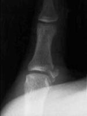
Anteroposterior radiograph displaying a gamekeeper's fracture.
-
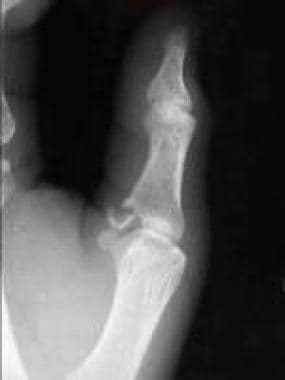
Lateral radiograph displaying a gamekeeper's fracture.
-
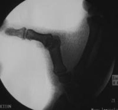
Radiograph displaying a stress test of a torn ulnar collateral ligament.
-
Stress testing of the metacarpophalangeal joint of the thumb in flexion.
-
Stress testing of the metacarpophalangeal joint of the thumb in extension.
-
Ruptured ulnar collateral ligament.
-
Completed repair using suture anchors for fixation.
-
Anterior view of a hand in a thumb spica splint.
-
Lateral view of a hand in a thumb spica splint.


