Continually Updated Clinical Reference
 
 
  All Sources     eMedicine     Medscape     Drug Reference     MEDLINE
 
eMedicine - Sacroiliac Joint Injury : Article by

Quick Find
Authors & Editors
Introduction
Clinical
Differentials
Workup
Treatment
Medication
Follow-up
Miscellaneous
Multimedia
References

Related Articles
Ankylosing Spondylitis and Undifferentiated Spondyloarthropathy

Hip Fracture

Hip Overuse Syndrome

Iliotibial Band Syndrome

Lumbosacral Discogenic Pain Syndrome

Lumbosacral Facet Syndrome

Lumbosacral Radiculopathy

Piriformis Syndrome

Sacroiliac Joint Infection

Seronegative Spondyloarthropathy

Superior Cluneal Nerve (Iliac crest) Syndrome

Trochanteric Bursitis


Patient Education
Bone Health Center

Back Pain Overview

Back Pain Causes

Back Pain Symptoms

Back Pain Treatment


Author: Andrew L Sherman, MD, Assistant Professor, Departments of Neurological Surgery, Orthopedics, and Rehabilitation, University of Miami

Andrew L Sherman is a member of the following medical societies: American Academy of Physical Medicine and Rehabilitation, American Association of Neuromuscular and Electrodiagnostic Medicine, American College of Sports Medicine, and American Medical Association

Coauthor(s): Robert Gotlin, DO, Director of Orthopaedic and Sports Rehabilitation, Assistant Professor, Department of Physical Medicine and Rehabilitation, Beth Israel Medical Center, Albert Einstein College of Medicine

Editors: Craig C Young, MD, Professor, Departments of Orthopedic Surgery and Community and Family Medicine, Medical Director of Sports Medicine, Sports Medicine Fellowship Director, Medical College of Wisconsin; Francisco Talavera, PharmD, PhD, Senior Pharmacy Editor, eMedicine; Henry T Goitz, MD, Chief, Sports Medicine, Associate Professor, Department of Orthopaedic Surgery, Medical College of Ohio; Jon B Whitehurst, MD, Clinical Instructor of Surgery, University of Illinois College of Medicine; Partner and Executive Board Member, Rockford Orthopedic Associates; Orthopedic Chairman, Rockford Memorial Hospital; Sherwin SW Ho, MD, Associate Professor, Department of Surgery, Section of Orthopedic Surgery and Rehabilitation Medicine, University of Chicago

Author and Editor Disclosure

Synonyms and related keywords: sacroiliitis, SIJ injury, lower back pain, low back pain, LBP, back pain, low back injuries, lower back injuries

Background

Lower back pain is one of the most prevalent sports maladies, affecting athletes in nearly every sport. Diagnosing the cause of a back injury is quite difficult and challenging because multiple structures in the lower back region can cause pain. However, an accurate diagnosis is paramount to providing successful treatment of the spine injury.

Although still somewhat controversial, the sacroiliac joint (SIJ) is generally accepted as an anatomic structure within the lumbar complex that if injured can be a cause of lower back pain. Mechanical dysfunction, inflammation, infection, trauma, and degeneration all have been attributed to the SIJ. Once the diagnosis of SIJ injury is established, specifically directed treatment can lead to satisfying results. This article discusses the diagnosis, management, and rehabilitation of sacroiliac injuries and pain.

For excellent patient education resources, visit eMedicine's Osteoporosis and Bone Health Center and Back, Neck, and Head Injury Center. Also, see eMedicine's patient education articles Back Pain and Lumbar Disc Disease.

Related eMedicine topics:
Lumbar Degenerative Disk Disease
Lumbar Disk Problems in the Athlete
Lumbosacral Spine Sprain/Strain Injuries
Mechanical Low Back Pain

Related Medscape topics:
Resource Center Exercise and Sports Medicine
Resource Center Spinal Disorders
CME/CE Best Evidence Review - Sciatica and Low Back Pain: Does Physical Therapy Provide Long-Term Benefits? A Best Evidence Review
CME Chronic Back Pain: Costs, Mechanisms, and Therapeutic Approaches (Slides With Transcript)
CME/CE NSAIDs May Not Be Best Bet for Low Back Pain

Frequency

United States

The incidence of lower back pain in humans parallels the incidence of the common cold, with a lifetime rate approaching 95%. Goldwaith and Osgood first discussed the possibility that SIJ injury could cause low back pain as early as 1905.1 In the decades since then, several attempts have been made to establish the prevalence of SIJ syndrome in persons with back pain, and the results of these reports vary widely.

Schwarzer et al remarked that "the prevalence of sacroiliac pain would appear to be at least 13% and perhaps as high a 30%" in patients with low back and buttock pain.2 Bernard and Kirkaldy-Willis reported the prevalence rate to be 22.5% in 1293 patients with back pain.3

Functional Anatomy

The SIJ is a true diarthrodial joint that joins the sacrum to the pelvis.4, 5, 6 In this joint, hyaline cartilage on the sacral side moves against fibrocartilage on the iliac side. The joint is generally C shaped with 2 lever arms that interlock at the second sacral level. The joint contains numerous ridges and depressions, indicating its function for stability more than motion. However, studies have documented that motion does occur at the joint; therefore, slightly subluxed and even locked positions can occur.2, 7

Stability is provided by the ridges present in the joint and by the presence of generously sized ligaments. The ligamentous structures offer resistance to shear and loading. The deep anterior, posterior, and interosseous ligaments resist the load of the sacrum relative to the ilium. More superficial ligaments (eg, sacrotuberous ligament) react to dynamic motions (eg, straight-leg raising during physical motion). The long dorsal sacroiliac ligament can become stretched in periods of reduced lumbar lordosis (eg, pregnancy).

Many large and small muscles have relationships with these ligaments and the SIJ, including the piriformis, biceps femoris, gluteus maximus and minimus, erector spinae, latissimus dorsi, thoracolumbar fascia, and iliacus. Any of these muscles can be involved with a painful SIJ. As a true joint, the SIJ is a pain-sensitive structure richly innervated by a combination of unmyelinated free nerve endings and the posterior primary rami of L2-S3. The wide possibility of innervation may explain why pain emanation from the joint can manifest in so many various ways, with different and unique referral patterns for individual patients.

Related Medscape topics:
Resource Center Back Pain
Resource Center Joint Disorders
Resource Centers Neurology & Neurosurgery

Sport-Specific Biomechanics

The function of the SIJ is to dissipate loads of the torso through the pelvis to the lower extremities and vice versa. The pelvis acts as a central base through which large forces are accepted and dissipated. Although the main role of the joint is to provide stability, the SIJ has limited motion that allows it to dissipate and transfer significant loads and stresses. Studies by Weisel indicate that most movement occurs when rising from the sitting to the standing position. However, the amount of motion is small, making assessment of sacroiliac motion during physical examination quite difficult. Selvik suggested that hyperextension produces the greatest degree of motion (2° on average, with only minimal translation of 0.5-1.6 mm).

If the motion in the pelvis is asymmetric, then dysfunction can occur. Some conditions that cause asymmetric motion include leg-length inequalities, a unilaterally weak lower limb (eg, polio), tight myofascial structures (eg, iliopsoas), and scoliosis. Hip osteoarthritis can lead to leg-length shortening and SIJ pain.

Women may be at increased risk for SIJ problems because their broader pelvises, greater femoral neck anteversion, and shorter limb lengths lead to different, possibly predisposing, biomechanics. In addition, pregnancy often leads to stretching of the pelvis, specifically targeting the sacroiliac ligaments and possibly leading to dysfunction, hypermobility syndromes, and chronic pain.

Innervation

The nerve supply of the SIJ originates from multiple lumbosacral root levels with partial innervation from L2 (anterior joint) to S3 (posterior joint). Because the root innervation can vary so widely, the pain referral patterns from primary sacroiliac pain can also vary. Fortin et al interviewed multiple patients documented to have sacroiliac pain by anesthetizing the joint with lidocaine injections under fluoroscopic guidance.8, 9 He found referral patterns ranging from localized buttocks pain to frank radicular leg pain and many other descriptions in between.


History

The key element in the diagnosis of sacroiliac dysfunction is pain. Many authors have attempted to define a typical pain pattern associated with the SIJ. Several of these reports describe patients reporting pain in one or both buttocks at or near the posterior superior iliac spine (PSIS). However, pain radiating to the hip, posterior thigh, or even calf has been described.

Patients often relate that pain especially worsens when they have been sitting for long periods or when they perform twisting or rotary motions.

  • Pain quality: The pain is described as a dull ache or sharp, stabbing, or knifelike.
  • Pain distribution: Reported distributions are the buttocks, back of the thigh, and upper back; it can be unilateral or bilateral.
  • History: Importantly, exclude a history of inflammatory disorders (eg, inflammatory bowel disease, Reiter syndrome).
  • Fevers, weight loss, and pain in the night with night sweats: These are potential red flags for a systemic illness.
  • Pain that is worse in the morning (morning stiffness) and resolves with exercise: This pattern is consistent with an inflammatory disease.

Physical

The reliability of the physical examination findings to diagnose SIJ dysfunction has been addressed in several articles. The usual pattern of examination is discussed, as follows:

  • Inspection often reveals a pelvis with asymmetric height. This finding can be an indication of unilateral restriction in motion of one or both SIJs. Standing flexion testing involves the comparison of the symmetry of motion between the PSIS on the tested side and the S2 spinous process (Gillet test). However, Freburger and Riddle questioned the reliability of examinations between testers.10
  • Of paramount importance is to measure the limb lengths to look for inequality, inspect the lumbar spine to look for scoliosis, and rotate the hips to look for motion restriction.
  • Palpation may be the most reliable indication of SIJ pain. The patient usually places a thumb directly onto one particular spot in the dimple of the PSIS (sacral sulcus). The patient can usually precisely reproduce the pain over that one spot (Fortin finger sign).9 More diffuse back or buttock and leg pain should prompt the clinician to question the diagnosis of SIJ dysfunction (see Differentials and Other Problems to Be Considered).
  • Upon neurologic examination, motor strength, sensory examination, and reflexes in the lower extremities should all prove normal. However, sometimes, strength examination proves challenging, and the patient may exhibit weaknesses because of pain inhibition or frank muscle imbalance that developed during episodes of pain and relative inactivity. True neurogenic weakness, numbness, or loss of reflex should alert the clinician to consider nerve root injury or pathology other than a mechanical dysfunction.
  • Perform pain provocation tests.9, 11, 12, 13, 14 Distraction can be performed to the anterior sacroiliac ligaments by applying pressure to the anterior superior iliac spine (iliac gapping test). Apply compression to the joint with the patient lying on his or her side. Pressure is applied downward to the uppermost iliac crest (iliac compression test).

    The goal of the Gaenslen test is to apply torsion to the joint. With one hip flexed onto the abdomen, the other leg is allowed to dangle off the edge of the table. Pressure should then be directed downward on the leg in order to achieve hip extension and stress the SIJ.

    For the flexion, abduction, and external rotation (FABER or Patrick) test, the examiner externally rotates the hip while the patient lies supine. Then, downward pressure is applied to the knee.

    In all tests, pain in the typical area raises suspicion for an SIJ lesion.

  • Unfortunately, although systematic, these tests have not proven reliable in controlled studies. Dreyfuss and colleagues studied 12 SIJ tests in relation to fluoroscopically guided SIJ injection.15 They were unable to find even one of these tests to be highly sensitive or specific for diagnosing SIJ pain. Hancock et al also published a review on physical examination testing reliability to diagnose SIH syndrome.12

    Stuber conducted a systematic review of the literature to determine the specificity, sensitivity, and predictive values of such clinical tests of the sacroiliac joint.11 According to the author, "the search was conducted using several online databases: Medline, Embase, Cinahl, AMED, and the Index to Chiropractic Literature. Reference and journal searching and contact with several experts in the area was also employed." Stuber concluded that the distraction test, compression test, thigh thrust/posterior shear, sacral thrust, and resisted hip abduction were the only tests to have specificity and sensitivity greater than 60% in at least one study, and that further investigation is warranted to determine which tests or combinations of these tests are the best for diagnosing SIJ dysfunction. 

Causes

Many patients state that their pain began spontaneously, whereas others can cite a specific inciting event. Bernard and Kirkaldy-Willis reported that 58% of patients diagnosed with SIJ pain based on clinical examination findings had some inciting traumatic injury.3

Many risk factors are associated with lower back pain, and many are directly associated with lumbar disk injury. These include, but are not limited to, smoking, poor physical condition, positive family history, and occupational lifting.

Factors that specifically increase the likelihood of mechanical injury to the SIJ have not been identified. Pregnancy is one particular condition attributed to SIJ dysfunction.

In the authors’ experience, certain biomechanical or muscle length imbalances may ultimately predispose a person to sacroiliac dysfunction and pain. Likely, this is a result of altered gait patterns and repetitive stress to the SIJ and related structures. These conditions exist in persons with leg-length inequality, scoliosis, a history of polio, poor-quality footwear, and hip osteoarthritis.

Related eMedicine topics:
Lumbar Degenerative Disk Disease
Lumbar Disk Problems in the Athlete
Lumbosacral Spine Sprain/Strain Injuries
Mechanical Low Back Pain

Related Medscape topics:
Resource Center Spinal Disorders
Resource Center Trauma
CME/CE Best Evidence Review - Sciatica and Low Back Pain: Does Physical Therapy Provide Long-Term Benefits? A Best Evidence Review
CME Chronic Back Pain: Costs, Mechanisms, and Therapeutic Approaches (Slides With Transcript)
CME/CE NSAIDs May Not Be Best Bet for Low Back Pain


Ankylosing Spondylitis and Undifferentiated Spondyloarthropathy
Hip Fracture
Hip Overuse Syndrome
Iliotibial Band Syndrome
Lumbosacral Discogenic Pain Syndrome
Lumbosacral Facet Syndrome
Lumbosacral Radiculopathy
Piriformis Syndrome
Sacroiliac Joint Infection
Seronegative Spondyloarthropathy
Superior Cluneal Nerve (Iliac crest) Syndrome
Trochanteric Bursitis

Other Problems to Be Considered

Crohn disease
Infection
Inflammatory spondyloarthropathies
Malignancy/metastasis
Psoriatic arthritis
Reiter syndrome
Stress fracture


Lab Studies

In a patient with sacroiliitis, inflammatory origins (eg, ankylosing spondylitis [AS], psoriatic arthritis) must be considered. The following laboratory studies are appropriate when an inflammatory disorder is suspected in a young patient:

  • Complete blood cell (CBC) count
  • Erythrocyte sedimentation rate (ESR)
  • C-reactive protein (CRP) level
  • Antinuclear antibody (ANA) profile
  • Human leukocyte antigen (HLA)-B27 status
  • Rheumatoid factor (RF) value

In patients in whom the pain has become chronic, clinicians need to consider whether the patients may have secondary reactive depression. Testing for hypothyroidism, cortisol abnormalities, or other metabolic or endocrine imbalances may be appropriate as part of the workup for a patient with depression.

Prostate-specific antigen (PSA) testing, serum protein electrophoresis/urine protein electrophoresis, and other laboratory tests used to investigate for malignancy are appropriate for older patients who have not improved with initial care and may have symptoms warranting further investigation.

Related Medscape topics:
Resource Centers Diabetes & Endocrinology 
Resource Centers Hematology-Oncology
Resource Centers Pathology & Lab Medicine

Imaging Studies

  • Use of imaging studies when evaluating sacroiliac pathology is a source of controversy among clinicians because whether normal and abnormal radiographic studies can help differentiate symptomatic versus nonsymptomatic patients is unclear. This is probably due to the great variability in joint anatomy among patients. Additional disagreement exists on the significance of inflammatory findings and degenerative findings (sclerosis) being diagnostic of pain within the joint.
  • The usual SIJ examination is performed using anteroposterior pelvis/lumbar spine radiography. Sclerosis or obliteration of the SIJ can be observed in older patients.
  • Patients with AS usually have normal radiographic findings; in older patients with this disease, the joint can appear fused.
  • Specific sacroiliac views superimpose the anterior and posterior joint margins, which may increase the sensitivity for detecting abnormalities. These radiographs are taken at a 25-30° angle to the anteroposterior plane. Joint widening with erosive and sclerotic changes at the bony margins may be suggestive of inflammatory sacroiliitis.
  • Computed tomography (CT) scanning can often be used to document reactive spurring, sclerosis, or even subluxation. Many clinicians believe reactive spurring is due to prolonged abnormal motion within the joint.
  • In persons with inflammatory conditions (eg, AS), bone scanning can show enhancement within the SIJ (often bilaterally).
    • Nuclear medicine bone scanning with single photon emission computed tomography (SPECT) can also be used to rule out femur and pelvic stress fractures and most bony metastatic disease.
    • Some clinicians view enhancement observed in the SIJ unilaterally in a patient with suspected SIJ conditions as an indicator of SIJ dysfunction or inflammation. Slipman et al found nuclear imaging under these circumstances to be very low in sensitivity but high in specificity for sacroiliac-mediated pain.16 Painful SIJs were confirmed with an intra-articular injection of anesthetic. Therefore, bone scanning was of little value in the diagnostic algorithm for SIJ pain.
    • Adding SPECT scanning may increase the sensitivity of nuclear imaging for SIJ injuries; however, this has not been studied.
  • Magnetic resonance imaging (MRI) is not generally used for evaluating the SIJ, although it can be a valuable tool to help exclude disc herniation (especially at L5-S1) as part of the SIJ dysfunction differential diagnosis (see Differentials and Other Problems to Be Considered). MRI can occasionally show inflammatory signs within the SIJ. MRI can also be used to investigate pelvic stress fracture, femoral neck stress fracture, or femoral head avascular necrosis.

Related eMedicine topics:
Acetabulum Fractures

Femoral Head Avascular Necrosis
Femoral Neck Stress and Insufficiency Fractures
Pelvic Fractures
Stress Fractures

Procedures


Acute Phase (1-10 d):

Subacute phase (if acute phase does not resolve; 10-180 d)

Chronic phase (>6 mo of pain)

Rehabilitation Program

Physical Therapy

Physical therapy focuses on pain control in the acute phase. Modalities such as ultrasonography with or without phonophoresis, deep and superficial heat, and superficial cold treatments can reduce pain. Neural therapies such as deep tissue massage, myofascial release, and muscle energy stretching techniques can also help. Pelvic myofascial stretching in the neutral spine position can be used for immediate, short-term relief of discomfort. By identifying activities that aggravate the condition, the physician or therapist can have the patient avoid these activities.

Related eMedicine topics:
Massage, Traction, and Manipulation
Superficial Heat and Cold

Osteopathic/chiropractic treatment

Although in the acute-phase muscle spasms may prevent frank manipulation, less aggressive techniques such as muscle energy stretching can be very helpful.

Medical complications

Patients may experience difficulty or even worsening symptoms with physical therapy treatments in certain cases. In these patients, reevaluate the diagnosis and consider other diagnostic possibilities (eg, infection, inflammatory disease, malignancy, neural [lumbosacral root] injury). Patients with acute inflammatory disorders or infections should not usually be administered heat treatments. Patients who cannot perform physical therapy may also have a functional component to their disorder or an underlying psychologic disorder, which needs to be addressed.

Medical interventions

Often, oral medications can be quite effective in the acute phase. Nonsteroidal anti-inflammatory drugs (NSAIDs) are used judiciously in this phase, often with good results. In the first 24-72 hours, a muscle relaxant can be quite effective if a myofascial component to the pain is present. Ice can be considered in the first 48-72 hours; then, the typical switch to heat or contrast treatments is warranted.

Oral medication management may change if the pain persists into the subacute and especially the chronic phase. Chronic lower back pain from any source often leads to the development of a cognitive/behavioral component. In such cases, the use of antidepressants, anticonvulsants, and antiarrhythmic topical and oral medications has been reported to benefit certain selected patients. Because the potential benefits of any of these medications is uncertain, their use must be balanced against their potential adverse effects.

Related Medscape topics:
Resource Center Pain Management: Advanced Approaches to Chronic Pain Management
Resource Center Pain Management: Pharmacologic Approaches

Surgical intervention

Surgical intervention is rarely used for nontraumatic SIJ pain.4, 17 Surgery is considered only in patients with chronic pain that has lasted for years, has not been effectively treated by other means, and has led to an extremely poor quality of life. The procedure is a fusion across the joint; however, although the surgery has been reported to result in benefit in selected cases or small case series, no randomized controlled study has shown reliable pain reduction with SIJ fusion.

Consultations

Consultation with a rheumatologist is necessary when the possibility of an underlying inflammatory disorder exists. Consultation with a musculoskeletal specialist is often helpful. The musculoskeletal specialist should provide each patient with a functional assessment, can direct nonoperative treatment, and can communicate with the entire treatment team (eg, physical therapists, trainers). Often, a physiatrist (specialist in the field of physical medicine and rehabilitation) can provide a unique, functional-based history and examination that can lead to an accurate diagnosis and a holistic treatment program.

Related eMedicine topic:
Adult Physiatric History and Examination

Related Medscape topics:
Resource Center Spinal Disorders
Resource Centers Rheumatology 

Other Treatment (injection, manipulation, etc.):

In the immediate acute phase, treatment consists of pain reduction through pain medications, rest, and avoidance of the inciting activity. Anti-inflammatory treatment with NSAID medications and externally applied ice is often helpful. Recommend the patient return to usual activities as soon as possible, usually within 24-48 hours. Sometimes a local trigger point injection into the muscle can relieve symptoms.

If the pain does not resolve well in the first 2-3 weeks, an intra-articular injection under fluoroscopic guidance should be considered. SIJ injection is frequently performed with a mixture of anesthetic and steroid, as described by Fortin in 1994 and others.8, 18, 19, 20, 21 When the actual source of the patient’s discomfort is unclear, postinjection pain reduction offers significant diagnostic information. Fluoroscopic guidance is important because, although a local blind injection into the area of maximal pain can be temporarily effective, the needle rarely enters the joint. CT scanning or MRI can also be used to guide injections into the SIJ, with excellent reliability.

Günaydin and colleagues reported that 20 of 31 patients with spondylarthropathy reported subjective improvement after the first SIJ injection of MRI-guided corticosteroid, and 9 of 15 patients reported subjective improvement after the second injection.20 The improvement lasted for a mean of 8.7 for the first group and 16.1 months for the second group.

Luukkainen and colleagues reported that periarticular injection of methylprednisolone may be effective in the treatment of pain in the region of the SIJ in nonspondyloarthropathy patients from a study of 24 consecutive patients.22

Although these studies are promising, they are not randomized, placebo-controlled studies. Therefore, before efficacy can be established, randomized, placebo-controlled studies must be undertaken.

Even if the injections do relieve the patient's pain, the relief from the injections alone is very often short-lived. Therefore, using the injections only as part of an interdisciplinary rehabilitation program is important. The pain relief offers a window of opportunity to increase the rehabilitation. The point in the course of recovery when a second or even third injection should be attempted is unclear. Most clinicians wait 2-4 weeks before proceeding with a repeat injection.

Manipulation has been reported in multiple studies as effective treatment for acute lower back pain. However, studies specifically on SIJ syndrome are less abundant. The SIJ is accessible to manipulation treatments and these may be extremely effective. As with other passive modalities, these treatments should be coupled with an extensive active rehabilitative program. Manipulation following intra-articular injection has been reported anecdotally to be beneficial in selected cases.

In chronic conditions, some practitioners believe that SIJ pain is due to hypermobility of the joint, which occurs because of laxity in the ligamentous complex. Prolotherapy is a series of saline and glucose injections applied to the SIJ ligaments to cause an inflammatory reaction, which results in scarring and tightening of the ligaments and a reduction in pain. However, no satisfactory outcome investigations have been performed on prolotherapy for this condition.

A relatively more recent procedure, radiofrequency denervation, has been advocated for the treatment of especially recalcitrant sacroiliac dysfunction.23, 24 The procedure was thought to be ineffective for SIJ pain because the innervation to the joint is so diffuse. However, in a study by Gevargez and colleagues, the authors reported that 3 months after the procedure, 13 patients (34.2%) were completely free of pain. Twelve patients (31.6%) reported substantial pain reduction, 7 patients (18.4%) had slight pain reduction, and 3 patients (7.9%) had no pain reduction. No longer-term follow-up data are available; further study regarding this procedure is needed.

Recovery Phase

Rehabilitation Program

Physical Therapy

The recovery phase cannot proceed without an active, aggressive rehabilitation program. Often, SIJ injury leaves patients with significant deconditioning and muscle imbalances. These functional muscular deficits were sometimes present before the injury and may have predisposed the patient to injury. Some muscles are known to be functioning in a tight or shortened position, such as the hip flexors, hamstrings, tensor fascia lata, obturator internus, and rectus femoris. Other muscles are weak or inhibited, such as the gluteal and abdominal muscles.

Begin physical therapy by correcting any mechanical or leg-length asymmetries (eg, orthotic/shoe lift), stretching overly tight lumbopelvic muscles, and strengthening weak and inhibited muscles. All of this should begin in the neutral spine position or a pelvic position, which minimizes acute discomfort.

The patient is asked to take on more challenging tasks while progressing through the program. Stabilization exercises are performed with the patient in a more dynamic, functional position and often include balance and proprioceptive activities. Strengthening of the core muscles surrounding the spine can be achieved in various ways. In the past several years, Pilates training has become very popular for this purpose. Finally, the patient should graduate to sport- or work-specific training designed to return the patient to his or her previous level of functioning.

Braces and belts

In patients who develop chronic injuries, an SIJ belt can provide compression and feedback to the gluteal muscles. Patients with ligamentous hypermobility can especially benefit from this apparatus because the belt can reduce SIJ rotation. The belt differs from a generalized lumbar orthosis because it is much thinner and thus secures across the anterior superior iliac spines.

Orthotics can decrease leg-length inequalities; these items include custom-fitted orthotics, internal shoe lifts, and external shoe lifts.

Related eMedicine topic:
Lower Limb Orthotics

Related Medscape topic:
Resource Center Exercise and Sports Medicine

Medical Issues/Complications

SIJ dysfunction usually improves significantly, relatively quickly. Reexamine patients whose pain persists, despite treatment, for longer than 4 weeks and consider other diagnostic possibilities.

Other Treatment

Perform injection under fluoroscopic guidance (see Image 1). SIJ injection is frequently performed with a mixture of anesthetic and steroid, as described by Fortin in 1994 and others.8 Postinjection pain reduction offers significant diagnostic information when the actual source of the patient’s discomfort is unclear. Although a local blind injection into the area of maximal pain can be temporarily effective, the needle rarely enters the joint. CT scanning or MRI can also be used to guide injections into the SIJ, with excellent reliability.18, 19, 20, 21

Unfortunately, injections usually offer only temporary relief. Therefore, couple injections with physical therapy and exercise to achieve more durable pain relief. The point in the course of treatment when a second or even third injection should be attempted is unclear. Most clinicians wait at least 2-4 weeks before proceeding with a repeat injection.

In a subset of patients who had temporary relief, Vallejo and coauthors performed pulsed radiofrequency denervation (PRFD) of lateral branches from L3-S2 and found good or excellent results in 16 of 22 subjects for 6-32 weeks.23

Manipulation has been reported in multiple studies as effective treatment for acute lower back pain. However, studies specifically on SIJ syndrome are less abundant. The SIJ is accessible to manipulation treatments, and these may be extremely effective. As with other passive modalities, these treatments should be coupled with an extensive active rehabilitative program. Manipulation following intra-articular injection has been reported anecdotally to be beneficial in selected cases.

In chronic conditions, some practitioners believe that SIJ pain is due to hypermobility of the joint, which occurs because of laxity in the ligamentous complex. Prolotherapy is a series of saline and glucose injections applied to the SIJ ligaments to cause an inflammatory reaction, which results in scarring and tightening of the ligaments and a reduction in pain. However, no satisfactory outcome investigations have been performed on prolotherapy for this condition.

Maintenance Phase

Rehabilitation Program

Physical Therapy

After the patient's pain resolves and he or she has regained sufficient strength, therapy should be transitioned from the therapy office to the gym or home gym. The therapist should teach the patient a home gym or gym program, and the patient should perform stabilization and general training at least 3 times per week to prevent recurrence.

Related Medscape topic:
Resource Center Exercise and Sports Medicine


As in most conditions involving acute and even chronic musculoskeletal pain, many oral medications can provide initial pain relief. NSAIDs are a mainstay and can be combined with acetaminophen for added effect. When the injury is acute and associated with secondary muscle spasm, muscle relaxants, light narcotics (eg, hydrocodone), or benzodiazepines are reasonable options. However, these medications should be administered cautiously and only for the initial acute phase of pain because dependence and tolerance can quickly occur.

Chronic SIJ dysfunction is more difficult to treat. Numerous medications are dispensed in a generic manner. Antidepressants, antiseizure agents, and antiarrhythmic agents are thought to be effective in neuropathic or nerve-related pain (radicular pain) and are not usually indicated for SIJ dysfunction pain.

Drug Category: Analgesics

Drug NameAcetaminophen (Tylenol, FeverAll)
DescriptionDOC for pain in patients with documented hypersensitivity to aspirin or NSAIDs, with upper GI disease, or who are taking oral anticoagulants.
Adult Dose375-650 mg PO q4-6h prn or 1000 mg PO q6-8h prn; not to exceed 4 g/d
Pediatric Dose<12 years: 10-15 mg/kg/dose PO q4-6h prn; not to exceed 2.6 g/d

>12 years: 325-650 mg PO q4h; not to exceed 5 doses in 24 h
ContraindicationsDocumented hypersensitivity; known G6PD deficiency
InteractionsRifampin can reduce analgesic effects; coadministration with barbiturates, carbamazepine, hydantoins, and isoniazid may increase hepatotoxicity.
PregnancyB - Fetal risk not confirmed in studies in humans but has been shown in some studies in animals
PrecautionsHepatotoxicity is possible in people with long-term alcoholism following various dose levels; severe or recurrent pain or high or continued fever may indicate a serious illness; acetaminophen is contained in many OTC products, and combined use with these products may result in cumulative doses exceeding the recommended maximum dose.

For most episodes of SIJ pain, oral outpatient analgesics can achieve adequate pain control.

Drug Category: Nonsteroidal anti-inflammatory drugs (NSAIDs)

NSAIDs work by decreasing inflammatory reactions and providing direct pain relief. Numerous choices are available, and they are separated into different families of agents. If an NSAID is ineffective, another agent from a different family can often provide relief. Efficacy and adverse effect profiles differ among agents and families.

Cyclooxygenase (COX)–2 inhibitors had been shown to reduce certain adverse effects (eg, gastrointestinal [GI] bleeding) and provide similar efficacy to standard agents. Unfortunately, 2 of the 3 agents (ie, rofecoxib [Vioxx], valdecoxib [Bextra]) were voluntarily removed from the market by their parent companies when an increased potential risk of adverse cardiovascular events was identified in an increased number of patients taking the drugs. Dosing requirements are usually individualized, based on the individual patient and patient response.

Related eMedicine topic:
Toxicity, Nonsteroidal Anti-inflammatory Agents

Related Medscape topic:
Resource Center Adverse Drug Events Reporting

Drug NameCelecoxib (Celebrex)
DescriptionDOC for patients with mild to moderate pain. Inhibits inflammatory reactions and pain by decreasing prostaglandin synthesis.
Adult Dose200 mg PO qd or bid
Pediatric DoseNot usually recommended for pain control
ContraindicationsDocumented hypersensitivity to aspirin, sulfa-based drugs, or other NSAIDS
InteractionsProbenecid may increase the concentrations and possibly the toxicity of NSAIDs; may decrease the effect of concurrently administered loop diuretics; PT duration may increase when administered concurrently with anticoagulants; closely monitor PT duration and instruct patients to watch for signs and symptoms of bleeding
PregnancyC - Fetal risk revealed in studies in animals but not established or not studied in humans; may use if benefits outweigh risk to fetus
PrecautionsCaution in patients with renal impairment, hepatic or cardiac dysfunction, and decreased hemoglobin values; monitor serum electrolytes in long-term use (>3 mo); caution in the presence of peptic ulcer disease, recent GI bleeding or perforation, or renal insufficiency

Drug NameIbuprofen (Motrin, Ibuprin)
DescriptionDOC for patients with mild to moderate pain. Inhibits inflammatory reactions and pain by decreasing prostaglandin synthesis.
Adult Dose400-600 mg PO q4-6h prn
Pediatric DoseNot usually recommended for pain control; however, often taken by adolescents in OTC form
ContraindicationsDocumented hypersensitivity to aspirin, iodides, or other NSAIDS
InteractionsProbenecid may increase the concentrations and possibly the toxicity of NSAIDs; may decrease the effect of concurrently administered loop diuretics; PT duration may increase when administered concurrently with anticoagulants; closely monitor PT duration and instruct patients to watch for signs and symptoms of bleeding; H2 antagonists and Carafate (active ingredient sucralfate) may decrease the risk of GI ulcer when administered concurrently with NSAIDs
PregnancyC - Fetal risk revealed in studies in animals but not established or not studied in humans; may use if benefits outweigh risk to fetus
D - Fetal risk shown in humans; use only if benefits outweigh risk to fetus
PrecautionsCaution in patients with renal impairment, hepatic or cardiac dysfunction, and decreased hemoglobin; monitor serum electrolytes in long-term use (>3 mo); caution in the presence of peptic ulcer disease, recent GI bleeding or perforation, renal insufficiency, and high risk of bleeding

Drug NameNaproxen (Anaprox, Naprelan, Naprosyn)
DescriptionCommon NSAID family used for relief of mild to moderate pain.
Adult Dose250-500 mg PO q8-12h; not to exceed 1.25 g/d
Pediatric Dose<2 years: Not established

>2 years: 2.5 mg/kg/dose PO; not to exceed 10 mg/kg/d
ContraindicationsDocumented hypersensitivity; peptic ulcer disease; recent GI bleeding; renal insufficiency; high risk of bleeding
InteractionsProbenecid and lithium may increase the concentrations and possibly the toxicity of NSAIDs; PT duration may increase when administered concurrently with anticoagulants
PregnancyB - Fetal risk not confirmed in studies in humans but has been shown in some studies in animals
D - Fetal risk shown in humans; use only if benefits outweigh risk to fetus
PrecautionsAcute renal insufficiency, hyperkalemia, hyponatremia, interstitial nephritis, and renal papillary necrosis may occur; low WBC counts occur rarely and usually return to the reference range in ongoing therapy.

Drug NameKetorolac tromethamine (Toradol)
DescriptionUsed primarily for control of hyperacute severe pain. Potency is higher than other NSAIDs, and use results in more marked GI upset, platelet inhibition, and renal effects.
Adult Dose30 mg IV q6h prn; alternatively, 30-60 mg IM q6h prn
Pediatric DoseNot established
ContraindicationsDocumented hypersensitivity; peptic ulcer disease; recent GI bleeding or perforation; renal insufficiency; high risk of bleeding
InteractionsMay prolong the PT duration when administered concurrently with anticoagulants; may increase the risk for methotrexate toxicity (eg, stomatitis, bone marrow suppression, nephrotoxicity); phenytoin levels may be increased when administered concurrently
PregnancyC - Fetal risk revealed in studies in animals but not established or not studied in humans; may use if benefits outweigh risk to fetus
D - Fetal risk shown in humans; use only if benefits outweigh risk to fetus
PrecautionsAcute renal insufficiency, hyperkalemia, hyponatremia, interstitial nephritis, and renal papillary necrosis may occur.

Drug Category: Muscle relaxants

Muscle relaxants can provide adjunctive pain relief in the acute setting. These agents usually should not be used in protracted courses.

Drug NameCyclobenzaprine hydrochloride (Flexeril)
DescriptionCentrally acting relaxant of skeletal muscle. Usually gains most of its analgesic effect indirectly as a general relaxant and sedative. Structurally related to TCAs.
Adult Dose10 mg PO up to tid prn
Pediatric DoseNot established
ContraindicationsDocumented hypersensitivity; administration of MAOIs within previous 14 d; hyperthyroidism
InteractionsMay enhance the effects of alcohol, barbiturates, or CNS depressants; may decrease the effectiveness of antihypertensives
PregnancyB - Fetal risk not confirmed in studies in humans but has been shown in some studies in animals
PrecautionsExercise caution in patients with angle-closure glaucoma and urinary hesitance; may impair consciousness, reactions, and ability to operate machinery

Drug NameMetaxalone (Skelaxin)
DescriptionPrescribed for use as a muscle relaxant. Mechanism of action not firmly established but may act as a CNS depressant and direct pain reliever. No direct action on the contractile mechanism of striated muscle. Can be used short term as an adjunct pain reliever in situations of severe myofascial strain.
Adult Dose800 mg (2 tab) PO tid/qid
Pediatric Dose<12 years: Not recommended

>12 years: Administer as in adults
ContraindicationsDocumented hypersensitivity; drug-induced anemia, hemolytic anemia, or other anemias
InteractionsNone reported
PregnancyC - Fetal risk revealed in studies in animals but not established or not studied in humans; may use if benefits outweigh risk to fetus
PrecautionsCaution in patients with known liver disease; perform serial LFTs in extended use


Return to Play

Similar to most conditions of mechanically related low back injury, an athlete's return to competition is a complex issue. In most SIJ injury cases, the athlete does not have a condition that can anatomically worsen with competition. However, pain may be exacerbated by the extreme motion and pelvic stress many athletes experience in their sport. Additionally, SIJ pain often leads to myofascial guarding and muscle imbalances, which, if not addressed before return to play, can lead to secondary injury in another part of the body. For example, a baseball pitcher with an SIJ strain may not be able to generate the support base or hip rotation needed to support the shoulder on overhead throws. Added stress to the shoulder can result in strains and even tears to the intrinsic elbow or shoulder muscles and ligaments.

Complications

Complications arise more from missed alternative causes of back pain than from any mechanical damage to the joint. Systemic conditions (eg, AS, Crohn-related arthritis) can cause future problems. Missed stress fractures to the hip could progress to a complete fracture. Finally, overlooked malignancy is a rare but real possibility.

Other complications can occur in athletes not fully rehabilitated. Muscle imbalances may persist and put the athlete at risk for reinjury or future injury to another structure. Finally, with any back injury, an inherent risk exists that the pain may become chronic. Excessive rest can often lead to adaption of a deconditioned state or sick role. These mechanical spine conditions must be identified early and rehabilitated aggressively to reduce this complication.

Prevention

Prevention of lower back injuries, including those to the SIJ, is multifaceted and relies on patient education concerning the back. Excessive lifting with a rotatory component can injure the SIJ in a manner similar to lumbar disk injuries. Using accessory muscles in forceful activities and training them for these activities can prevent injury. Sport-specific training after rehabilitation and before return to play is most important to prevent future injury.

Related eMedicine topics:
Lumbar Degenerative Disk Disease
Lumbar Disk Problems in the Athlete
Lumbosacral Spine Sprain/Strain Injuries
Mechanical Low Back Pain

Related Medscape topics:
Resource Center Exercise and Sports Medicine
Resource Center Spinal Disorders

Prognosis

Sacroiliac injury has an excellent prognosis for full recovery. While most studies suggest 80% of people with a lower back injury significantly improve within 2 weeks, no scientific studies show any stratification into diagnostic groups (ie, SIJ injury vs disk injury vs piriformis injury).

Education

Patient education is essential to achieving good outcomes. Patients can be informed that their SIJ pain is considered a benign condition, which, in most cases, improves with time and conservative treatments. Encourage them to resume physical activity as soon as possible to prevent deconditioning. Also encourage them to immediately enlist the help of a physical therapist to assist with therapeutic exercise. Home exercise programs are essential to help prevent reinjury and can be provided by a physician, chiropractor, or physical therapist.


Medical/Legal Pitfalls

  • Legal issues primarily occur when underlying malignant diagnoses are overlooked. Patients with pain that persists unchanged for longer than 1 month should have their diagnosis reconsidered (see Differentials and Other Problems to Be Considered) and should undergo a proper workup.
  • Injured workers often have injuries similar to those of athletes. These patients require a similar aggressive rehabilitation approach to minimize time away from work and to prevent reinjury. Worker's compensation can often interfere and delay the patient recovery. When providing work excuses, patient confidentiality must be protected. In some states, the only information entitled to the employer is whether the work excuse is legitimate and whether the employee has any medical problems that may prevent the employee from doing their job.

Related Medscape topic:
Resource Center Medical Malpractice and Legal Issues

Special Concerns

  • Back pain is less common in pediatric age groups. In such groups, nonmechanical causes (eg, infection, malignancy, spondylolisthesis) must be considered.
  • When pain persists in an adult for more than 1 month, a more aggressive diagnostic workup should be considered.


Media file 1:  
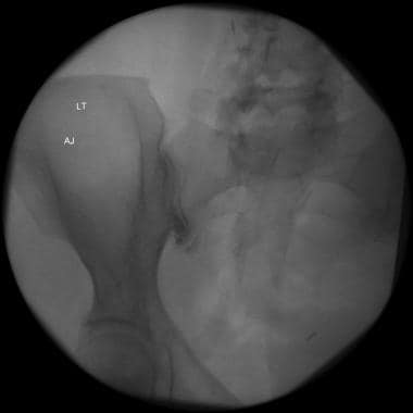
Flouroscopically guided sacroiliac joint injection. Contrast seen throughout the joint.
Click to see larger pictureClick to see detailView Full Size Image
Media type:  Radiograph


  1. Goldwaith JH, Osgood RB. A consideration of the pelvic articulations from an anatomical pathological and clinical standpoint. Boston Med Surg J. 1905;152(21):593-601.
  2. Schwarzer AC, Aprill CN, Bogduk N. The sacroiliac joint in chronic low back pain. Spine. Jan 1 1995;20(1):31-7. [Medline].
  3. Bernard TN Jr, Kirkaldy-Willis WH. Recognizing specific characteristics of nonspecific low back pain. Clin Orthop Relat Res. Apr 1987;217:266-80. [Medline].
  4. Edge-Hughes L. Hip and sacroiliac disease: selected disorders and their management with physical therapy. Clin Tech Small Anim Pract. Nov 2007;22(4):183-94. [Medline].
  5. Foley BS, Buschbacher RM. Sacroiliac joint pain: anatomy, biomechanics, diagnosis, and treatment. Am J Phys Med Rehabil. Dec 2006;85(12):997-1006. [Medline].
  6. Frieberg AH, Vinke TH. Sciatica and the sacroiliac joint. Clin Orthop Relat Res. 1974;16:126-34.
  7. Hungerford B, Gilleard W, Hodges P. Evidence of altered lumbopelvic muscle recruitment in the presence of sacroiliac joint pain. Spine. Jul 15 2003;28(14):1593-600. [Medline].
  8. Fortin JD, Dwyer AP, West S, Pier J. Sacroiliac joint: pain referral maps upon applying a new injection/arthrography technique. Part I: Asymptomatic volunteers. Spine. Jul 1 1994;19(13):1475-82. [Medline].
  9. Fortin JD, Falco FJ. The Fortin finger test: an indicator of sacroiliac pain. Am J Orthop. Jul 1997;26(7):477-80. [Medline].
  10. Freburger JK, Riddle DL. Using published evidence to guide the examination of the sacroiliac joint region. Phys Ther. May 2001;81(5):1135-43. [Medline][Full Text].
  11. Stuber KJ. Specificity, sensitivity, and predictive values of clinical tests of the sacroiliac joint: a systematic review of the literature. JCCA J Can Chiropr Assoc. Mar 2007;51(1):30-41. [Medline].
  12. Hancock MJ, Maher CG, Latimer J, et al. Systematic review of tests to identify the disc, SIJ or facet joint as the source of low back pain. Eur Spine J. Oct 2007;16(10):1539-50. [Medline].
  13. Slipman CW, Sterenfeld EB, Chou LH, Herzog R, Vresilovic E. The predictive value of provocative sacroiliac joint stress maneuvers in the diagnosis of sacroiliac joint syndrome. Arch Phys Med Rehabil. Mar 1998;79(3):288-92. [Medline].
  14. Dreyfuss P, Dryer S, Griffin J, Hoffman J, Walsh N. Positive sacroiliac screening tests in asymptomatic adults. Spine. May 15 1994;19(10):1138-43. [Medline].
  15. Dreyfuss P, Michaelsen M, Pauza K, McLarty J, Bogduk N. The value of medical history and physical examination in diagnosing sacroiliac joint pain. Spine. Nov 15 1996;21(22):2594-602. [Medline].
  16. Slipman CW, Sterenfeld EB, Chou LH, Herzog R, Vresilovic E. The value of radionuclide imaging in the diagnosis of sacroiliac joint syndrome. Spine. Oct 1 1996;21(19):2251-4. [Medline].
  17. Prather H, Hunt D. Conservative management of low back pain, part I. Sacroiliac joint pain. Dis Mon. Dec 2004;50(12):670-83. [Medline].
  18. Friedly J, Chan L, Deyo R. Increases in lumbosacral injections in the Medicare population: 1994 to 2001. Spine. Jul 15 2007;32(16):1754-60. [Medline].
  19. Murakami E, Tanaka Y, Aizawa T, Ishizuka M, Kokubun S. Effect of periarticular and intraarticular lidocaine injections for sacroiliac joint pain: prospective comparative study. J Orthop Sci. May 2007;12(3):274-80. [Medline].
  20. Günaydin I, Pereira PL, Fritz J, König C, Kötter I. Magnetic resonance imaging guided corticosteroid injection of sacroiliac joints in patients with spondylarthropathy. Are multiple injections more beneficial?. Rheumatol Int. Mar 2006;26(5):396-400. [Medline].
  21. Pulisetti D, Ebraheim NA. CT-guided sacroiliac joint injections. J Spinal Disord. Aug 1999;12(4):310-2. [Medline].
  22. Luukkainen RK, Wennerstrand PV, Kautiainen HH, Sanila MT, Asikainen EL. Efficacy of periarticular corticosteroid treatment of the sacroiliac joint in non-spondylarthropathic patients with chronic low back pain in the region of the sacroiliac joint. Clin Exp Rheumatol. Jan-Feb 2002;20(1):52-4. [Medline].
  23. Vallejo R, Benyamin RM, Kramer J, Stanton G, Joseph NJ. Pulsed radiofrequency denervation for the treatment of sacroiliac joint syndrome. Pain Med. Sep-Oct 2006;7(5):429-34. [Medline].
  24. Gevargez A, Groenemeyer D, Schirp S, Braun M. CT-guided percutaneous radiofrequency denervation of the sacroiliac joint. Eur Radiol. Jun 2002;12(6):1360-5. [Medline].
  25. Akbas M, Yegin A, Karsli B. Superior cluneal nerve entrapment eight years after decubitus surgery. Pain Pract. Dec 2005;5(4):364-6. [Medline].
  26. Smuck M, Christensen S, Lee SS, Sagher O. An unusual cause of S1 radicular pain presenting as early phantom pain in a transfemoral amputee: a case report. Arch Phys Med Rehabil. Jan 2008;89(1):146-9. [Medline].
  27. Yeoman W. The relation of arthritis of the sacroiliac joint to sciatica with analysis of 100 cases. Lancet. 1928;2:1119-22.
  28. Ziran BH, Heckman D, Smith WR. CT-guided stabilization for chronic sacroiliac pain: a preliminary report. J Trauma. Jul 2007;63(1):90-6. [Medline].

Sacroiliac Joint Injury excerpt

Article Last Updated: Apr 28, 2008