Practice Essentials
Rotator cuff injuries are a common cause of shoulder pain in people of all age groups. They represent a spectrum of disease, ranging from acute reversible tendinitis to massive tears involving the supraspinatus, infraspinatus, and subscapularis. Diagnosis is usually made through detailed history, physical examination, and often, imaging studies. [1, 2, 3, 4, 5]
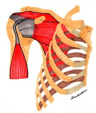
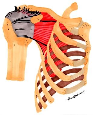
A normal rotator cuff and rotator cuff tear are shown below.
Often, younger individuals with rotator cuff injuries relate a history of repetitive overhead activities involving the rotator cuff or, less commonly, a history of trauma preceding clinical onset of symptoms. In contrast, older individuals usually present with a gradual onset of shoulder pain and, ultimately, after radiographic testing is shown to have significant partial or full rotator cuff tears without a clear history of predisposing trauma. Nonoperative or conservative treatment is usually sufficient to heal the problem in the vast majority of individuals, with a few exceptions that are discussed. [1, 2, 3, 4, 5]
Epidemiology
United States statistics
The frequency of full-thickness rotator cuff tears ranges from 5-40%, with an increasing incidence of cuff pathology in advanced age. Cadaveric studies by Bigliani et al found that 39% of individuals older than 60 years had full-thickness rotator cuff tears with an even higher incidence of partial tears. [6]
Functional Anatomy
Normal shoulder motion
The shoulder complex is comprised of several joints, including the sternoclavicular joint, acromioclavicular joint, glenohumeral (GH) joint, and scapulothoracic (ST) joint or pseudoarticulation. These articulations work together to carry out normal shoulder motion. The majority of motion occurs at the GH and ST joints. A rhythm between these 2 areas of motion has been described. [1, 3, 7, 8, 9, 10, 11, 12]
The GH–to–ST motion ratio of total shoulder motion is 2:1 (ie, 180° of abduction, consisting of 120° of GH motion and 60° of ST motion). The 2:1 ratio is an average over the entire arc of motion. This ratio changes through the arc of motion (ie, the 2:1 ratio is not constant throughout the entire range of motion [ROM]). In the initial portion of abduction, GH motion predominates and the ratio is 4:1 (GH:ST). As the shoulder moves above 90° of abduction, this ratio becomes 1:1° GH to 1° ST motion.
The importance of the scapula in normal shoulder motion cannot be overstated. The scapula, with the glenoid as its contact point, forms the platform for humeral head articulation and motion. A stable platform is essential for normal shoulder biomechanics in everyday activities and is crucial for high-demand activities (eg, overhead sports or work). [13]
The scapula must glide along the chest wall as it protracts and retracts during normal shoulder movements. Scapular winging results in glenoid antetilting, which results in functional elevation of the humeral head and impingement of the rotator cuff. In addition, without scapular motion, the origin and insertion of the deltoid approximate each other, resulting in a decreased optimal length-tension relationship and a decrease in force as the shoulder abducts. Normal scapular motion allows the deltoid to maintain its length-tension relationship and generate adequate force.
Stabilizers of the shoulder
The shoulder is considered a ball-in-socket joint, although the glenoid fossa is flat. In addition, the surface area of the glenoid is much smaller than that of the contacting humeral head (25-30%). The cartilaginous labrum provides much of the socket function and increases the surface area of contact for the humeral head.
Together, these components provide a great amount of shoulder mobility with limited stability. Shoulder stabilizers can be grossly categorized as static or dynamic. Dynamic stabilizers require an intact neuromuscular system to function, whereas static stabilizers help maintain congruity.
The static stabilizers have been studied well in cadaver specimens to understand their stabilizing effects. Static stabilizers continue to function in the setting of neurologic or intrinsic muscle pathology in conditions such as hemiplegia, spinal cord injury, brachial plexus injury, suprascapular nerve injury, and myopathies. This is not true for the dynamic stabilizers (eg, rotator cuff muscles). With neuromuscular injury or intrinsic muscle damage, the dynamic stabilizers lose their ability to exert dynamic motor control of the humeral head, ultimately leading to GH laxity and shoulder pain.
Static stabilizers
Static stabilizers include the bony structures, labrum, GH ligaments, and joint capsule. Unlike the hip joint, the bony articulation of the shoulder offers little stability. This is due to the limited contact area of the glenoid with the humeral head, flattened architecture, and retroverted positioning. The labrum is a fibrous structure that attaches to the glenoid to increase the contact area and deepen the socket of the glenoid up to 50%, forming a concave surface. Three GH ligaments exist, as follows: superior, middle, and inferior. The inferior GH ligament is the most important for shoulder stability and has 3 components, anterior, inferior, and posterior, therefore, it is more appropriately referred to as the inferior GH complex.
Dynamic stabilizers
Dynamic stabilizers [14] include the rotator and scapular stabilizers (ie, teres major, rhomboids, serratus anterior, trapezius, levator scapula). The rotator cuff is composed of 4 muscles: the supraspinatus, infraspinatus, subscapularis, and teres minor. The supraspinatus is the principal supporting and kinetic muscle of the shoulder. The primary function of the rotator cuff muscles is to stabilize the GH joint so that the larger shoulder movers (eg, deltoid, latissimus dorsi) can carry out their function without significant motion of the humeral head on the glenoid. Increased movement results in shearing forces across the joint (to the labrum, in particular) and may result in humeral head migration and impingement upon the rotator cuff muscles and tendons.
The rotator cuff muscles are associated and assist with some shoulder motion; however, their main function is to provide stability to the joint by compressing the humeral head on the glenoid. The supraspinatus assists in shoulder abduction by maintaining the humeral head centered on the glenoid, with the middle deltoid acting as the primary mover. These muscles act as force couples, because they work synergistically to carry out a particular movement.
Electromyography (EMG) studies have demonstrated a high degree of supraspinatus activity during the initial 30° of abduction. This has been misinterpreted to imply that the supraspinatus initiates shoulder abduction and acts to abduct the shoulder in the first 30°. In actuality, the supraspinatus fires to stabilize the GH joint as the deltoid abducts the arm. [15, 16, 17, 18, 19]
Increased EMG activity in the supraspinatus during the initial 30° is a reflection of increased firing requirements of this muscle to stabilize the GH joint as the deltoid is activated. The infraspinatus and teres minor muscles assist in external rotation of the shoulder and also provide an inferior pull upon the humeral head, assisting in its centering during overhead activity. The subscapularis muscle participates in this centering but also acts with the pectoralis muscles and latissimus dorsi as an internal rotator of the shoulder, serving as the main internal rotators of the shoulder.
Weakness or insufficiency of the rotator cuff muscles results in increasing demands on the static stabilizers. If these demands are long term or recurrent, static stabilizers may begin to fail. This can result in stretching or attenuation of the capsule, which results in even greater shoulder laxity and greater demands on the already weak rotator cuff muscles. Humeral head migration may occur with capsule laxity and result in rotator cuff impingement and pain. Pain may inhibit rotator cuff muscle firing, leading to disuse and further weakening of the dynamic stabilizers with greater demands placed on the static stabilizers.
Increased humeral head translation can also lead to shearing and injury to the glenoid labrum. Rotator cuff impingement, tendinitis, and labral pathology are commonly encountered injury patterns in athletes and workers who perform overhead motions. Focusing solely on the static stabilizers in treatment neglects the dynamic structures that probably initiate and perpetuate the cycle.
Sport-Specific Biomechanics
A similar type of motion is involved in a number of overhead sports activities (eg, serving in tennis, spiking in volleyball, throwing a football or baseball). The baseball throwing motion has been studied in detail and can be divided into 5 stages.
-
Stage 1 is the wind-up phase. EMG studies have determined that the rotator cuff muscles are inactive during this initial stage.
-
Stage 2 is the early cocking stage and involves shoulder external rotation and abduction supplied primarily by the deltoid.
-
Stage 3 is the late cocking stage, which continues until maximal external rotation is achieved. The rotator cuff muscles are very active during this stage, especially the subscapularis, which eccentrically contracts and acts as a dynamic stabilizer.
-
Stage 4 is the acceleration stage, which begins with internal rotation of the humerus and ends with release of the baseball. During this phase, the pectoralis major and the latissimus dorsi are very active, whereas the muscles of the rotator cuff are inactive.
-
Stage 5 is the follow-through of the baseball pitch, where deceleration takes place. During this phase, the rotator cuff muscles and the posterior deltoid are most active. The supraspinatus eccentrically contracts to decelerate internal rotation of the limb.
Proper balance between the concentrically contracting muscles that generate force and the eccentrically contracting muscles that control movement is important. Imbalance between these opposing muscle groups results in overuse of muscles and, ultimately, overuse injuries of the shoulder. Note that a great deal of the force generated in overhead sports occurs in the trunk and lower extremity, and these areas should be targeted in any conditioning program for athletes who throw.
Etiology
Several primary causes of rotator cuff pathology have been described, including age-related degeneration, compromised microvascular supply, and primary outlet impingement. Secondary factors (eg, GH instability) also appear to be related to rotator cuff injuries.
Age-related degeneration
Intrinsic tendinopathy is an age-related degenerative process. [20]
Uhthoff and Ozaki found an increase in frequency of partial-thickness and full-thickness tears with increasing age.
Increased degenerative changes are observed in athletes and workers who perform overhead motions.
Compromised microvascular supply
In 1934, Codman first described a critical zone in the supraspinatus tendon where a tenuous blood supply exists. [1]
A decrease in vascularity is noted with aging.
In 1970, Rathburn and Macnab showed that shoulder position is important for proper vascular supply to the rotator cuff. [21]
The term "wringing out" was coined to describe the reduced blood flow that occurs upon shoulder adduction.
The microvascular pattern of the supraspinatus tendon is thought to be nonhomogenic in cadavers. [22]
In 1990, Lohr and Uhthoff found that the bursal side of the supraspinatus tendon has a higher blood supply compared to the articular surface. [22] This difference in blood supply is thought to contribute to the increased incidence of articular surface tears compared with bursal tears.
Outlet impingement
The rotator cuff is surrounded by the coracoacromial arch, which comprises the supraspinatus outlet and consists of the acromion, coracoacromial ligament, and coracoid process.
The shape of the acromion has been implicated in rotator cuff pathology.
Bigliani and Morrison classified 3 types of acromions based on cadaveric examination, as follows [6] :
-
Type I – Flat
-
Type II – Curved
-
Type III – Hooked
Bigliani noted a significant increase in rotator cuff tears in curved (type II) and hooked (type III) acromions. This work has led to the belief that rotator cuff pathology occurs secondary to the type of acromion and that treatment should be directed toward correcting pathoanatomic changes by making the acromion smoother and flatter. However, debate exists concerning whether the acromion shape causes pathology to the rotator cuff or is a result of a diseased rotator cuff that secondarily causes bony changes to the acromion.
Neer proposed that acromial changes are secondary to rotator cuff tendinopathy. [3] According to this view, the initial process is migration of the humeral head superiorly with repeated impingement, followed by secondary bony changes to the undersurface of the anterior acromion. This view was further corroborated by the work of Yamanaka and Fukuda, who found a greater incidence of partial rotator cuff tears on the articular surface of the rotator cuff rather than the bursal surface. [20] If acromial changes actually caused rotator cuff tendinopathies, one would expect the opposite (ie, higher incidence of pathology on the bursal surface).
The rotator cuff contacts the coracoacromial arch undersurface in the normal shoulder.
The coracohumeral ligament is often resected in order to decompress the supraspinatus outlet, which can lead to increased superior translation of the humeral head, particularly in the young athlete.
Rotator cuff abrasions and fiber failure occur when repeated and excessive compression from humeral head migration is present.
This occurs secondary to underlying muscular imbalance and loss of rotator cuff depressor effects.
Instability
Most people with ligamentous laxity are functionally stable. In patients with inherent shoulder or generalized laxity, instability may develop with minimal or no injury.
Ligamentous laxity may be acquired by repetitive stretching of the joint, as observed in swimmers, gymnasts, and tennis players.
Dynamic stability may be lost if the shoulder becomes deconditioned. As a result, a vicious self-perpetuating cycle of instability, less use, more muscle weakness, and more instability is present.
These patients frequently have relative rotator cuff muscle weakness, particularly the external rotators and scapular stabilizers.
Subtle instability patterns may contribute to the impingement development.
Increased anterior and superior translation of the humeral head, as observed in athletes with generalized laxity and multidirectional instability of the shoulder, may predispose to impingement along the coracoacromial arch, resulting in rotator cuff injury.
Prognosis
Most athletes with primary outlet impingement without full-thickness rotator cuff tears respond well to nonoperative treatment. Rehabilitation is also effective in the majority of athletes with rotator cuff pathology due to other causes (eg, instability), except when instability is caused by trauma. When surgery is performed for rotator cuff injuries not responding to conservative treatments, results vary depending upon patient age, size and pattern of the tear, degree of retraction, tissue quality, and quality of repair.
One study evaluated 51 patients, aged 60 years or younger, with nonoperatively treated rotator cuff tears and found that full-thickness rotator cuff tears tended to increase in size in about half of the patients. The study suggests that surgery be considered to prevent an increase in size tear, and those treated nonoperatively should be monitored for tear size increase. [23]
Complications
When treatment is delayed in rotator cuff injuries and shoulder discomfort persists, the patient can develop symptomatic stiffness of the GH joint, which is called adhesive capsulitis. In this condition, the patient consciously or subconsciously limits the use of the shoulder because of pain, leading to the development of soft-tissue tightness or stiffness in one or more planes. The chance of developing adhesive capsulitis can be minimized through prompt diagnosis of painful problems in the shoulder, such as rotator cuff injuries, and the institution of early shoulder ROM as part of the rehabilitation program.
Severe supraspinatus and infraspinatus muscle atrophy is often associated with massive rotator cuff tears, but an underlying entrapment of the suprascapular nerve should always be considered. Symptoms of suprascapular nerve entrapment include shoulder pain that is described as a deep dull ache localized to the posterolateral aspect of the shoulder. Weakness of the shoulder and arm is common, with visible wasting and atrophy of the supraspinatus and infraspinatus and normal bulk in the deltoid. Clinical differentiation of suprascapular nerve entrapment from rotator cuff injuries may be difficult, especially if both are present simultaneously. EMG is the single most helpful test for diagnosing this condition.
Patient Education
Proper sport technique can be of great importance in the prevention and rehabilitation of rotator cuff injuries. This includes proper hand position on water entry in swimming, changes in paddling technique in canoeing and kayaking, and evaluation of pitching mechanics by coaches and trainers in throwing athletes. Encourage the importance of maintaining proper trunk and lower extremity strengthening in athletes performing overhead motions, because these muscles generate significant force during overhead activities and serve to reduce stresses on the shoulder stabilizers.
-
Rotator cuff, normal anatomy.
-
Rotator cuff tear, anterior view.
-
The acromioclavicular arch and the subacromial bursa.
-
Neer impingement test. The patient's arm is maximally elevated through forward flexion by the examiner, causing a jamming of the greater tuberosity against the anteroinferior acromion. Pain elicited with this maneuver indicates a positive test result for impingement.
-
Hawkins test. The examiner forward flexes the arms to 90° and then forcibly internally rotates the shoulder. This movement pushes the supraspinatus tendon against the anterior surface of the coracoacromial ligament and coracoid process. Pain indicates a positive test result for supraspinatus tendonitis.
-
Rotator cuff injury.


