Practice Essentials
Low back pain (LBP) is one of the most common reasons for missed playing time in professional athletics, as well as a leading reason for healthcare provider visits. When evaluating patients with LBP, lumbar disc problems should be considered (see image below).
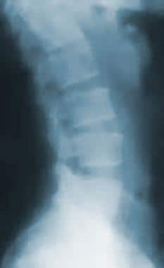 Radiograph of the lumbar spine. This image demonstrates L5-S1 disk space narrowing (the most common location).
Poor training technique and overtraining are frequent causes of disc herniation. Activities that involve chronic combined flexion and rotation with compression can lead to radial tears of the annulus fibrosis and gradual posterior disc bulging. Chronic changes such as calcification and osteophyte formation of the vertebrae can also lead to disc problems.
See Back Pain: Find the Cause, Watch for the Comeback, a Critical Images slideshow, to help diagnose and manage this common problem.
Epidemiology
United States statistics
At-risk sports for lumbar disc problems include activities that require frequent flexion, extension, and rotation of the spine. An example is gymnastics, in which disc degeneration is seen in up to 75% of participants that present with back pain. [1] Disc degeneration is significantly more common in elite athletes (75%) compared with nonathletes (31%). [2]
Lumbar disc herniation accounts for only 4% of back pain cases [3] ; symptomatic disc herniations are more common in adults (48%) than adolescent athletes (11%). Acute disc herniations commonly occur in individuals between the ages 30 and 55 years [4] ; however, athletes between ages 20 and 35 years are at the greatest risk for disc injury, [5] and the L4-L5 and L5-S1 levels are most commonly affected.
International statistics
A study by Morimoto et al found that the most common cause of LBP among Japanese professional baseball players in their 20s was lumbar disc herniation (57%). Additionally, the incidence of lumbar intervertebral disc degeneration was significantly higher among players in their 30s (91%) than among those in their 20s (14%). [6]
In another Japanese study, Enoki et al reported that the prevalence of lumbar intervertebral disc degeneration among male pole vaulters was 38%. All of the athletes in the study had a history of LBP. [7]
Functional Anatomy
The vertebrae are separated by vertebral discs that are composed of a gel substance (nucleus pulposus) surrounded by outer collagen fibers, which are arranged in a crossed manner (annulus fibrosis). These discs are further supported by the anterior and posterior longitudinal ligaments. Together, the vertebral disc complex resists spinal compression.
During axial rotation of the spine, the annular fibers are placed at a mechanical disadvantage. Furthermore, in forward flexion, the anterior vertebral endplates approximate, increasing the pressure of the disc posteriorly. The most common disc herniation is directed posteriorly toward the foraminal window, where the nerve roots exit the spinal canal. As such, a common mechanism of herniation in athletes is combined flexion, rotation, and compression of the spine. Football, wrestling, hockey, gymnastics, tennis, and golf are some sports in which this injury mechanism commonly occurs.
In the presence of a disc herniation, forward flexion worsens the herniation. In extension, the opposite occurs. The posterior vertebral endplates approximate, forcing the disc anteriorly, to reduce the herniation.
The anatomic structures that have been implicated as pain generators include the vertebral discs, nerve roots, ligaments, zygapophysial joints (z-joints), sacroiliac joints, and the musculature. Some studies suggest that discogenic pain secondary to annular disruption is the most common cause of LBP [8] ; vascularized granulation tissue with innervation along a torn annulus fibrosis is thought to be the cause. Inflammatory factors caused by the leakage of nuclear material from annular tears can delay intradiscal tissue healing. These factors include matrix metalloproteinases (MMPs), phospholipase A2 (PLA2), cyclooxygenase (COX), prostaglandins, nitric oxide (NO), cytokines, interleukins, and macrophages.
Classifications of disc herniations
Disc protrusion describes a bulged annulus that has not ruptured. In this scenario, there is no contact between the nucleus and the extradiscal space.
Disc extrusion describes a ruptured annulus with some expelled nucleus that remains attached to the disc.
Sequestered disc or complete prolapse describes a nucleus that is expelled from the disc and is no longer attached.
Sport-Specific Biomechanics
In cycling, an incorrect seat position may predispose an individual to disc herniation. Running can cause wear and tear to the vertebral discs secondary to the repetitive trauma that is involved. Well-cushioned shoes and more forgiving training surfaces are thought to protect against disc injury.
Former elite weight lifters and soccer players have been noted to have a higher incidence of degenerative disc disease (DDD)—as noted on magnetic resonance imaging (MRI) studies—but these findings have not been correlated with increased pain in the affected athletes. [5]
(See also the Medscape Drugs & Diseases articles Lumbar Degenerative Disk Disease [in the Physical Medicine and Rehabilitation section], Degenerative Disk Disease [in the Orthopedic Surgery section], and Degenerative Lumbar Disc Disease in the Mature Athlete [in the Sports Medicine section].)
Prognosis
About 95% of athletes have symptom resolution within 4 weeks, and 95% of those with lumbar disc problems return to play fully recovered.
A history of LBP is the greatest predictor for future episodes by up to a 3-fold increased risk; active LBP increases the risk of recurrence by 6 fold.
Complications
Complications of lumbar disc problems include prolonged or permanent nerve symptoms, chronic pain, and inability to return to sports.
Patient Education
Coaches, trainers, gym classes, pamphlets, and team and/or primary care physicians should provide education on how to avoid lumbar disk injuries.
For excellent patient education resources, see eMedicineHealth's patient education articles Slipped Disk and Back Pain.
-
Radiograph of the lumbar spine. This image demonstrates L5-S1 disk space narrowing (the most common location).
-
Magnetic resonance image of the lumbar spine. This image demonstrates a herniated nucleus pulposus at multiple levels.


