Practice Essentials
With an increase in public interest in physical fitness, clinical practitioners are diagnosing stress fractures with greater frequency. [1, 2, 3] First described by Aristotle in 200 BC, stress fractures were initially recorded in the medical literature in 1855 by the Prussian military physician Breithaupt, who described what is now known as a march fracture, or stress fracture of the metatarsals. See the images below.
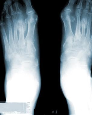 Radiograph of the feet. This image depicts a stress fracture of the left second metatarsal with exuberant callus.
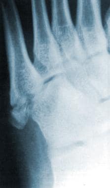 Radiograph of the left foot. This image depicts a stress fracture of the fifth metatarsal.
Metatarsal stress fractures are not limited to high-level athletes or military recruits. This type of injury is seen in runners of all levels, as well as ballet dancers and gymnasts and patients with rheumatoid arthritis (RA), metabolic bone disease, and neuropathic conditions. [4, 5, 6] Metatarsal stress fractures are also seen with increasing frequency in patients who engage in aerobics activities, particularly high-impact aerobics.
Signs and symptoms
Initially, dull pain occurs only with exercise, then the condition progresses to pain at rest.
Pain starts diffusely, then localizes to the site of the fracture.
See Presentation for more detail.
Diagnosis
Imaging studies
Stress fracture changes may not be evident on plain films until 3 months after the onset of symptoms. Up to 50% of stress fractures are never observed on plain films.
Bone scanning is nearly 100% sensitive for the diagnosis of stress fractures, although the specificity of this modality is considerably lower.
Magnetic resonance imaging (MRI) and single-photon emission computed tomography (SPECT) may also be used to image stress fractures; however, MRI has become the study of choice because it has the same sensitivity as a bone scan but with a much higher specificity.
See Workup for more detail.
Management
The patient should rest from the offending activity, and immobilization is recommended for comfort. It is important to apply ice and elevate the foot to minimize pain and swelling.
Stress fractures of the second or third metatarsals rarely require surgical intervention. Most of these fractures heal uneventfully, and nonunion is rare. However, stress fractures of the fifth-metatarsal base are more problematic. Displacement of these fractures tends to increase with continued weight bearing.
See Treatment and Medication for more detail.
Etiology
Causes of metatarsal stress fractures include the following:
-
Increased intensity, duration, or frequency of exercise
-
New footwear
-
Insufficient rest periods
-
Continuing to train despite pain
-
Osteopenia/osteoporosis
-
Rheumatoid arthritis
-
Neuropathic foot
-
Female athletic triad
Epidemiology
United States statistics
The incidence of stress fractures in the general population is unknown, as virtually all literature on the subject is derived from a military population or advanced-level athletes. Stress fractures are estimated to constitute up to 16% of all injuries that are related to athletic participation; running is the cause in most of these cases. Most stress fractures (up to 95%) involve the lower extremities, particularly the metatarsals. The incidence of stress fractures among female athletes may be as high as 13%. [7]
A study by Waterman et al reported the incidence rate for lower extremity stress fractures in the US military (not adjusted for sex, race, age, rank, and service branch), including of the metatarsals, to be 5.69 per 1000 person-years, although tibial/fibular fractures were the most common. The highest fracture risk was found in service members under age 20 years or age 40 years or above, with the risk also higher in White service members than in Black service members. [8]
Functional Anatomy
The second and third metatarsals are relatively fixed in position within the foot; the first, fourth, and fifth metatarsals are relatively mobile. More stress is placed on the second and third metatarsals during ambulation; thus, these bones are at increased risk for stress fractures.
The fifth metatarsal, which is approximately 1.5 cm from the proximal pole of the bone, bears greater stress in those who oversupinate when they walk or run. The fifth metatarsal also has a diminished blood supply and, thus, a decreased ability to heal. [9, 10]
Stress fractures of the proximal fifth metatarsal must be distinguished from proximal avulsion fractures ("pseudo-Jones" fractures) and Jones fractures. The proximal avulsion fracture is usually associated with a lateral ankle strain and occurs at the insertion of the peroneus brevis tendon. The true Jones fracture is an acute fracture of the proximal diametaphyseal junction.
Sport-Specific Biomechanics
Queen et al investigated whether foot type (flat or normal) resulted in loading differences during four sport-specific tasks (cross-cut, side-cut, shuttle run, and landing from a simulated lay-up). [11] Of 22 healthy individuals, 12 had normal feet and 10 had flat feet, and each completed 5 trials per condition. In-shoe pressure data were collected at 50 Hz, and analyses of the entire foot and 8 regions of the foot were carried out on contact area, maximum force, and the force time integral. The investigators' findings included the following statistically significant (P< 0.05) findings [11] :
-
During the cross-cut task, there was an increase in medial midfoot contact area.
-
During the side-cut task, an increase in contact area, force time integral, and maximum force in both the medial and lateral midfoot were demonstrated.
-
During the shuttle run task, an increase in force time integral in the lateral midfoot and increases in maximum force in both the medial and lateral midfoot were present
-
During the landing task, an increase in maximum force in the medial midfoot was present. However, flat feet showed a decrease in middle forefoot maximum force.
Queen et al concluded that individuals with a normal foot may have a lower risk for medial and lateral midfoot injuries such as metatarsal stress fractures. Thus, foot type should be assessed when determining an individual's risk for metatarsal stress fractures. [11]
A case-control study that included 51 NFL players reported increased risk for fifth metatarsal fractures in players with long, narrow, and straight fifth metatarsals with an adducted forefoot. [12]
Fujitaka et al studied a cohort of 273 collegiate male soccer players which included 16 who developed a fifth metatarsal stress fracture. Analysis of various history, physical, and equipment variables suggested that stress fracture may be a result of a weak toe-grip that leads to an increase in the load applied onto the lateral side of the foot. [13]
Prognosis
Stress fractures in the first 4 metatarsals routinely heal without complication.
Complications
Nonunion is the primary complication of metatarsal stress fractures. Stress fractures at the base of the fifth metatarsal have a nonunion rate of 35-50%. For other metatarsal stress fractures, the nonunion rate is low.
Patient Education
The key to preventing stress fractures lies in the education of athletes, parents, coaches, trainers, and doctors.
Properly selected and fitted equipment, particularly running shoes, is important in deterrence of metatarsal stress fractures.
Training needs to be performed in a slow cyclical progression that allows the body to adapt. Adequate rest and recovery time needs to be incorporated into the participant's training regimen.
The quality of physical activity, rather than quantity, should be stressed in any exercise program.
The athlete, coach, trainer, and physician must recognize that exercise regimens are not "one size fits all." Tailor the training to the participant's baseline ability, previous experience, and current level of physical activity.
-
Radiograph of the feet. This image depicts a stress fracture of the left second metatarsal with exuberant callus.
-
Radiograph of the left foot. This image depicts a stress fracture of the fifth metatarsal.
-
Bone scan of the lower extremities. This image depicts a right fifth metatarsal stress fracture.


