Practice Essentials
Kawasaki disease is an acute febrile illness associated with multiorgan vasculitis of unknown etiology that primarily affects infants and children. The disease mainly affects children younger than 5 years. During the acute phase, children may develop aseptic meningitis, hyperemic tympanic membrane, or uveitis. Neurologic complications, which include facial nerve palsy, seizures, and ataxia cerebral infarctions, are rare. Other common features include diarrhea, vomiting, abdominal pain, and pneumonitis. Gallbladder hydrops (acute acalculous distention of the gallbladder; see the image below) may occur in the first 2 weeks of illness; it may be the result of the extension of periportal inflammation to the cystic duct; and it is typically self-limited. Arthritis and arthralgia are common in the acute phase. Findings that include erythema and induration at the site of recent Bacille Calmette-Guérin vaccination, testicular swelling, and peripheral gangrene also have been reported in patients with this disease. [1, 2, 3, 4, 5]
The diagnosis of Kawasaki disease is ideally made by clinical criteria according to the American Heart Association. An algorithm was created by the AHA to help identify patients at risk. Clinical decision making for Kawasaki disease relies on the measurements of the coronary Z-score obtained by 2-dimensional echocardiography. Patients with large or giant aneurysms (ie, Z score ≥10) are at the highest risk for thrombosis and stenosis. Echocardiography should be performed at the time of diagnosis and then again at 1-2 weeks and 4-6 weeks after treatment in patients with uncomplicated cases who do not have significant coronary artery involvement. For patients with evolving abnormalities, more frequent assessment is necessary. When performed by skilled imagers, 2-dimensional echocardiography has 100% sensitivity and approximately 90% specificity for detecting aneurysms of the proximal coronary arteries. [6, 7, 8, 9, 10, 3, 11, 12, 5, 13]
Chest radiography is not routinely performed to evaluate for Kawasaki disease, and the results are often normal. [14] If coronary aneurysm and calcification of the coronary artery aneurysm wall are present, they may be detected as cystic calcification in the region of the coronary vessels, overlying the heart shadow.
CT is not routinely performed for the evaluation of Kawasaki disease; however, CT is more sensitive than chest radiography in detecting coronary calcifications. Contrast-enhanced CT may demonstrate enhancement of the vessel walls in the coronary arteries, particularly in the acute phase. [15, 16]
MRI is not performed routinely to investigate Kawasaki disease; however, MRI may depict coronary aneurysms. [17, 18] In addition, MRI is considered useful for the long-term follow-up of patients with Kawasaki disease. [19]
Liver and gallbladder ultrasonography may be necessary if liver or gallbladder dysfunction is suspected. Acute distention of the gallbladder (hydrops) is identified on abdominal ultrasonography in 15% of patients.
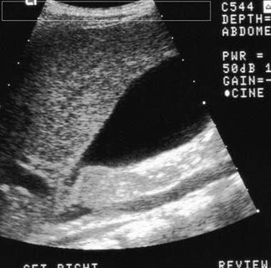 Kawasaki disease. Sonogram of the right upper quadrant shows hydrops of the gallbladder. Note the size of the gallbladder compared with that of the inferior vena cava. Courtesy of Dr S. Methratta, UMDNJ-New Jersey Medical School.
See Kawasaki Disease: Do You Know the Signs?, a Critical Images slideshow, to help identify the specific criteria for diagnosis.
Computed Tomography
CT is not routinely performed for the evaluation of Kawasaki disease; however, CT is more sensitive than chest radiography in detecting coronary calcifications. Contrast-enhanced CT may demonstrate enhancement of the vessel walls in the coronary arteries, particularly in the acute phase. [20]
Although no published data describe the use of CT in detecting Kawasaki-related coronary aneurysms, advances in multihelical technologies, as well as electron-beam CT, may be useful in detecting and sizing coronary artery aneurysms. CT is also useful in evaluating the size of the gallbladder and in detecting gallstones.
Retropharyngeal lymphadenopathy and retropharyngeal edema have been found to be relatively common features of Kawasaki disease on CT. [21]
In selected high-risk patients with Kawasaki disease, coronary CT angiography may identify a subset of patients at increased risk for future coronary pathology who may benefit from medical therapy. [15, 16]
Magnetic Resonance Imaging
MRI is not performed routinely to investigate Kawasaki disease; however, MRI may depict coronary aneurysms. [17, 18] MRI with gadopentetate dimeglumine can be use to noninvasively and simultaneously evaluate myocardial thinning and the presence of circulation. In addition, MRI is considered useful for the long-term follow-up of patients with Kawasaki disease. [19] Recent advances in cardiac MRI may enable the detection of early changes in Kawasaki disease. Acute-phase disease may be detected on MRI with or without gadolinium enhancement as an area of inflammation characterized by a high signal intensity on T2-weighted images; this area may be enhancing. [20, 22, 23]
Gadolinium-based contrast agents have been linked to the development of nephrogenic systemic fibrosis (NSF) or nephrogenic fibrosing dermopathy (NFD). The disease has occurred in patients with moderate to end-stage renal disease after being given a gadolinium-based contrast agent to enhance MRI or MRA scans. NSF/NFD is a debilitating and sometimes fatal disease. Characteristics include red or dark patches on the skin; burning, itching, swelling, hardening, and tightening of the skin; yellow spots on the whites of the eyes; joint stiffness with trouble moving or straightening the arms, hands, legs, or feet; pain deep in the hip bones or ribs; and muscle weakness.
Ultrasonography
Clinical decision making for Kawasaki disease relies on the measurements of the coronary Z-score obtained by 2-dimensional echocardiography. Patients with large or giant aneurysms (ie, Z score ≥10) are at the highest risk for thrombosis and stenosis. Echocardiography should be performed at the time of diagnosis and then again at 1-2 weeks and 4-6 weeks after treatment in patients with uncomplicated cases who do not have significant coronary artery involvement. For patients with evolving abnormalities, more frequent assessment is necessary. When performed by skilled imagers, 2-dimensional echocardiography has 100% sensitivity and approximately 90% specificity for detecting aneurysms of the proximal coronary arteries. [6, 7, 8, 9, 10, 3, 11, 12, 5, 13]
Liver and gallbladder ultrasonography may be necessary if liver or gallbladder dysfunction is suspected. Acute distention of the gallbladder (hydrops) is identified on abdominal ultrasonography in 15% of patients. [8]
(See the image below.)
Kawasaki disease. Sonogram of the right upper quadrant shows hydrops of the gallbladder. Note the size of the gallbladder compared with that of the inferior vena cava. Courtesy of Dr S. Methratta, UMDNJ-New Jersey Medical School.
Echocardiography should be performed to evaluate for CAAs during the acute stage. In order of highest to lowest frequency, the involvement of the coronary arteries is as follows:
Proximal left anterior descending and right coronary arteries
Left main coronary artery
Left circumflex artery
Distal right coronary artery
Posterior descending artery
In addition to evaluating the coronary arteries for dilation and thrombosis, the baseline echocardiogram is also performed to evaluate for other signs of cardiac involvement. This includes aortic root dilation, depressed contractility, ventricular and valvular dysfunction, and pericardial effusion.
Diffuse dilatation of coronary lumina can be observed in 50% of patients by the 10th day of illness. In children, pediatric cardiologists should ideally perform this study, because they are familiar with coronary artery diameter normal values. Coronary artery dimensions must be adjusted for body surface area to accurately identify dilation. A basic rule is that if the internal diameter of a segment is greater than 1.5 times that of an adjacent segment, then dilation probably exists.
The echocardiogram should be repeated at 1-2 weeks and then 5-6 weeks after disease onset; echocardiograms may need to be performed more frequently in high-risk patients. Frequency of subsequent echocardiography and/or additional cardiac imaging is dependent on the disease severity and the expert opinion of pediatric cardiologists.
Angiography
Angiography may be used to evaluate for coronary aneurysms (see the image below). [24] Although it is considered to be the criterion standard for evaluating coronary aneurysms, ultrasonography provides an alternative method of detecting coronary aneurysms, particularly proximal ones.
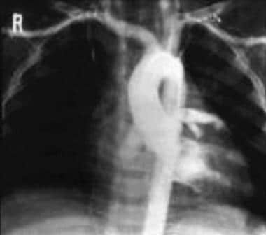 Kawasaki disease. Angiogram of the ascending aorta and coronary vessels shows aneurysmal dilatation of the coronary vessels. Courtesy of Dr Chong Hyun Yoon, Professor of Radiology, University of Ulsan, Seoul, Korea.
-
Kawasaki disease. Sonogram of the right upper quadrant shows hydrops of the gallbladder. Note the size of the gallbladder compared with that of the inferior vena cava. Courtesy of Dr S. Methratta, UMDNJ-New Jersey Medical School.
-
Kawasaki disease. Angiogram of the ascending aorta and coronary vessels shows aneurysmal dilatation of the coronary vessels. Courtesy of Dr Chong Hyun Yoon, Professor of Radiology, University of Ulsan, Seoul, Korea.


