Practice Essentials
Fractures of the foot are common, and the metatarsals account for 35% of foot fractures. About 80% of metatarsal fractures are nondisplaced or minimally displaced. In adults and children older than 5 years, fractures of the fifth metatarsal are most common, followed by fractures of the third metatarsal. The fifth metatarsal is divided into 3 anatomic zones: zone 1 is the tuberosity; zone 2 is the metaphyseal-diaphyseal junction; and zone 3 is the diaphyseal area within 1.5 cm of the tuberosity. Acute foot fractures of normal bones are usually caused by the dropping of heavy objects on the foot or by stress associated with abnormal repetitive trauma. In deficient bones, insufficiency fractures may result from normal stress. The main goal of treatment is to restore the alignment of all of the metatarsals and thereby maintain the arches of the forefoot and allow distribution of a normal weight under the head of metatarsals. [1, 2, 3, 4, 5]
Imaging modalities
Radiography is the first and often the only investigation required for the diagnosis of fractures. Radiographs may be used to diagnose all acute fractures, dislocations, and established stress fractures. Small avulsions may be missed on radiographs. In the early stages of stress fracture, radiographs may be normal, or they may show only subtle periosteal reaction, which can be easily missed. Radiography cannot be used to assess soft-tissue and ligamentous disruption. [6, 7]
Bone scanning is more sensitive than plain radiography; it is indicated when a stress fracture or an acute fracture is suspected and radiographs are negative. Bone scanning is not a specific investigation. Although bone scanning is sensitive, some stress fractures may go undetected in the early stages of these injuries.
Although magnetic resonance imaging (MRI) is more sensitive than radiography and bone scanning, it is used only for the assessment of soft-tissue structures and ligamentous injuries. MRI is the most sensitive technique for imaging stress fractures of the foot; MRI may depict bone marrow edema even before increased uptake is seen on bone scans.
Computed tomography (CT) scanning is useful for finding avulsion fractures and comminuted fractures and in assessing for intra-articular extension. Although CT and MRI are more sensitive than radiography, they are not cost-effective and are not indicated for the diagnosis of fractures. In the case of multiple fractures of the base of metatarsals, CT is recommended to rule out Lisfranc fracture-dislocation. [2]
High-resolution ultrasound is useful in fracture assessment because of its availability, ease of use, and multiplanar capabilities. Its indications include injury assessment for the presence of a fracture when radiography is not immediately available; detecting occult fractures not revealed on radiographs; and diagnosing bone stress injury before radiographic changes. Sonographic evaluation of bone, however, has limitations and should be coupled with radiographs and possibly CT and MRI when clinically indicated. [8]
Radiologic anatomy
The metatarsal bones and tarsal bones are connected by strong ligaments. Soft-tissue support for the joints in the plantar aspect of the foot is better than that in the dorsal aspect. The distance between the base of the first and second metatarsals and the medial and intermediate cuneiform is more than the distance between other corresponding joints. If a lateral image is obtained, a line drawn through the long axis of the talus bone and the long axis of the first metatarsal bone should be straight if there is no dislocation. [9, 7, 10]
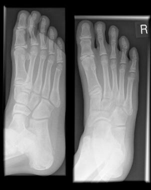
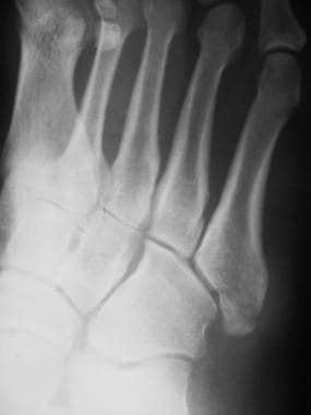
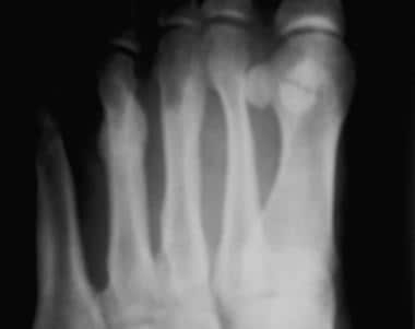
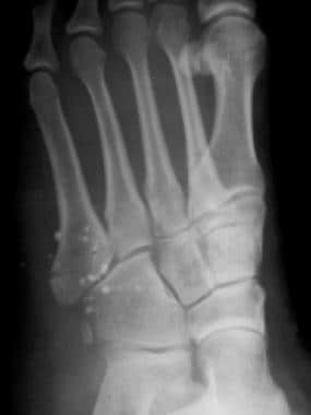
On the anteroposterior view (see the image below), the lateral border of the first metatarsal should be aligned with the lateral border of the medial cuneiform. The medial border of the second metatarsal should be aligned with the medial border of the intermediate cuneiform bone.
 Fractured metatarsals. Normal anteroposterior view of the foot. Note the alignment of (1) the lateral border of the first metatarsal with the lateral border of the medial cuneiform and (2) the medial border of the second metatarsal with the medial border of the middle cuneiform.
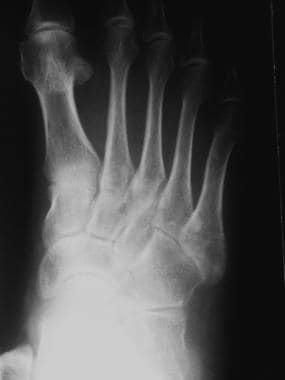
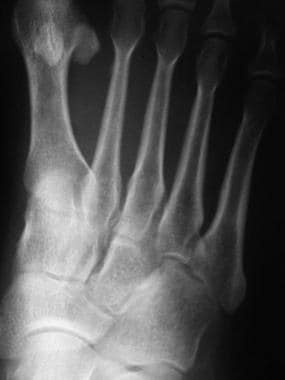
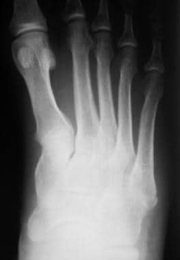
On the oblique view (see the image below), the medial and lateral borders of the third metatarsal should be aligned with the medial and lateral borders of lateral cuneiform bone. The medial border of the fourth metatarsal should be aligned with the medial border of the cuboid bone. The fourth and fifth metatarsals are aligned with the cuboid bone, but the lateral part of the fifth metatarsal may project beyond the margin of the cuboid bone by up to 3 mm.
 Fractured metatarsals. Oblique view of a normal foot shows that the medial and lateral borders of the third metatarsal are aligned with the corresponding borders of the lateral cuneiform bone. The medial border of the fourth metatarsal is aligned with the medial border of the cuboid bone. The lateral border of fifth metatarsal projects a few centimeters beyond the cuboid bone.
Classifications
In a simple classification for fractures of the proximal end of the fifth metatarsal, fractures are classified as zone 1, the tuberosity; zone 2, the metaphyseal-diaphyseal junction; and zone 3, the diaphyseal area within 1.5 cm of the tuberosity. Fractures through zone 1 are called pseudo-Jones fractures, and fractures through zone 2 are called Jones fractures. [1] Acute fractures, Jones fractures, and stress fractures may be described as (1) early fractures, (2) delayed union fractures, or (3) nonunion fractures.
The Torg classification is used for fractures within 1.5 cm of the metatarsal tuberosity. Type I includes fractures with sharp margins and no widening, sclerosis, periosteal reaction, or cortical hypertrophy. Type II includes fractures with widening, periosteal reaction, sclerosis, or both. Type III fractures involve widening, periosteal reaction, or complete sclerosis at the fracture line.
The Stewart classification of fifth metatarsal fractures is as follows: type I, extra-articular fracture between the metatarsal base and diaphysis; type II, intra-articular fracture of the metatarsal base; type III, avulsion fracture of the base; type IV, comminuted fracture with intra-articular extension; and type V, partial avulsion of the metatarsal base with or without a fracture.
The zonal classification, reported by Dameron, Lawrence, and Botte, categorizes metatarsal fractures by the region affected: zone 1 corresponds to the tuberosity, zone 2 corresponds to Jones fractures, and zone 3 is the diaphysis.
Acute fractures
Acute fractures may be transverse, oblique, or comminuted (as seen in the images below); they are easily recognized. [11]
 Fractured metatarsals. Transverse fracture at the base of the fifth metatarsal in a male adolescent.
 Fractured metatarsals. Oblique fracture of the metaphysis of the distal shaft of the fifth metatarsal.
 Fractured metatarsals. Comminuted fracture of the base of the fifth metatarsal bone.
Jones and pseudo-Jones, or tennis, fractures
A Jones fracture (see the image below) is caused by inversion of the foot, which produces tension on the peroneus brevis tendon and on the lateral cord of the plantar aponeurosis. [12, 9, 13] In this type of fracture, significant displacement is absent. Jones fractures are more prone to nonunion.
 Fractured metatarsals. Transverse fracture at the base of the fifth metatarsal; this is a Jones fracture.
A fracture of the metatarsal tuberosity (see the image below) is an avulsion fracture. This is also called a pseudo-Jones fracture or a tennis fracture. The mechanism of injury is forcible inversion of the foot in plantar flexion, which may occur when one steps on a curb or when one falls while climbing stairs. A direct blow to the tuberosity can cause a comminuted fracture.
 Fractured metatarsals. Avulsion fracture of the tuberosity of the fifth metatarsal.
Distal, or dancer's, fractures
Distal fractures, also called dancer's fractures, are caused by a rotational force resulting from axial loading with the foot in a plantigrade position.
Lisfranc dislocation
The Lisfranc joints are the tarsometatarsal joints. A Lisfranc fracture-dislocation (see the image below) is caused by falling from a height, falling down stairs, or stepping off a curb. [14] Lisfranc fracture-displacements are relatively rare, with a reported incidence of one per 55,000 people in the United States annually. [15] Because of their rarity and often subtle findings on radiography, Lisfranc injuries may be missed. Lisfranc fracture-dislocation should be suspected in cases of substantial foot pain after minor trauma. [16] They have been classified as homolateral and divergent. In homolateral fractures, there is lateral displacement of the first to fifth metatarsals or the second to fifth metatarsals where the first metatarsophalangeal joint remains congruent. In divergent fractures, there is lateral displacement of the second to fifth metatarsals with medial displacement of the first metatarsal. [2]
 Fractured metatarsals. Image shows a Lisfranc fracture-dislocation: a fracture of the base of the second metatarsal and a lateral dislocation of the second metatarsal.
Mechanisms of injury are (1) rotation around a fixed forefoot (eg, falling from a horse with the foot caught in the stirrup) or (2) longitudinal compression of the foot. In this second mechanism, the metatarsal head is fixed. The weight of the body is on the hindfoot against the base of the metatarsals during rotation; these forces result in a distal dorsal dislocation of the metatarsal.
Stress fractures
Stress fractures are the result of abnormal stress on a normal bone (see the image below). Stress fractures of the foot are also called marcher's foot, because of the high incidence of occurrence in military recruits and in persons who engage in heavy exercise for prolonged periods. This fracture is also common in ballet dancers, gymnasts, and athletes. [17] Other predisposing factors include surgery, stress fractures in adjacent bones, neuropathic disease, and rheumatoid arthritis.
 Fractured metatarsals. Image shows a stress fracture more florid than that shown in the previous image, with extensive periosteal reaction on either side of the third and fourth metatarsals.
When a normal step is initiated, maximum force is placed on the head of the second or the third metatarsal. With increased activity, microinfarction takes place in the bones, resulting in a fracture.
Stress fractures are difficult to recognize in the early stages (as demonstrated in the image below), when they are manifested only by a periosteal reaction. Bone scans are helpful in this situation. Recognition of fracture is crucial for guiding appropriate management and for preventing complications.
 Fractured metatarsals. Image shows a thin layer of subtle, solid periosteal reaction on the medial side of the shaft of the second metatarsal bone. This is an early stage of a stress fracture.
Insufficiency fractures
Insufficiency fractures are the result of normal stress on a weakened bone. Such injuries are seen in people with osteoporosis; postmenopausal women are commonly affected.
Radiography
Radiography is sensitive in the diagnosis of acute fractures. An acute fracture is seen as a linear lucency and a break in the cortical surface. Nondisplaced, impacted fractures may appear as an opaque line; such fractures may be confirmed on a different view.
The presence of an accurate text-based localizing history and/or a graphic indicating the site of pain have been found to improve diagnostic accuracy. In a study of 126 subtle foot fractures, diagnostic accuracy improved from 79% without a graphic to 82% with a graphic. Across experience level and specialization, use of the graphic increased sensitivity for the presence or absence of subtle fracture from 67% to 73% (P< 0.001), improved degree of confidence from 8.1 to 8.4 (P< 0.0001), and decreased the mean interpretation time by 6%, from 53 seconds to 50 seconds (P=0.006). Specificity changed from 93% without a graphic to 94% with a graphic (P=0.33). [18]
Fractures may affect any metatarsal, but the fifth metatarsal is most commonly affected (see the images below). The fracture may be transverse, oblique, or comminuted. Longitudinal linear fractures are extremely rare.
 Fractured metatarsals. Spiral fracture through the distal shaft of the fifth metatarsal.
 Fractured metatarsals. A fracture of the fifth metatarsal, oblique, in the shaft.
Fractured metatarsals. Oblique fracture of the metaphysis of the distal shaft of the fifth metatarsal.
Fractured metatarsals. Transverse fracture at the base of the fifth metatarsal in a male adolescent.
Fractured metatarsals. A fracture of the fifth metatarsal, oblique, in the shaft.
Fractured metatarsals. Oblique fracture of the metaphysis of the distal shaft of the fifth metatarsal.
Fractured metatarsals. Transverse fracture at the base of the fifth metatarsal in a male adolescent.
 Fractured metatarsals. Transverse fracture of the base of the fifth metatarsal bone and associated features, including radiopaque foreign bodies in the soft tissue and the accessory ossicle lateral to the cuboid bone.
Fractured metatarsals. Comminuted fracture of the base of the fifth metatarsal bone.
Fractured metatarsals. Transverse fracture of the base of the fifth metatarsal bone and associated features, including radiopaque foreign bodies in the soft tissue and the accessory ossicle lateral to the cuboid bone.
Fractured metatarsals. Comminuted fracture of the base of the fifth metatarsal bone.
The 2 most common fractures in the fifth metatarsal are a fracture at the tip of the tuberosity and a transverse fracture 1.5-2 cm from the tuberosity; the latter is called a Jones fracture. Small avulsions derived from the tip of the base of the fifth metatarsal may be seen only in the oblique projection of the ankle. [19] (See the images below.)
Fractured metatarsals. Transverse fracture at the base of the fifth metatarsal; this is a Jones fracture.
 Fractured metatarsals. Fracture of the fifth metatarsal tuberosity with lateral displacement of the fracture fragment.
Stress fractures
The radiographic findings of a stress fracture depend on the bone involved and the stage of disease. Radiographs are normal in the early stages of the disease (see the first image below); stress fractures appear as well-defined linear lucency or fluffy periosteal reactions by 7-10 days. The periosteal reaction is variable and occasionally florid (as in the second image below).
Fractured metatarsals. Image shows a thin layer of subtle, solid periosteal reaction on the medial side of the shaft of the second metatarsal bone. This is an early stage of a stress fracture.
Fractured metatarsals. Image shows a stress fracture more florid than that shown in the previous image, with extensive periosteal reaction on either side of the third and fourth metatarsals.
The head of the second metatarsal and, occasionally, the third metatarsal are commonly affected. The first metatarsal is injured in 10% of metatarsal stress fractures; such fractures involve a different kind of reaction (the endosteal variety), with liner sclerosis. Periosteal reaction is not common in this type of injury. One third of such fractures heal with only an intramedullary callus.
The base of second metatarsals may be affected in ballet dancers. The proximal aspect of the shaft of the fourth and fifth metatarsals is affected; the pattern is that of a linear lucency, which is slow to heal. Fractures in the sesamoid bones are also seen in ballet dancers.
Lisfranc fracture-dislocation
A Lisfranc fracture-dislocation (seen in the images below) is a dislocation of the tarsometatarsal joints. Two types of Lisfranc dislocation have been described: homolateral and divergent.
Fractured metatarsals. Image shows a Lisfranc fracture-dislocation: a fracture of the base of the second metatarsal and a lateral dislocation of the second metatarsal.
 Fractured metatarsals. Image shows a Lisfranc dislocation with a fracture of the base of the third and fourth metatarsals.
In the homolateral type, all of the metatarsals are dislocated to one side. Usually, the second to fifth metatarsals are dislocated, but occasionally, all of the metatarsals are affected. Lateral displacement is more common than medial displacement.
A divergent dislocation is medial displacement of the first metatarsal and lateral displacement of the second to fifth metatarsals. A variant of this type is an isolated medial dislocation of the first metatarsal.
Lisfranc dislocations are associated with fractures of the base of the second metatarsal, fractures of the cuboid bone, fractures of the shaft of the other metatarsal bones, dislocations of the middle and medial cuneonavicular joints, and fractures of the navicular bone. The base of the second metatarsal is relatively fixed, as compared to the other metatarsal bones. Therefore, it is involved in both types.
This dislocation is overlooked in as many as 20% of cases if the alignment is not carefully evaluated. [15] Lisfranc dislocations should be suspected if a gap of more than 5 mm is present between the bases of first and second metatarsals or between the medial and middle cuneiforms.
Radiographs may not show stress fractures in the early stages of these injuries and in as many as 50% of patients. In addition, nondisplaced fractures may be difficult to visualize. Associated ligamentous injuries and soft-tissue changes are not depicted on radiographs. Even on weight-bearing radiographs, Lisfranc injuries may not be visible initially and may take up to 6 weeks to become apparent. [15]
If there are clinical signs of a Lisfranc injury, anteroposterior, lateral, and 30° internally rotated oblique weight-bearing radiographs are indicated to better assess Lisfranc joint complex alignment. The inclusion of both feet on anteroposterior radiographs can help in the detection of subtle malalignment on the injured side. [15]
Computed Tomography
CT scanning is not essential for diagnosing metatarsal fractures. If CT is planned, it should be performed in at least 2 planes: the coronal plane (perpendicular to the sole of foot) and the axial plane (parallel to the sole). With modern multisection scanners, images may be acquired in 1 plane and reconstructed in other planes with a fairly high degree of resolution.
Images are usually acquired with 5-mm sections, but 3- and 1.5-mm sections may also be acquired; the thinner sections have better resolution. Coronal images are acquired with the patient in the supine position, with knees flexed and feet flat on the table. The heels of both feet are superimposed in the lateral position. Longitudinal images are also acquired with the patient in the supine position, but with knees extended and feet perpendicular to the table.
CT scans often depict fractures that are not visualized on plain radiographs. They are useful in evaluating the following: comminuted fractures; intra-articular extension; soft-tissue trapping, including that of tendons and muscle slips; underlying pathologic lesions, if any; displacement of the fragment in the axial plane; and bony complications.
CT is more sensitive than plain radiography in the detection of stress fractures. CT findings may also show stress fractures and early degenerative changes. Although CT allows for diagnosis of stress fracture earlier than conventional radiographs, CT has lower sensitivity than bone scanning and MRI. [20]
Magnetic Resonance Imaging
Although MRI is sensitive for the diagnosis of fractures, it is not required, because plain radiographic findings are fairly sensitive and specific. MRI is useful in the assessment of fractures and dislocations, soft tissue, the plantar plate, structures of the capsule, the extent of marrow hyperemia, the exact number of bones involved, and small chip fractures.
The American College of Radiology recommends MRI for follow-up imaging in cases of persistent posttraumatic foot pain with negative or equivocal radiographic findings. [21]
MRI is more sensitive than radiography and even scintigraphy in the early diagnosis of stress fractures, because it shows bone marrow edema exquisitely. MRI may be used to differentiate stress fractures from early degenerative changes and early stress fractures from synovitis. MRI is the gold standard for diagnosis of stress fractures: it allows for concurrent evaluation of soft tissue structures; it is noninvasive; it does not expose patients to ionizing radiation; and the low rate of false negatives eliminates the need for further testing, resulting in overall decreased health care costs. [20]
MRI scans of the foot should include T1-weighted, T2-weighted, and short-tau inversion recovery (STIR) images in the axial, sagittal, and coronal planes. [22, 23, 24, 25, 26, 27, 28]
The fracture line is visualized as a linear hypointensity in T1- and T2-weighted images, whereas STIR images may show hyperintensity. Edema of the bone has low signal intensity on T1-weighted images and high signal intensity on T2-weighted images. Soft-tissue swelling, ligamentous injuries, and plantar-plate injuries are better visualized with MRI than with other modalities.
MRI is accurate for detecting traumatic injury of the Lisfranc ligament and for predicting Lisfranc joint complex instability when the plantar Lisfranc ligament bundle is used as a predictor, according to a study by Raikin et al. In this study, manual stress radiographic evaluation under anesthesia, along with surgical findings, were used as the reference standard in 21 feet of 20 patients. [23] Intraoperatively, 17 unstable and 4 stable Lisfranc joints were identified. Of the 21 Lisfranc joint complexes, 19 were correctly classified on MRI. In 1 case, a stable Lisfranc joint complex was interpreted as unstable on MRI, and in another, an unstable joint complex was interpreted as stable.
Ultrasonography
When radiographic findings are normal, ultrasonography is indicated in the diagnosis of metatarsal bone stress fractures, according to a study by Banal et al. They recommended the use of ultrasonography in the diagnosis of metatarsal bone stress fractures because it is a low-cost modality; it is noninvasive, rapid, and easily performed; and it has good sensitivity and specificity. The authors evaluated the sensitivity and specificity of ultrasonography versus those of dedicated MRI (0.2 Tesla) in the early diagnosis of metatarsal bone stress fractures in 37 patients (41 feet). Ultrasonography had a sensitivity of 83%, a specificity of 76%, a positive predictive value of 59%, and a negative predictive value of 92%. The positive likelihood ratio was 3.45, and the negative likelihood ratio was 0.22. [22, 29]
Nuclear Imaging
Bone scanning is performed with the use of technetium-99m (99mTc) methylene diphosphonate. Vascular flow and delayed images are obtained. [30] Fractures become evident on bone scans before they become evident on radiographs.
Acute fractures are seen as foci of increased uptake in the affected bone. However, scintigraphy is not routinely indicated for the diagnosis of acute fractures. This study is performed if the clinical findings suggest a fracture but the plain radiographs are negative.
Bone scanning is highly sensitive and allows for much earlier diagnosis than plain films; its sensitivity is surpassed only by that of MRI in certain instances. For instance, MRI and CT scanning are more sensitive than bone scanning for evaluating stress fractures, because MRI and CT scanning can depict bone marrow edema.
Bone scans are also nonspecific, and other conditions such as tumors or infections often mimic stress injury. [20] Hence, its results should not be reported in isolation. A hot spot may be seen in fractures, degenerative areas, or neoplasms. Nuclear medicine images must be correlated with plain radiographs.
-
Fractured metatarsals. Normal anteroposterior view of the foot. Note the alignment of (1) the lateral border of the first metatarsal with the lateral border of the medial cuneiform and (2) the medial border of the second metatarsal with the medial border of the middle cuneiform.
-
Fractured metatarsals. Oblique view of a normal foot shows that the medial and lateral borders of the third metatarsal are aligned with the corresponding borders of the lateral cuneiform bone. The medial border of the fourth metatarsal is aligned with the medial border of the cuboid bone. The lateral border of fifth metatarsal projects a few centimeters beyond the cuboid bone.
-
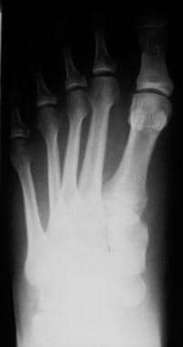
Fractured metatarsals. Image shows a bone fragment parallel to the base of the fifth metatarsal bone. This is not a fracture; rather, it is the apophysis of the base of the fifth metatarsal bone. It occurs in association with an ossification center. This center is always parallel to the long axis of metatarsal and has smooth margins, unlike a fracture.
-
Fractured metatarsals. Spiral fracture through the distal shaft of the fifth metatarsal.
-
Fractured metatarsals. A fracture of the fifth metatarsal, oblique, in the shaft.
-
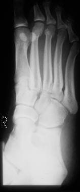
Fractured metatarsals. Fracture at the base of the first metatarsal in a child.
-
Fractured metatarsals. Transverse fracture at the base of the fifth metatarsal in a male adolescent.
-
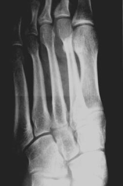
Fractured metatarsals. Fracture of the midshaft of the third metatarsal.
-
Fractured metatarsals. Magnified view of the foot shows a fracture with callus formation in the third metatarsal bone.
-
Fractured metatarsals. Fracture of the distal shaft of the third metatarsal.
-
Fractured metatarsals. Transverse fracture at the base of the fifth metatarsal; this is a Jones fracture.
-
Fractured metatarsals. Avulsion fracture of the tuberosity of the fifth metatarsal.
-
Fractured metatarsals. A fracture of the tuberosity of the fifth metatarsal.
-
Fractured metatarsals. Oblique fracture of the metaphysis of the distal shaft of the fifth metatarsal.
-
Fractured metatarsals. Avulsion fracture at the base of the fifth metatarsal; this was the result of the action of the peroneus brevis tendon.
-
Fractured metatarsals. Fracture of the metatarsal tuberosity.
-
Fractured metatarsals. Fracture of the fifth metatarsal tuberosity with lateral displacement of the fracture fragment.
-
Fractured metatarsals. Transverse fracture of the base of the fifth metatarsal bone and associated features, including radiopaque foreign bodies in the soft tissue and the accessory ossicle lateral to the cuboid bone.
-
Fractured metatarsals. Comminuted fracture of the base of the fifth metatarsal bone.
-
Fractured metatarsals. Fracture of the distal shaft of the third metatarsal.
-
Fractured metatarsals. Fracture of the proximal shaft of the first metatarsal bone.
-
Fractured metatarsals. Image shows a thin layer of subtle, solid periosteal reaction on the medial side of the shaft of the second metatarsal bone. This is an early stage of a stress fracture.
-
Fractured metatarsals. Image shows a stress fracture more florid than that shown in the previous image, with extensive periosteal reaction on either side of the third and fourth metatarsals.
-
Fractured metatarsals. Image shows a Lisfranc fracture-dislocation: a fracture of the base of the second metatarsal and a lateral dislocation of the second metatarsal.
-
Fractured metatarsals. Image shows a Lisfranc dislocation with a fracture of the base of the third and fourth metatarsals.


