Practice Essentials
Transient tachypnea of the newborn (TTN) represents persistence of fetal lung fluid at birth and generally presents within a few hours of birth. [1] The prominent clinical feature of TTN is tachypnea, but neonates with the condition may also present with chest retractions; mild to moderate respiratory distress, including expiratory grunting and nasal flaring; or occasionally cyanosis. [1, 2]
Common risk factors for TTN include cesarean deliveries, prolonged labor, maternal diabetes or asthma, male sex, macrosomia, and late preterm gestation. [1] Studies have shown that TTN accounts for 33-50% of breathing difficulties in neonates, with an incidence of 5-6 cases per 1000 births. [1, 3]
TTN is caused by delayed fetal lung fluid reabsorption and clearance of alveolar fluid. [1, 2] This phenomenon occurs because of a variety of factors, namely a lack of epinephrine during labor to activate epithelial Na+ channels to promote fluid absorption and the absence of a vaginal delivery to provide the mechanical expulsion of lung fluid. [2] This results in engorged lymphatics and a buildup of interstitial fluid, which leads to expiratory obstruction and hyperinflation. [2]
A TTN diagnosis is made after assessing an infant’s clinical presentation, a physical examination, and often chest radiographs. [2] Common differential diagnoses include surfactant deficiency disease, meconium aspiration, and neonatal pneumonia. Other conditions to be considered are congenital lymphangiectasia, congenital heart disease, polycythemia, cerebral hyperventilation, and anemia/hypovolemia.
The 2 main imaging modalities for TTN are radiography and ultrasonography. Chest radiography is the continued standard method of radiologic examination. [1, 2, 4] The clinical course of transient tachypnea is relatively benign, and radiographic severity may not correlate with the clinical picture. [4]
The overall prognosis for TTN is excellent, with most symptoms resolving within 48-72 hours. [1, 4] TTN is a self-limited condition that may need supportive treatment. [1, 2] The condition typically resolves without the need for medical interventions, and as such, mechanical ventilation is not usually required. [1, 2, 5] However, fluid restriction can be beneficial, cyanosis can be relieved with minimal oxygen, and continuous positive airway pressure will likely improve respiratory function. [1, 2] Malignant TTN has been reported in some cases, where newborns develop persistent pulmonary hypertension from the possible increase in pulmonary vascular resistance as a result of retained lung fluid. [4, 6, 7]
Radiography
The spectrum of radiographic findings can vary from normal to pleural effusions. Neonates with TTN may have engorged lymphatics and capillaries. Chest radiographs will show this retained lung fluid through diffuse, streaky, interstitial, strandlike opacities commonly accompanied by mild hyperinflation and small pleural effusions. [1, 8, 9, 10, 11, 12] Fluid in fissures and edema of the interlobar septae may also be present, but fluid may also appear at costophrenic angles associated with widening of intercostal spaces. [2] The “sunburst pattern” may be observed, which describes prominent perihilar vascular markings. [2]
The radiographic appearance of TTN may mimic the diffuse, granular appearance of surfactant deficiency disease but usually without pulmonary underaeration. [1] Another distinguishing factor between these 2 conditions is that neonates with transient tachypnea are usually at term, whereas surfactant deficiency disease is a cause of respiratory distress generally in premature infants. [5] TTN radiographic lung changes also may resemble the interstitial pattern of other causes of pulmonary edema, and echocardiography may be useful if there is an atypical course or abnormal cardiac size (cardiomegaly) and/or contour is suspected. [2, 13, 14]
Initially, it may be difficult to distinguish transient tachypnea from other causes of respiratory distress. A notable characteristic and differential feature of TTN, compared to other conditions, is that radiographic resolution of TTN occurs within 48-72 hours, and significant improvement by 24 hours is typical. [1, 2] If radiographic resolution does not occur in 48-72 hours, an alternative diagnosis should be sought. [4] However, it may rarely take up to 7 days for the complete disappearance of radiographic TTN features. [2]
The clinicoradiologic correlation and timeline should be used to corroborate the diagnosis.
(See the images below.)
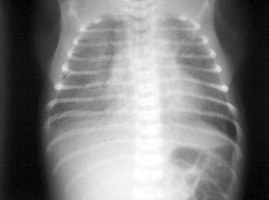 Transient tachypnea of the newborn. Radiograph of a neonate at age 6 hours. Overaeration, streaky, bilateral, pulmonary interstitial opacities and prominent perihilar interstitial markings. Mild cardiomegaly.
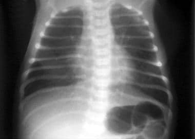 Transient tachypnea of the newborn. Chest radiograph of a neonate at age 2 days. Moderate parenchymal abnormalities with perihilar, streaky markings. No cardiomegaly.
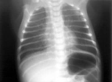 Transient tachypnea of the newborn. Radiograph of a neonate at age 4 days. Normal heart size and clear lungs are seen.
Ultrasonography
Lung ultrasound has become an attractive diagnostic tool in neonatal settings and is increasingly being used for diagnosing several neonatal respiratory morbidities. [15] Ultrasound may permit differentiation between surfactant deficiency disease, TTN, and neonatal pneumonia. [16, 17, 18, 19] It has also been proposed for helping predict interventions such as NICU admission, as well as surfactant treatment or mechanical ventilation in preterm infants. [15]
On lung ultrasound, the linear pleural interface beneath the thoracic wall musculature will move continuously with respiration. [2] A-lines are a series of echogenic lines deep to pleura, which are normally visualized and are equidistant to each other. [2] The typical features of TTN on ultrasound are thickening/fuzziness of the pleural line and minor or complete disappearance of A-lines. B-lines are defined as "comet tail" hyperechoic artifacts arising from the pleural line. [2, 16] The presence of more than 3 B-lines in every region, known as interstitial syndrome, is also observed in patients with TTN. [2]
Copetti and Cattarossi conducted a study evaluating the ultrasonographic appearance of TTN by comparing 32 newborn infants with radiologic and clinical findings of TTN to 60 healthy infants, 29 with respiratory distress syndrome (RDS), 6 with pneumonia, and 5 with atelectasis. [20] They reported a sensitivity and specificity of 100% for the diagnosis of TTN because of the “double lung point” observed exclusively on infants with this condition. The interface between very compact B-lines in the inferior pulmonary field, as compared to less compact B-lines in the superior lung field, is known as the "double lung point." [20, 21] Further studies of this sign have described sensitivities of 38-100%, with specificities close to 100%. [21, 22] Approximately 46% of patients with TTN will present with the double lung point. [22]
According to the POCUS Working Group of the European Society of Paediatric and Neonatal Intensive Care, point-of-care ultrasound is helpful in distinguishing between RDS and TTN. [23] RDS is characterized by a poorly aerated lung, with the absence of A-lines, the presence of small subpleural consolidations, and diffuse white lung (confluent B-lines). [23] In TTN, however, the interstitial pattern alternates with areas of near-normal lung (with A-lines). [23]
Ultrasonography may also be helpful in differentiating COVID-19 from TTN. In a study of 27 infants, the lung sonograms in 2 asymptomatic COVID-19–positive neonates revealed several coalescent B lines, pleural thickening, and areas of opacity. [24]
At this point, ultrasound has not replaced radiography, and the clinicoradiologic correlation remains fundamental for an accurate diagnosis.
-
Transient tachypnea of the newborn. Radiograph of a neonate at age 6 hours. Overaeration, streaky, bilateral, pulmonary interstitial opacities and prominent perihilar interstitial markings. Mild cardiomegaly.
-
Transient tachypnea of the newborn. Chest radiograph of a neonate at age 2 days. Moderate parenchymal abnormalities with perihilar, streaky markings. No cardiomegaly.
-
Transient tachypnea of the newborn. Radiograph of a neonate at age 4 days. Normal heart size and clear lungs are seen.