Practice Essentials
Toxic megacolon (or toxic colitis) is defined as a severe episode of colitis with segmental or total dilatation of the colon. It is typically a complication of ulcerative colitis, but it may be a complication of Crohn disease, antibiotic-related pseudomembranous colitis, and other colitides. [1] Pathologically, acute fulminant colitis is associated with neuromuscular degeneration and a rapid and extensive colonic dilatation. [2, 3, 4, 5, 6, 7, 8, 9]
Patients with toxic megacolon often present in the emergency department as having abdominal distention superimposed on chronic or acute diarrhea. The diagnosis should be considered in all such patients. The diagnosis is usually based on thorough clinical history taking and physical examination combined with plain abdominal radiography. [10, 11, 12, 13, 14]
On radiographs, the transverse colon is often identified as dilated because of the anterosuperior position. Diameter greater than 6 cm is considered a highly suspicious finding. Images may show a coarse, irregular mucosal pattern of the large bowel known as thumbprinting, which is caused by mucosal edema due to inflammatory infiltration. [15, 1] CT findings can highlight the extent of involvement (eg, edema), inflammation, and other features such as ascites and abscesses. [15] Ultrasonography and radionuclide studies may have a limited role.
Technetium-99m (99mTc) hexamethyl-propyleneamine oxime (HMPAO)–labeled WBC scanning can be used as an alternative to colonoscopy to assess the extent and severity of the disease in critically ill patients with ulcerative colitis. This technique decreases the number and severity of complications that may occur in these patients. However, the role of this method of scintigraphy is limited in the diagnosis of toxic megacolon and in the determination of its severity. [16]
Chagas disease, Hirschsprung disease, and intestinal pseudo-obstruction may superficially resemble toxic megacolon on plain radiographs. [17] However, because they occur in totally different clinical settings, they are unlikely to be confused with toxic megacolon.
(See the images below.)
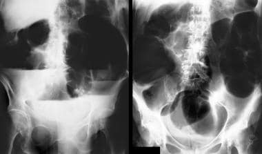
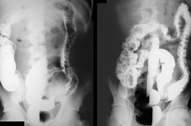 Double-contrast barium enema studies in a 44-year-old man known to have long history of ulcerative colitis. Images show total colitis and extensive pseudopolyposis.
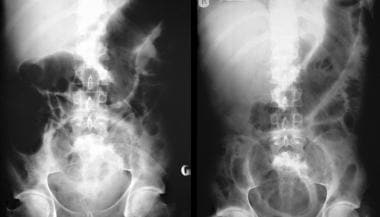 A 22-year-old man presented with abdominal pain, passage of blood and mucus per rectum, abdominal distention, fever, and disorientation. Findings from sigmoidoscopy confirmed ulcerative colitis. Abdominal radiographs obtained 2 days apart show mucosal edema and worsening of the distention in the transverse colon. The patient's clinical condition deteriorated over the next 36 hours despite steroid and antibiotic therapy, and the patient had to undergo a total colectomy and ileostomy.
Radiography
Toxic megacolon is a clinical diagnosis, one based on thorough history taking and physical examination and supported by plain abdominal radiographic findings. A diagnosis of toxic megacolon can be made fairly confidently by using plain radiography in the appropriate clinical setting, although a series of radiographs may be required.
If toxic megacolon is clinically suspected, patients are usually followed up with plain abdominal radiography every 12-24 hours, depending on the patient's clinical condition. A single abdominal radiograph may not be sufficient and should be combined with a horizontal-beam radiograph, which may better depict large, dilated bowel loops with fluid levels. Also, abdominal perforation is less likely to be missed. (See the images below).
Double-contrast barium enema studies in a 44-year-old man known to have long history of ulcerative colitis. Images show total colitis and extensive pseudopolyposis.
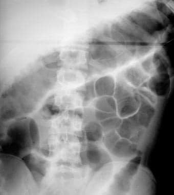
 Plain abdominal radiograph. The patient presented with an acute exacerbation of symptoms. Image shows thumbprinting in the region of the splenic flexure of the colon.
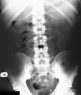
 Plain abdominal radiograph that shows distention of the transverse colon associated with mucosal edema. The maximum transverse diameter of the transverse colon is 7.5 cm.
A 22-year-old man presented with abdominal pain, passage of blood and mucus per rectum, abdominal distention, fever, and disorientation. Findings from sigmoidoscopy confirmed ulcerative colitis. Abdominal radiographs obtained 2 days apart show mucosal edema and worsening of the distention in the transverse colon. The patient's clinical condition deteriorated over the next 36 hours despite steroid and antibiotic therapy, and the patient had to undergo a total colectomy and ileostomy.
Plain abdominal radiograph that shows distention of the transverse colon associated with mucosal edema. The maximum transverse diameter of the transverse colon is 7.5 cm.
A 22-year-old man presented with abdominal pain, passage of blood and mucus per rectum, abdominal distention, fever, and disorientation. Findings from sigmoidoscopy confirmed ulcerative colitis. Abdominal radiographs obtained 2 days apart show mucosal edema and worsening of the distention in the transverse colon. The patient's clinical condition deteriorated over the next 36 hours despite steroid and antibiotic therapy, and the patient had to undergo a total colectomy and ileostomy.
 A 72-year-old woman presented with vomiting and abdominal distention. The supine (right) and erect (left) plain abdominal radiographs show gross dilatation of the colon with multiple air-fluid levels. On further questioning, the patient revealed that she was taking diuretics for hypertension. Blood biochemical tests revealed markedly lowered potassium levels. After potassium replacement therapy, the patient's pseudo-obstruction completely resolved.
Megacolon is considered to be present if the diameter of the colon is 5.5 cm or more, with apparent edema of the bowel wall on plain abdominal radiographs. Rarely, the toxic dilatation may extend to the terminal ileum.
Toxic megacolon is almost always a complication of pancolitis, with occasional sparing of the rectum. Therefore, changes such as strictures and mucosal abnormalities may be seen in association with toxic megacolon.
Toxic megacolon in the setting of Crohn disease is less common, but the plain radiographic findings of toxic megacolon in ulcerative colitis and those of Crohn disease overlap. However, with Crohn disease, the colonic wall tends to be thicker; thus, a thicker colonic wall in the setting of toxic megacolon in a patient with no previous disease should suggest Crohn disease rather than ulcerative colitis.
Marked dilatation is observed in the transverse colon; the upper range of normal for the transverse diameter is 5.5-6.5 cm. This finding has led to the belief that the transverse colon is the area most severely affected. However, if a prone radiograph is obtained, the greatest distention is observed in the ascending colon and descending colon. The apparent prominent involvement simply reflects the movement of the retained gas to the least dependent part of the colon. Serial radiographs may show increasing dilatation of the transverse diameter of the colon.
Images may show a coarse, irregular mucosal pattern of the large bowel. This thumbprinting is caused by mucosal edema due to inflammatory infiltration. The normal haustral pattern is absent in the involved segments, and pseudopolyps often extend into the lumen. [18] These represent mucosal islands in denuded ulcerated colonic wall in ulcerative colitis. Pneumatosis coli is an occasional finding. If perforation occurs, radiographic signs of a pneumoperitoneum may be apparent on the supine and/or lateral decubitus radiographs. [13]
Clinical examination is not accurate in the detection of perforation in the setting of toxic megacolon. The first hint of a colonic perforation may be provided on a plain abdominal radiograph.
The clinical or radiographic features of a toxic megacolon are an absolute contraindication to barium enema examination or the administration of laxatives. Contrast-enhanced studies of the colon should be considered only after the acute symptoms subside and the patient's condition is stabilized.
Dilatation in toxic megacolon may fluctuate or resolve, leaving the patient with toxic colitis. A perforated large bowel in association with a toxic megacolon may be missed on a plain abdominal radiograph.
Computed Tomography
The large bowel appears distended, with associated fluid levels. The haustral pattern may show edema. In toxic megacolon associated with ulcerative colitis, the bowel wall may be thin. Intramural air in association with small pericolonic fluid collections may be observed. Extraluminal air may be present if a perforation is present as a complication of toxic megacolon. [19]
CT scanning provides better anatomic detail of transmural disease, mesenteric involvement, and intraperitoneal complications of inflammatory bowel disease. [19, 20] Extraluminal air associated with bowel perforation is better seen with CT than with other techniques. [21]
Distinguishing severe acute colitis from toxic megacolon is important in clinical decision making. CT is useful in distinguishing patients with toxic megacolon from patients with severe acute colitis, but not toxic megacolon as a complication. The association of air-filled colonic distension greater than 6 cm, abnormal haustral pattern, and segmental colonic parietal thinning seems pathognomonic of toxic megacolon and should lead to rapid surgery. [20]
None of the CT scan findings is specific; they may also be found in severe forms of colitides.
Questions & Answers
-
Double-contrast barium enema studies in a 44-year-old man known to have long history of ulcerative colitis. Images show total colitis and extensive pseudopolyposis.
-
Plain abdominal radiograph. The patient presented with an acute exacerbation of symptoms. Image shows thumbprinting in the region of the splenic flexure of the colon.
-
Plain abdominal radiograph that shows distention of the transverse colon associated with mucosal edema. The maximum transverse diameter of the transverse colon is 7.5 cm.
-
A 22-year-old man presented with abdominal pain, passage of blood and mucus per rectum, abdominal distention, fever, and disorientation. Findings from sigmoidoscopy confirmed ulcerative colitis. Abdominal radiographs obtained 2 days apart show mucosal edema and worsening of the distention in the transverse colon. The patient's clinical condition deteriorated over the next 36 hours despite steroid and antibiotic therapy, and the patient had to undergo a total colectomy and ileostomy.
-
A 72-year-old woman presented with vomiting and abdominal distention. The supine (right) and erect (left) plain abdominal radiographs show gross dilatation of the colon with multiple air-fluid levels. On further questioning, the patient revealed that she was taking diuretics for hypertension. Blood biochemical tests revealed markedly lowered potassium levels. After potassium replacement therapy, the patient's pseudo-obstruction completely resolved.