Practice Essentials
Subdural hematoma (SDH) is a type of bleeding in which a collection of blood gathers between the inner layer of the dura mater and the arachnoid mater of the meninges surrounding the brain. [1] Acute SDH is a devastating neurologic injury with significant morbidity and mortality. In patients with large SDH resulting in compression of underlying brain and lateral brain shift, severe neurologic deficits and coma can occur. Emergent neurosurgical decompression is a life-saving intervention that improves mortality and neurologic function. Persistent coma despite SDH evacuation is often the result of persistent midline shift, cerebral infarctions related to initial elevated intracranial pressure and herniation, nonconvulsive seizures, and other metabolic and infectious events; however, a subset of patients remain comatose without a discernible etiology. [2]
SDH is among the 3 types of extra-axial intracranial hemorrhages (along with subarachnoid and epidural hemorrhages) that usually occur as a result of trauma. Deceleration injuries are often the cause of subdural bleeding from rupture of veins via a shearing mechanism. Other entities such as child abuse and ventricular decompression can result in subdural bleeding, and spontaneous hemorrhage may occur in patients receiving anticoagulants or in those with coagulopathy. Compression of a dural sinus does not directly cause SDH, although compression may result in a venous infarction.
Acute recurrent SDH is a rare entity in the absence of trauma. Atraumatic SDH may be due to vascular disorders, coagulopathies, or intracranial hypotension. It is a rare complication of disseminated intravascular coagulation, with no prior reports in patients with intracranial metastatic colon cancer. [3]
SDH primarily occurs in elderly patients. Although most patients have a good prognosis, some do not. Hematoma recurrence is one of the factors influencing prognosis. [4] Chronic SDH is more common among elderly individuals because of an age-associated decrease in brain volume and increased venous fragility. The elderly population can be particularly vulnerable to tearing of cortical bridging veins. [5]
Xian and colleagues reported that acute SDH has both a high incidence and a high mortality and may cause high intracranial pressure, cerebral blood circulation disorders, and brain bulge, which may aggravate these circulation disorders. [6] Chronic SDH is common, especially among those older than 50 years, and represents about 10% of all intracranial hematomas. [7]
Traumatic brain injury (TBI) is a frequent cause of mortality and acquired neurologic impairment in children. Pediatric SDH is a rare but serious condition. In a retrospective study, Binder and associates confirmed that despite a poor prognosis, most infants and children treated at a level one trauma center had good outcomes (70% overall) when treated appropriately via surgical or conservative means. [8]
Some SDHs are clinically silent; others cause symptoms as a result of mass effect on adjacent brain. Some hematomas can grow large enough to cause herniation of cerebral tissue. Before computed tomography (CT) scanning and magnetic resonance imaging (MRI) technologies became available, SDH was diagnosed only on the basis of this mass effect, which was depicted as displacement of blood vessels on angiography or as a calcified pituitary gland on skull radiography.
CT is now the first step in diagnosis of SDH. MRI is considered an excellent imaging tool for evaluating such patients. Symptomatic SDH requires emergent treatment to identify and control sites of bleeding conservatively or by surgery. SDH with no history of trauma should be treated emergently and evaluated strictly. Follow-up is essential in patients with neural symptoms. Massive symptomatic SDH should be treated with surgery to control bleeding. [1]
Endoscope-assisted surgery is becoming a more common modality for surgical treatment of subdural collections. [9] Adoption of middle meningeal artery embolization in the management of chronic SDH has led to renewed interest in dural vascular anatomy. Fortunately, microvascular aspects of dural anatomy, previously limited to ex vivo investigations, are becoming increasingly accessible to in vivo visualization, setting the stage for synthesis of the old and the new, and providing a rationale for the endovascular approach to management of subdural collections in particular. [10]
Chronic SDH can result from metastatic tumors of the dura mater. In cases of spontaneous nontraumatic hematoma around the dura mater, making the precise diagnosis is sometimes doubtful and confusing. The stream of diagnostic thinking should be open to possibilities such as medical diseases, including liver and kidney disease, drug history, history of cancer, and other possible clues. A detailed and purposeful systematic medical history review and physical examination is important to the selection of appropriate management strategies for SDH. [11]
Imaging modalities
CT scanning is usually the first evaluation in patients with suspected acute SDH because CT depicts acute hemorrhage and skull fractures well, it is relatively fast to obtain, and CT scanning is more readily available than MRI. Smaller hemorrhages may be missed on CT in the nonacute setting, where MRI is the study of choice because of its high sensitivity and specificity. [12, 13, 14, 15]
CT scanning may fail to depict small hemorrhages because of similarity in attenuation between blood and adjacent bone and because of streak artifacts in the posterior fossa and the inferior middle cranial fossa. MRI aids in detection of small hematomas through its multiplanar capabilities. [16, 17, 18, 19, 20, 21]
(See the image below.)
 Axial head computed tomography scan shows a skull fracture with an adjacent, small subdural hematoma. Window and level values are widened over standard values, which aids in detection of small hemorrhages.
Computed Tomography
Computed tomography (CT) findings in SDH depend on the age of the hemorrhage. [22, 23, 24] Differentiating subdural from epidural hematoma may be difficult when the hemorrhage is small, because images of the blood may not reveal a typical shape in either condition. Follow-up imaging to ensure that the hematoma is not expanding and to check for an adjacent skull fracture is typical.
The underlying mechanism of chronic SDH after minor head injury is complex, probably due to mechanical injury of the arachnoid membrane, hematologic coagulopathy, and pathologic angiogenesis in the dura caused by inflammatory cytokines such as vascular endothelial growth factor (VEGF). To confirm whether VEGF might be a reliable predictive biomarker for the natural history of chronic SDH, including progression and recurrence, Takei and coworkers analyzed the correlation of VEGF concentration in subdural fluid with CT findings and clinical features, including interval from minor head injury, and found a significant association; however, investigators concluded that overall VEGF concentration in SDH is not a reliable predictive biomarker for the natural history of SD, including its recurrence. [25]
Small SDH may not be depicted because attenuation may be similar to the adjacent inner table of the skull. Viewing these images with a wider window and level (eg, 240 HU, 80 HU) assists in detection in these cases; however, CT fails to depict some small hemorrhages. [20, 26, 27] Measurement of white matter has been found to be a helpful predictor of outcomes in patients with SDH with cerebral edema. A cutoff value of 31.5 HU of white matter showed 80% sensitivity and 99.9% specificity for death. [16]
(See the image below.)
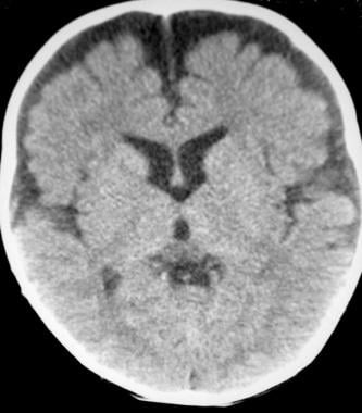
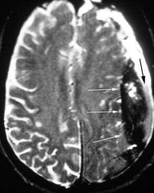
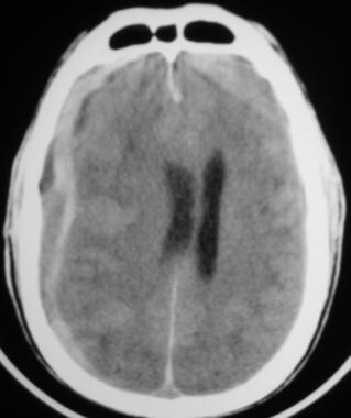 Computed tomography (CT) scan shows a patient with subdural hematomas of varying ages. This patient had a CT scan 1 week prior that revealed a chronic subdural hematoma (represented by low-density fluid in this study). Over the next week, the patient's clinical condition progressively declined; he collapsed shortly before this image was obtained. Gray blood represents subacute hemorrhage, whereas white blood represents acute hemorrhage.
In the acute phase, SDH appears as a crescent-shaped extra-axial collection with increased attenuation that, when large enough, causes effacement of adjacent sulci and midline shift. Attenuation changes as the hematoma ages. [28, 29]
(See the images below.)
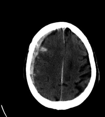
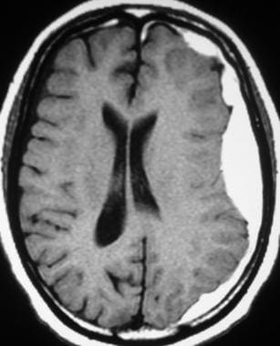
 Late subacute subdural hematoma has decreased attenuation compared with adjacent brain tissue. Attenuation of the hematoma remains higher than that of cerebrospinal fluid.
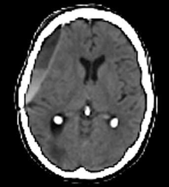
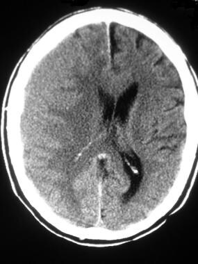 Computed tomography scan in a patient with a subacute right frontal subdural hematoma. The blood has the same attenuation as adjacent gray matter and is difficult to distinguish. Note that the gray matter–white matter junction is displaced medially and midline shift is seen, indicating the presence of a space-occupying extra-axial lesion.
Subacute SDH may be difficult to detect because of possible isoattenuation as compared to adjacent gray matter. Displacement of the gray matter–white matter junction is an important sign that indicates the presence of a space-occupying lesion. Although often administered in the past to help detect displacement of cortical vessels, contrast medium is no longer necessary in light of technological advances.
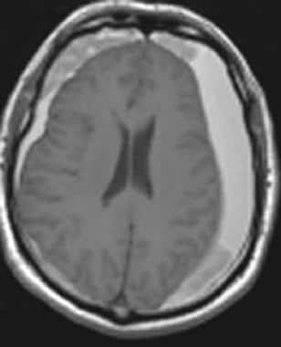
Chronic SDH shows isoattenuation relative to cerebrospinal fluid (CSF). In rare cases, such hematomas may calcify, resulting in an unusual appearance that can be mistaken for a calcified mass.
(See the image below.)
 Late subacute-to-chronic subdural hematoma with a blood-fluid level indicating acute hemorrhage into the chronic collection.
Unlike epidural hematoma, SDH is not restricted by dural tethering at cranial sutures; SDH can cross suture lines and continue along the falx and the tentorium. However, it does not cross the midline because of meningeal reflections.
(See the image below.)
 Tentorial subdural hematoma in an adult with trauma. In children with this pattern of injury, abuse should be considered.
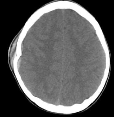
When SDH is discovered on CT, it is important to check for the presence of related injuries such as skull fracture (see the first image below), intraparenchymal contusions, and subarachnoid blood (see the second image below). The presence of adjacent parenchymal injury in patients with SDH is the most important factor in predicting clinical outcomes.
Axial head computed tomography scan shows a skull fracture with an adjacent, small subdural hematoma. Window and level values are widened over standard values, which aids in detection of small hemorrhages.
 Subdural hematoma with adjacent subarachnoid hemorrhage was the result of a ruptured middle cerebral artery aneurysm. Aneurysms are unusual causes of subdural hematoma.
Rebleeding into SDH may occur and is depicted as a layer of high-attenuation hemorrhage within a lower-attenuation hematoma.
In older patients with cerebral atrophy, bilateral frontal subdural hygromas may be seen when the patient is in the supine position. However, lack of mass effect and the presence of general atrophy suggest that this appearance is merely the result of settling of the atrophic brain, rather than a pathologic subdural collection. A similar finding can be seen in young children (benign enlargement of the subarachnoid space) and should resolve in the first few years of life.
(See the image below.)
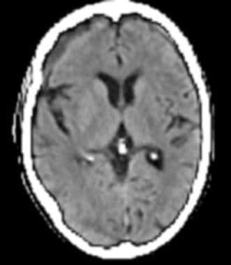
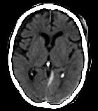
 Axial computed tomography scan shows benign enlargement of the subarachnoid space, which occurs in children. The extra-axial fluid does not cause mass effect and normally resolves within the first 2 years of life.
Posttraumatic subdural hygroma can be confused with chronic SDH. This develops days or weeks after trauma and results from tears in the arachnoid and resultant leakage of CSF into the subdural space. This condition is self-limited and usually resolves after several months.
Magnetic Resonance Imaging
Magnetic resonance imaging (MRI) is the most sensitive imaging test available for detection of SDH. Smaller hemorrhages may be missed on CT in the nonacute setting, where MRI is the study of choice because of its high sensitivity and specificity. [12, 13, 14, 15]
MRI is more sensitive than CT in detecting SDH because multiplanar capabilities and superior tissue differentiation of MRI make detection easier. In particular, sensitivity greater than 95% has been described with T2-weighted images of SDH because of marked differences in signal intensity between blood products and adjacent structures. [17, 18]
The presence of interleukin (IL)-6 and IL-8 on hyperintense T1-weighted images and evidence of beta-trace proteins on hyperintense T2-weighted images appear to be associated with rebleeding and CSF admixure in chronic SDH. [17]
Preoperative findings on MRI, particularly T1-weighted classification, have been found to be a significant indicator of recurrence of chronic SDH, with T1-iso/hypointensity determined to be a high indicator of risk (18.2% recurrence rate vs 5.2% for other indicators). [18] The middle meningeal artery has been shown to be larger on magnetic resonance angiography in patients who have developed chronic SDH. [19]
(See the images below.)
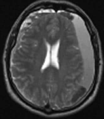
 Axial T1-weighted magnetic resonance imaging reveals bilateral subacute subdural hematoma with increased signal intensity. Areas of intermediate intensity represent more acute hemorrhage into subacute collections.
 T2-weighted magnetic resonance imaging in a patient with subdural hematoma shows blood products of differing ages.
SDH on axial images reveals the same crescent-shaped pattern that is seen on CT images. Coronal images are useful in evaluating the extent of SDH and in detecting temporal and tentorial hemorrhages—2 aspects that are poorly depicted on CT scans.
In SDH, the signal depends on the age of the hemorrhage and follows the pattern of intraparenchymal hematoma in acute and subacute cases. Chronic SDH, which appears as isoattenuation relative to CSF on CT scans, often shows increased signal intensity on T1-weighted images because of the presence of free methemoglobin, although intensity decreases over time. Hemosiderin usually is not present; this is believed to result from lack of a dural blood–brain barrier. [30]
When hemorrhages of differing ages are noted within a subdural collection, septae may separate the different blood products (see the first image below). In addition, a blood-fluid level may be observed. When blood products of various ages are depicted on MRI in a child, particularly when blood is at multiple sites, child abuse must be suspected (see the second image below). Posterior interhemispheric and tentorial subdural hematomas are also suggestive of child abuse because they are associated with shaken baby syndrome.
 Subacute subdural hematoma with extension into the anterior interhemispheric cistern. Note that the sutures do not contain the spread of these hemorrhages.
 T2-weighted magnetic resonance imaging in a patient with subdural hematoma and rebleeding clearly shows hemorrhages of 3 different ages; these are hyperintense, isointense, and hypointense relative to brain tissue.
Detection and severity assessment of SDH is a major step in the evaluation of TBI. Researchers have found that the combination of classical image processing and deep learning can outperform deep learning–only methods to achieve greater average performance and robustness. Such a system can aid critical care physicians in reducing time to intervention, thereby improving long-term patient outcomes. [31]
-
Axial head computed tomography scan shows a skull fracture with an adjacent, small subdural hematoma. Window and level values are widened over standard values, which aids in detection of small hemorrhages.
-
Subacute subdural hematoma with extension into the anterior interhemispheric cistern. Note that the sutures do not contain the spread of these hemorrhages.
-
Tentorial subdural hematoma in an adult with trauma. In children with this pattern of injury, abuse should be considered.
-
Subdural hematoma with adjacent subarachnoid hemorrhage was the result of a ruptured middle cerebral artery aneurysm. Aneurysms are unusual causes of subdural hematoma.
-
Late subacute subdural hematoma has decreased attenuation compared with adjacent brain tissue. Attenuation of the hematoma remains higher than that of cerebrospinal fluid.
-
Computed tomography scan in a patient with a subacute right frontal subdural hematoma. The blood has the same attenuation as adjacent gray matter and is difficult to distinguish. Note that the gray matter–white matter junction is displaced medially and midline shift is seen, indicating the presence of a space-occupying extra-axial lesion.
-
Late subacute-to-chronic subdural hematoma with a blood-fluid level indicating acute hemorrhage into the chronic collection.
-
Axial computed tomography scan shows benign enlargement of the subarachnoid space, which occurs in children. The extra-axial fluid does not cause mass effect and normally resolves within the first 2 years of life.
-
Axial T1-weighted magnetic resonance imaging reveals bilateral subacute subdural hematoma with increased signal intensity. Areas of intermediate intensity represent more acute hemorrhage into subacute collections.
-
T2-weighted magnetic resonance imaging in a patient with subdural hematoma shows blood products of differing ages.
-
T2-weighted magnetic resonance imaging in a patient with subdural hematoma and rebleeding clearly shows hemorrhages of 3 different ages; these are hyperintense, isointense, and hypointense relative to brain tissue.
-
Computed tomography (CT) scan shows a patient with subdural hematomas of varying ages. This patient had a CT scan 1 week prior that revealed a chronic subdural hematoma (represented by low-density fluid in this study). Over the next week, the patient's clinical condition progressively declined; he collapsed shortly before this image was obtained. Gray blood represents subacute hemorrhage, whereas white blood represents acute hemorrhage.


