Practice Essentials
The term scurvy is derived from the Nordic word skyrbjugr, meaning swelling or edema. It has also been suggested that the term is derived from the Old Icelandic words skyrbugr, scarby, or skurvic. Scurvy is caused by a lack of vitamin C and manifests as collagen defects, hemorrhagic diathesis, abnormalities in bone maturation, epiphyseal disease, lifting of the periosteum, hemarthroses, irritability, decreased appetite, delayed development, and pseudoparalysis related to bone pain. [1, 2, 3, 4] Comorbid anemia and gastrointestinal disorders have been reported. [5, 6, 7]
Radiography is the preferred imaging examination for diagnosis. Serum vitamin C levels can be obtained to confirm the diagnosis of scurvy. Vitamin C deficiency is characterized by cortical thinning, which is sometimes described as a “pencil-point” cortex. Metaphyseal “beaks” and transverse lines of increased or decreased opacity may be seen in scurvy. The “beaks,” known as Pelkan spurs, are associated with fractures of the Trummerfeld zone. [8]
Congenital syphilis and neuroblastoma produce findings similar to those of scurvy. Syphilis, often called the great imitator, produces metaphyseal beaking similar to that noted in scurvy; however, syphilis does not produce radiopaque metaphyseal lines. The same findings may also be seen in a limb with residual palsy as a result of polio.
T1-weighted and PD/SPIR (proton density/spectral presaturation with inversion recovery) images reveal focal areas of metaphyseal marrow edema attributed to focal hemorrhage or infarcts. Subperiosteal fluid and displacement of epiphyses has been reported. [9] Skull changes may be below the resolution of computed tomography (CT) or magnetic resonance imaging (MRI). A change in skull shape is not diagnostic, as this finding may also be seen with hemolytic anemia, other causes of bone marrow hyperplasia, and rickets. [10, 11, 12]
Scurvy is not a common condition, although in one study, vitamin C deficiency was present in up to 23% of respondents. [13, 14] Young children and older persons are predisposed to scurvy because of their diets or the overpreparation of food (cooking destroys vitamin C). Smokers, non-Hispanic black males, and individuals who do not use vitamin supplements have an increased risk of vitamin C deficiency. [13] In a series of 22 adults with scurvy, 54% were using proton pump inhibitors. [5] Other risk factors include low socioeconomic status, alcohol use disorder, and gastrointestinal malabsorption. [15] Gastric bypass surgery may be complicated by vitamin C deficiency within 29-90 days, if diet is not supplemented. [16]
Signs and symptoms of scurvy include fatigue, malaise, anemia, myalgia, bone pain, easy bruising, swelling, petechiae, gingivitis, perifollicular hemorrhages, corkscrew hairs, and poor wound healing. If left untreated, the disease can progress to jaundice, neuropathy, hemolysis, seizures, and death. Onset is after 60-90 days of a diet deficient in vitamin C, with patients initially experiencing fatigue and muscle aches in the legs and abdomen. In the second stage, patients will have dry skin, folliculitis, vascular purpura, and painful hematomas. The third stage manifests as osteoprososis, bone growth abnormality, and subperiosteal or intraosseous hemorrhagic lesions, with the gums bleeding profusely, along with severe muscle pain. Last-stage scurvy patients have high fevers, and death can occur from brain or cardiac hemorrhaging. [17, 18, 19, 20]
The National Institutes of Health recommend the following amounts of vitamin C depending on life stage [21] :
-
Birth to 6 months: 40 mg
-
Infants 7–12 months: 50 mg
-
Children 1–3 years: 15 mg
-
Children 4–8 years: 25 mg
-
Children 9–13 years: 45 mg
-
Teens 14–18 years (boys): 75 mg
-
Teens 14–18 years (girls): 65 mg
-
Adults (men): 90 mg
-
Adults (women): 75 mg
-
Pregnant teens: 80 mg
-
Pregnant women: 85 mg
-
Breastfeeding teens: 115 mg
-
Breastfeeding women:120 mg
If a patient is a smoker, 35 mg should be added to the above amount.
Radiography
Some authors have suggested that the most diagnostic radiologic finding of vitamin C deficiency is a large, fluctuant, parietal swelling, which is apparently caused by subperiosteal hemorrhage. This author, however, considers long-bone changes to be better clinical identifiers of vitamin C deficiency than parietal swelling.
The epiphyses and periosteum also become easily detachable because of hemorrhage below the periosteum. Separation of the metaphyseal plate from the diaphysis, epiphyseal clefts, and malalignment of the metaphysis may also occur. A circular, opaque radiologic shadow often surrounds epiphyseal centers of ossification. This ring of increased opacity formed around the ossification center of long bone epiphyses is known as the Wimberger sign, which may result from bleeding or attachment movement. [8]
Vitamin C deficiency is characterized by cortical thinning, which is sometimes described as a “pencil-point” cortex. Decreased trabeculae produce a decrease in radiopacity, resulting in a transparent aspect similar in appearance to ground glass (see the image below).
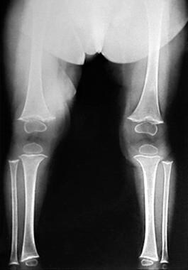 Anteroposterior radiograph of the lower extremities shows ground-glass osteopenia. Transverse metaphyseal lines of increased and decreased opacity (Trummerfeld zone) are associated with lateral growth of the metaphyseal calcification zone and periosteal elevation, which produces the characteristic metaphyseal beaks known as Pelkan spurs.
The increased opacity of distal diaphysis may be accompanied by a subjacent zone of decreased opacity. The thickening is known as a Frankel line, and the lucent zone on the diaphyseal side of the Frankel line (secondary to poorly formed trabeculae) is known as the Trummerfeld zone. [8] Its origin might be related to vascular compromise, similar to increases in bone density noted with avascular necrosis.
Costochondral junctions of the first 6 or 8 thoracic ribs may be expanded; this change may be related to fracturing of the zone of provisional calcification during normal respiration. The costochondral junctions are rounded and appear smooth, knobby, and steplike. The enlargement of the costochondral junctions simulates that seen in rickets.
The zone of proliferating cartilage cells is distorted, producing spicules from the metaphysis into the epiphyseal plate region. The zone of temporary calcification broadens, producing a wide, radiopaque metaphyseal band. Subjacent to this is a zone of poor-quality trabeculae, which appears radiolucent. A steplike lateral projection is found at the epiphyseal line in patients who are severely affected. Scorbutic changes are radiologically more severe in the lower extremities, whereas scorbutic changes seen in rickets are allegedly more severe in the upper extremities.
Metaphyseal “beaks” and transverse lines of increased or decreased opacity may be seen in scurvy. The “beaks,” known as Pelkan spurs, are associated with fractures of the Trummerfeld zone. [8] They may be produced by lateral growth of the metaphyseal calcification zone and are associated with periosteal elevation. Subepithelial marginal clefts may also be present.
Skull changes may produce a porotic hyperostosis (“hair-on-end” appearance) or crew-cut appearance secondary to marrow hyperplasia in response to anemia. No sphenoid changes are reported. Sphenoid porosity has not been shown to be caused by scurvy.
Subperiosteal hemorrhages are visualized only in the healing phase of scurvy, and these are almost invariably paraepiphyseal in distribution. Epiphyseal separation often results. Healing scurvy also appears with the loss of the scurvy line, in which the only residual manifestation is a double line of ossification at the original active site.
Degree of confidence
Periosteal elevation and epiphyseal separation both appear to be relatively specific for scurvy. Osteoporosis is a nonspecific finding.
The periosteal reaction of syphilis is more generalized than that of scurvy and is usually thick or multilaminated. Syphilis, often called the great imitator, produces metaphyseal beaking similar to that noted in scurvy; however, syphilis does not produce radiopaque metaphyseal lines. Although periosteal elevation may occur and produce a linear elevated area in patients, a spiculated periosteal reaction does not occur in scurvy.
Metaphyseal lesions caused by scurvy involve epiphyseal separation. Epiphyseal separation is a known complication of scurvy; however, it is also seen in cases of child abuse. The periosteal reaction resulting from child abuse is more generally distributed and is associated with a fracture.
Costochondral beading is more common with rickets than with scurvy.
Skull-marrow hyperplasia is more likely to result from hemolytic anemia or anemia related to parasitic infestation.
-
Anteroposterior radiograph of the lower extremities shows ground-glass osteopenia. Transverse metaphyseal lines of increased and decreased opacity (Trummerfeld zone) are associated with lateral growth of the metaphyseal calcification zone and periosteal elevation, which produces the characteristic metaphyseal beaks known as Pelkan spurs.


