Practice Essentials
Bone hemangiomas are benign, malformed vascular lesions, overall constituting less than 1% of all primary bone neoplasms. They occur most frequently in the vertebral column (30-50%) and skull (20%), [1, 2, 3, 4, 5] whereas involvement of other sites (including the long bones, [6] short tubular bones, and ribs) is extremely rare. A simple classification schema divides hemangiomas into capillary (small-vessel), cavernous (large-vessel), and mixed. Most hemangiomas of bone are cavernous. [7]
Bone hemangiomas are usually asymptomatic lesions discovered incidentally on imaging or postmortem examination and mostly encountered in middle-aged persons. The symptoms are largely nonspecific and depend on the site, size, and aggressiveness of the tumors. [8, 9, 10, 11]
Plain radiography is useful for evaluation as the first-line imaging modality in most cases. Radiographic appearances differ depending on the anatomic site and histologic variant of the lesion. However, the radiographic hallmark of bone hemangiomas is a prominent trabecular pattern. [7] Radiographic patterns may be nonspecific, necessitating further imaging or histology to achieve diagnosis. This is especially true in extraspinal hemangiomas occurring in an age group and location in which other more ominous diagnostic entities, such as myeloma or metastases, are more common.
When plain radiographs do not suffice and appearances remain equivocal, cross-sectional imaging is crucial for further characterization of these lesions. Computed tomography (CT) scanning is especially useful for assessing changes in bone trabeculae; the results support the plain radiographic findings and provide greater detail.
The superior soft-tissue and bone marrow contrast resolution of magnetic resonance imaging (MRI) allows for better evaluation of extraosseous extension and depiction of the characteristic fatty content in vertebral hemangiomas and flow patterns in general. The multiplanar capabilities of MRI are also crucial in defining the extent of neural involvement in the spine and planning therapeutic interventions. [12, 6]
Osseous hemangiomas usually show normal uptake on isotope bone scans, but they may also demonstrate photopenia and mildly to moderately increased activity. Scintigraphy with labeled red blood cells usually demonstrates focally increased activity. Single-photon emission CT (SPECT) scans are more sensitive than planar images in depicting abnormal activity. [13, 14]
Angiographic findings confirm the hypervascularity of the lesions. Angiography usually is performed in conjunction with embolization of symptomatic hemangiomas prior to surgery. [15]
Despite the added diagnostic information available with CT scanning and MRI, the angiomatous nature of many extraspinal lesions can be confirmed only with histologic analysis.
(See the images below.)
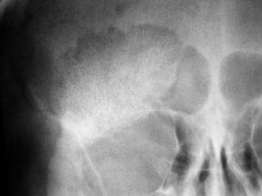 Bone hemangioma. Localized view of a frontal skull radiograph shows a well-demarcated lesion in the frontal bone with a characteristic sunburst appearance or a radiating, weblike trabecular pattern.
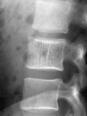
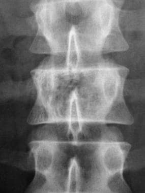 Bone hemangioma. View depicting the typical corduroy or accordion appearance of coarse, thickened vertical trabeculae in a hemangioma affecting the right side of the vertebral body at L2.
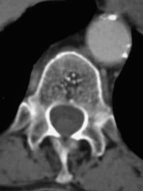 Bone hemangioma. Incidental finding of a small thoracic vertebral body hemangioma in a patient who had another lesion in the lumbar spine. Note the punctate sclerotic foci, or polka-dot appearance, which is a characteristic finding.
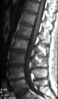 Bone hemangioma. Sagittal T1-weighted MRI of a spinal hemangioma affecting most of the body of L2. There is hyperintense change; hypointense thickened vertical trabeculae are also visible.
Radiography
Most vertebral hemangiomas are small and cannot be seen on plain radiographs. The characteristic radiographic appearance is of a sclerotic or ivory vertebra with coarse, thickened vertical trabeculae giving a corduroy, accordion, or honeycomb appearance. This appearance is due to resorption of horizontal trabeculae, caused by vascular channels and consequent reinforcement of vertical trabeculae. This finding can be differentiated from Paget disease, in which picture framing of the vertebral body is seen because of prominent horizontal trabeculae. Similar findings may occur with lymphoma and metastases. Bulging of the posterior cortex or expansion of the vertebral body is sometimes present.
Radiographic appearances of hemangiomas can be pathognomonic, especially with vertebral and calvarial hemangiomas. CT scanning and MRI increase diagnostic confidence in equivocal cases.
Calvarial hemangiomas are usually round, osteolytic lesions that may demonstrate the characteristic sunburst, radiating spoke-wheel, or weblike pattern of trabecular thickening. [16, 2] Radiographic appearances in craniofacial hemangiomas are often nonspecific. Mixed radiopacity, radiolucency, and honeycomb patterns are observed.
Long-bone hemangiomas are usually lytic, with a spiculated pattern creating a latticelike or Irish-lace appearance. A honeycomb structure can also result from bubbly bone osteolysis. Irregular bone destruction can occur, simulating malignant lesions. Reactive sclerosis may be seen at the margins of the lesions. Surface-based hemangiomas may mimic osteoid osteoma. Epithelioid hemangiomas characteristically demonstrate well-defined lysis, and they may also exhibit surrounding sclerosis, cortical expansion, or destruction. [17]
(See the images below).
Bone hemangioma. Localized view of a frontal skull radiograph shows a well-demarcated lesion in the frontal bone with a characteristic sunburst appearance or a radiating, weblike trabecular pattern.
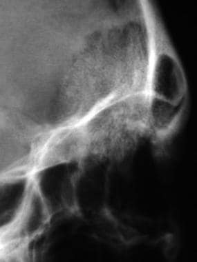 Bone hemangioma. Lateral projection in the same patient as in the previous image depicts the diagnostic appearance of a calvarial hemangioma well.
Bone hemangioma. View depicting the typical corduroy or accordion appearance of coarse, thickened vertical trabeculae in a hemangioma affecting the right side of the vertebral body at L2.
Bone hemangioma. Lateral projection in the same patient as in the previous image depicts the diagnostic appearance of a calvarial hemangioma well.
Bone hemangioma. View depicting the typical corduroy or accordion appearance of coarse, thickened vertical trabeculae in a hemangioma affecting the right side of the vertebral body at L2.
 Bone hemangioma. Lateral view in the same patient as in the previous image shows no obvious involvement of the posterior elements, though this is better assessed with CT and MRI. The trabecular pattern on plain images is usually better seen on this view.
Computed Tomography
CT scanning is more sensitive than plain radiography. Vertebral hemangiomas are typified by punctate sclerotic foci representing thickened vertical trabeculae seen in cross-section and giving a polka-dot appearance. [7] This finding may be absent in patients with symptomatic lesions. Bulging of the posterior cortex and paravertebral soft-tissue extension are readily assessed on CT scans, as is bone destruction with aggressive hemangiomas. CT findings in nonvertebral hemangiomas confirm plain radiographic results but give more detailed assessment of medullary, cortical bone, and extraosseous involvement.
(See the image below.)
Bone hemangioma. Incidental finding of a small thoracic vertebral body hemangioma in a patient who had another lesion in the lumbar spine. Note the punctate sclerotic foci, or polka-dot appearance, which is a characteristic finding.
Magnetic Resonance Imaging
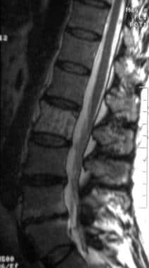
MRI features largely depend on the proportion of fat and vascularity of the lesions. With T1-weighted MRI, particularly in vertebral hemangiomas, areas of high fat content appear as areas of high signal intensity. On T2-weighted images, high signal intensity typically corresponds to the vascularity of hemangiomas.
Lesions in flat and long bones may show serpentine vascular channels. These demonstrate low signal intensity on T1-weighted images and high signal intensity on T2-weighted images with slow blood flow; they show low signal intensity on MRI obtained with all sequences in conditions of high blood flow. [18]
In a study by Qin et al of the imaging characteristics of long bone hemangiomas, 13 of 16 lesions were intramedullary, and 3 were cortical, subperiosteal, and mixed intracortical and intramedullary. Radiography and CT identified sclerotic margins in 8 cases, thickened trabeculae in 6, internal calcification in 1, cortical thickening in 1, and fracture in 4. Two lesions were confusing on radiography but clearly identifiable on MRI. All intraosseous hemangiomas showed high signal intensity on T2-weighted imaging and intermediate signal intensity on T1-weighted imaging. Four cystlike lesions showed peripheral and filling enhancement, whereas others exhibited diffuse enhancement with an intensity similar to adjacent vessels. [6]
Complicated symptomatic spinal hemangiomas may be difficult to differentiate from malignant lesions.
(See the images below.)
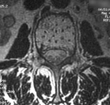 Bone hemangioma. Axial T2-weighted MRI shows the MRI equivalent of the CT polka-dot appearance. The hypointense foci correspond to coarsened, thickened trabeculae.
Bone hemangioma. Sagittal T1-weighted MRI of a spinal hemangioma affecting most of the body of L2. There is hyperintense change; hypointense thickened vertical trabeculae are also visible.
Bone hemangioma. Axial T2-weighted MRI shows the MRI equivalent of the CT polka-dot appearance. The hypointense foci correspond to coarsened, thickened trabeculae.
Bone hemangioma. Sagittal T1-weighted MRI of a spinal hemangioma affecting most of the body of L2. There is hyperintense change; hypointense thickened vertical trabeculae are also visible.
 Bone hemangioma. T2-weighted image in the same patient as in the previous image demonstrates the typically high signal intensity of marrow and the low signal intensity of the vertical trabeculae.
Low signal intensity on T1-weighted images indicates decreased marrow fat or a greater vascular component; such a finding may be correlated with more aggressive behavior and is also more characteristic in cases involving vertebral collapse.
Thickened trabeculae demonstrate low signal intensity on MRI obtained with all sequences. Extraosseous components tend not to show high signal intensity on T1-weighted images because of the paucity or absence of adipose tissue, but avid enhancement occurs with gadolinium enhancement because of the vascularity of the lesions.
Epidural extension and neural involvement are well depicted with MRI (see the images below).
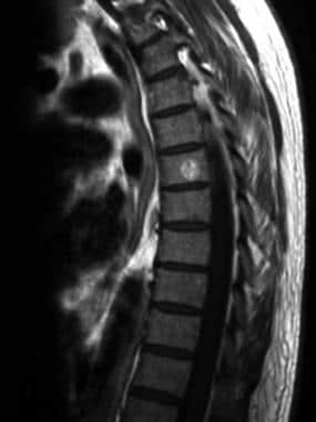 Bone hemangioma. Sagittal T1-weighted MRI of a typical example of a thoracic hemangioma involving only part of the vertebral body.
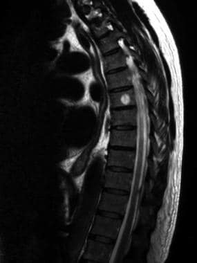 Bone hemangioma. Sagittal T2-weighted MRI in the same patient as in the previous image.
Gadolinium-based contrast agents have been linked to the development of nephrogenic systemic fibrosis (NSF) or nephrogenic fibrosing dermopathy (NFD). The disease has occurred in patients with moderate to end-stage renal disease after being given a gadolinium-based contrast agent to enhance MRI or MRA scans. NSF/NFD is a debilitating and sometimes fatal disease. Characteristics include red or dark patches on the skin; burning, itching, swelling, hardening, and tightening of the skin; yellow spots on the whites of the eyes; joint stiffness with trouble moving or straightening the arms, hands, legs, or feet; pain deep in the hip bones or ribs; and muscle weakness.
-
Bone hemangioma. Localized view of a frontal skull radiograph shows a well-demarcated lesion in the frontal bone with a characteristic sunburst appearance or a radiating, weblike trabecular pattern.
-
Bone hemangioma. Lateral projection in the same patient as in the previous image depicts the diagnostic appearance of a calvarial hemangioma well.
-
Bone hemangioma. View depicting the typical corduroy or accordion appearance of coarse, thickened vertical trabeculae in a hemangioma affecting the right side of the vertebral body at L2.
-
Bone hemangioma. Lateral view in the same patient as in the previous image shows no obvious involvement of the posterior elements, though this is better assessed with CT and MRI. The trabecular pattern on plain images is usually better seen on this view.
-
Bone hemangioma. Incidental finding of a small thoracic vertebral body hemangioma in a patient who had another lesion in the lumbar spine. Note the punctate sclerotic foci, or polka-dot appearance, which is a characteristic finding.
-
Bone hemangioma. Axial T2-weighted MRI shows the MRI equivalent of the CT polka-dot appearance. The hypointense foci correspond to coarsened, thickened trabeculae.
-
Bone hemangioma. Sagittal T1-weighted MRI of a spinal hemangioma affecting most of the body of L2. There is hyperintense change; hypointense thickened vertical trabeculae are also visible.
-
Bone hemangioma. T2-weighted image in the same patient as in the previous image demonstrates the typically high signal intensity of marrow and the low signal intensity of the vertical trabeculae.
-
Bone hemangioma. Sagittal T1-weighted MRI of a typical example of a thoracic hemangioma involving only part of the vertebral body.
-
Bone hemangioma. Sagittal T2-weighted MRI in the same patient as in the previous image.


