Practice Essentials
Gout is caused by the presence of monosodium urate monohydrate crystals in the joint space and soft tissue that can result in debilitating illness characterized by recurrent episodes of pain and joint inflammation. All patients with gout have hyperuricemia, but gout attacks are not caused by the level of uric acid but by acute changes in the uric acid level. [1, 2, 3, 4]
Primary gout is related to underexcretion or overproduction of uric acid, which is often associated with dietary excesses or overuse of alcohol and metabolic syndrome. Secondary gout is related to medications or conditions that cause hyperuricemia, such as myeloproliferative diseases and their treatment, hyperproliferative skin disorders, enzymatic defects, and renal failure. [1, 2, 3, 4]
The American College of Rheumatology/European League Against Rheumatism Collaborative Initiative has noted that gout is the most common form of inflammatory arthritis, with a prevalence of 3.9% in the United States, 0.9% in France, 1.4–2.5% in the United Kingdom, 1.4% in Germany, and 3.2% (European ancestry)–6.1% (Maori ancestry) in New Zealand. [4]
Hypertension is present in up to 75% of gout patients, and chronic kidney disease (stage 3 or greater) is present in many patients with gout. [5]
Imaging modalities
Various noninvasive imaging modalities such as radiography, ultrasonography, conventional (single‐energy) CT, dual energy CT, and magnetic resonance imaging (MRI), have all been used for the evaluation and diagnosis of gout. Dual-energy CT (DECT) and ultrasonography have been shown to accurately confirm the presence and extent of urate crystals. [1, 2, 3, 4, 6, 7, 8, 9, 10, 11, 12, 13, 14, 15, 16]
Plain film radiography may be used to evaluate gout; however, radiographic imaging findings generally do not appear until after at least 1 year of uncontrolled disease. The classic radiographic finding of gout late in disease is that of punched-out or rat-bite erosions with overhanging edges and sclerotic margins. [12]
Nuclear medicine studies can be used as a tool to measure the extent of gouty arthritis and to confirm clinically suspected disease. Characteristic findings include increased activity in the affected areas in all phases of a triple-phase bone scan.
Ultrasonography for the diagnosis of gout has advantages in that it is easily available and portable and doesn't require ionizing radiation. However, its limitation include being unable to image deep structures or joint and is highly operator dependent. [12] Findings include the double-contour sign (hyperechoic irregular enhancement over the surface of the hyaline cartilage) and can identify tophus deposition in and around joints, erosions, and tissue inflammation if power Doppler US is used. [17, 18, 3, 14] According to the Agency for Healthcare Research and Quality (AHRQ), 4 ultrasound studies on gout showed sensitivities that ranged from 37-100% and specificities that ranged from 68-97%. [13]
CT scanning can be used to study the effects of gout in areas that are hard to visualize with plain-film radiography. Many studies have been performed using dual-energy CT with good results, providing visualization, characterization, and quantification of monosodium urate crystals. [19, 20, 21, 12, 22, 4] DECT scanners are able to perform simultaneous acquisitions at 80 and 140 kVp using two separate sets of x‐ray tubes and detectors positioned 90 to 95 degrees apart, thereby differentiating materials based on their relative absorption of x‐rays at the different photon energy levels. [12] According to the AHRQ, DECT has shown good sensitivity and specificity for predicting gout compared with synovial fluid analysis for monosodium urate crystals, with 3 studies showing sensitivities that ranged from 85-100% and specificities that ranged from 83-92%. [13]
The use of MRI in the radiologic examination of gout has not been extensively studied. However, this modality has excellent potential in the future study of gout. It can be used to assess inflammation, bone erosion, and cartilage damage in gout. [12, 23]
Rettenbacher et al compared radiography with ultrasonography in diagnosing gout, and radiography suggested gout with a sensitivity of 31% (32/102) and a specificity of 93% (55/59), whereas US suggested gout with a sensitivity of 96% (98/102) and a specificity of 73% (43/59). Ultrasonography, according to the authors, often provided additional diagnostic information in patients with clinical suspicion of gout when laboratory findings and radiographic results were negative or inconclusive and should therefore be used in such cases. [24]
In a study by Perez-Ruiz et al, the ultrasound measurement of tophi appeared to be useful as an outcome measure for chronic gout. [25] However, the authors caution that further randomized trials should be conducted.
Choi et al found that dual-energy CT scanning (DECT) can produce obvious color displays for urate deposits and help identify subclinical tophus deposits and that tophus volume can be measured by DECT scans through automated volume estimation. [26] Further, after an initial retrospective assessment of 94 patients with suspected gout, Glazebrook et al determined that DECT is a sensitive, noninvasive, and reproducible method for spotting uric acid deposits in joints and periarticular soft tissues. [27]
Gruber et al studied 21 patients suspected of having gout in 37 joints by comparing the results of DECT with ultrasonography. They concluded that both imaging techniques had comparable sensitivity for detecting gout, but that DECT had some false-negative findings. [28]
Early radiologic findings in gout are limited to the soft tissues and involve asymmetric swelling in the affected joints. In the intermediate stage of disease, gout causes subtle changes in the bony structures on plain-film radiographs. In the periphery of affected joints, small punched-out lesions arise; obtaining 2 views is important to appreciate these subtle findings. The hallmark sign of late-phase gout is the appearance of large and numerous interosseous tophi on plain-film radiographs. Joint-space narrowing is also prominent in late-phase gout. (See the images below.)
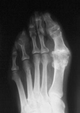 Radiograph of the foot in a patient with chronic gout. Podagra, or first metatarsophalangeal joint pain, can easily be understood when this radiograph is evaluated. Sclerosis and joint-space narrowing are seen in the first metatarsophalangeal joint, as well as in the fourth interphalangeal joint. Image courtesy of Larry Brent, MD.
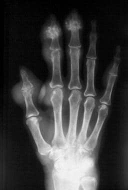 Radiograph of the hand. On this image of chronic tophaceous gouty arthritis, extensive bony erosions are noted throughout the carpal bones. Urate depositions may be present in the periarticular areas. Image courtesy of Larry Brent, MD.
EULAR recommendations
The European League Against Rheumatism (EULAR) published consensus recommendations for the diagnosis of gout, including the following [29] :
-
The search for crystals in synovial fluid (SF) or tophus aspirates is recommended in every person with suspected gout, because demonstration of monosodium urate (MSU) crystals provides a definitive diagnosis of gout.
-
Gout should be considered in the diagnosis of any acute arthritis in an adult. When SF analysis is not feasible, a clinical diagnosis of gout is supported by the following suggestive features: monoarticular involvement of a foot (especially the first metatarsophalangeal joint) or ankle joint; previous similar acute arthritis episodes; rapid onset of severe pain and swelling (at its worst in < 24 hr); erythema; male gender; and associated cardiovascular diseases and hyperuricemia.
-
It is strongly recommended that SF aspiration and examination for crystals is undertaken in any patient with undiagnosed inflammatory arthritis.
-
The diagnosis of gout should not be made on the presence of hyperuricemia alone.
-
When a clinical diagnosis of gout is uncertain and crystal identification is not possible, imaging should be used to search for MSU crystal deposition and features of any alternative diagnosis.
-
Plain radiographs are indicated for evidence of MSU crystal deposition but have limited value for the diagnosis of gout flares. Ultrasound can be more helpful in establishing a diagnosis in patients with suspected gout flares or chronic gouty arthritis by detection of tophi that are not evident on clinical examination, or a double contour (DC) sign at cartilage surfaces, which is highly specific for urate deposits in joints.
-
Typical radiographic features include bone erosions with overhanging edges and a sclerotic rim; bone proliferation; joint space narrowing, which occurs late in the disease course; and soft-tissue masses, sometimes calcified, corresponding to soft-tissue tophi.
-
Risk factors for chronic hyperuricemia should be searched for in every person with gout, specifically chronic kidney disease; overweight; medications (including diuretics, low-dose aspirin, cyclosporine, and tacrolimus); and excess consumption of alcohol (particularly beer and spirits), non-diet sodas, meat, and shellfish.
-
Systematic assessment for the presence of associated comorbidities in people with gout is recommended, including obesity, renal impairment, hypertension, ischemic heart disease, heart failure, diabetes, and dyslipidemia.
Radiography
In the early phase of gout, the clinical findings are limited to the soft tissues, of which an asymmetric swelling around the affected joint is typical. Another finding that may be evident is edema of the soft tissues around the joints. In a patient who has had multiple episodes of gouty arthritis in the same joint, a cloudy area of increased opacity may be seen on plain-film radiographs (see the images below).
Radiograph of the foot in a patient with chronic gout. Podagra, or first metatarsophalangeal joint pain, can easily be understood when this radiograph is evaluated. Sclerosis and joint-space narrowing are seen in the first metatarsophalangeal joint, as well as in the fourth interphalangeal joint. Image courtesy of Larry Brent, MD.
Radiograph of the hand. On this image of chronic tophaceous gouty arthritis, extensive bony erosions are noted throughout the carpal bones. Urate depositions may be present in the periarticular areas. Image courtesy of Larry Brent, MD.
In the intermediate phase of gout, the earliest bony changes appear, most commonly appearing initially in the first metatarsophalangeal joint area. These changes generally appear outside the joint or are in the juxta-articular area and are often described as punched-out lesions. Such lesions can progress to become sclerotic as they increase in size. In severe cases of intermediate-phase gout, fractures may be present in the affected areas.
In late-phase gout, the hallmark findings are numerous interosseous tophi. Another change that is evident on plain-film radiographs is joint-space narrowing, which can be severe and symptomatic. Marked deformities and subluxation may also be noted in affected areas, as well as calcium deposits in the soft tissues.
Computed Tomography
CT scanning can be used to study the effects of gout in areas that are hard to visualize with plain-film radiography. Many studies have been performed using dual-energy CT with good results, providing visualization, characterization, and quantification of monosodium urate crystals. [19, 20, 21, 12, 22, 4] DECT scanners are able to perform simultaneous acquisitions at 80 and 140 kVp using two separate sets of x‐ray tubes and detectors positioned 90 to 95 degrees apart, thereby differentiating materials based on their relative absorption of x‐rays at the different photon energy levels. [12] According to the AHRQ, DECT has shown good sensitivity and specificity for predicting gout compared with synovial fluid analysis for monosodium urate crystals, with 3 studies showing sensitivities that ranged from 85-100% and specificities that ranged from 83-92%. [13]
Choi et al found that dual-energy CT scanning (DECT) can produce obvious color displays for urate deposits and help identify subclinical tophus deposits and that tophus volume can be measured by DECT scans through automated volume estimation. [26] Further, after an initial retrospective assessment of 94 patients with suspected gout, Glazebrook et al determined that DECT is a sensitive, noninvasive, and reproducible method for spotting uric acid deposits in joints and periarticular soft tissues. [27]
Gruber et al studied 21 patients suspected of having gout in 37 joints by comparing the results of DECT with ultrasonography. They concluded that both imaging techniques had comparable sensitivity for detecting gout, but that DECT had some false-negative findings. [28]
Ultrasonography
Ultrasonography for the diagnosis of gout has advantages in that it is easily available and portable and doesn't require ionizing radiation. However, its limitation include being unable to image deep structures or joint and is highly operator dependent. [12] Findings include the double-contour sign (hyperechoic irregular enhancement over the surface of the hyaline cartilage) and can identify tophus deposition in and around joints, erosions, and tissue inflammation if power Doppler US is used. [17, 18, 3, 14] According to the Agency for Healthcare Research and Quality (AHRQ), 4 ultrasound studies on gout showed sensitivities that ranged from 37-100% and specificities that ranged from 68-97%. [13]
Rettenbacher et al compared radiography with ultrasonography in diagnosing gout, and radiography suggested gout with a sensitivity of 31% (32/102) and a specificity of 93% (55/59), whereas US suggested gout with a sensitivity of 96% (98/102) and a specificity of 73% (43/59). Ultrasonography, according to the authors, often provided additional diagnostic information in patients with clinical suspicion of gout when laboratory findings and radiographic results were negative or inconclusive and should therefore be used in such cases. [24]
In a study by Perez-Ruiz et al, the ultrasound measurement of tophi appeared to be useful as an outcome measure for chronic gout. [25] However, the authors cautioned that further randomized trials should be conducted.
-
Radiograph of the foot in a patient with chronic gout. Podagra, or first metatarsophalangeal joint pain, can easily be understood when this radiograph is evaluated. Sclerosis and joint-space narrowing are seen in the first metatarsophalangeal joint, as well as in the fourth interphalangeal joint. Image courtesy of Larry Brent, MD.
-
Radiograph of the hand. On this image of chronic tophaceous gouty arthritis, extensive bony erosions are noted throughout the carpal bones. Urate depositions may be present in the periarticular areas. Image courtesy of Larry Brent, MD.


