Practice Essentials
Croup is a generic term that encompasses a heterogeneous group of relatively acute conditions (mostly infectious) that are characterized by a syndrome of distinctive brassy coughs. Croup includes laryngotracheitis, laryngotracheobronchitis, and laryngotracheobronchopneumonitis. These conditions may be accompanied by inspiratory stridor, hoarseness, and signs of respiratory distress as a result of laryngeal obstruction. [1, 2, 3, 4, 5, 6, 7] Croup typically occurs in the fall and winter months. The word croup derives from an old Scottish term roup, which means "to cry out in a shrill voice."
Croup accounts for 7% of hospitalizations in children younger than 5 years and affects about 3% of children per year, usually between 6 months and 3 years of age. [8, 9]
The most common form of croup is acute laryngotracheobronchitis or viral croup, an infection of both the upper and lower respiratory tracts. A reactive inflammatory response causes subglottic edema. Narrowing of the airway can be life threatening in infants and young children because of their small airway. Viral croup may be complicated by bacterial tracheitis that is caused by Staphylococcus aureus, Haemophilus influenzae, Streptococcus pneumoniae, or Moraxella catarrhalis.
Failure to correctly differentiate croup from epiglottitis is a special concern. Epiglottitis is a life-threatening medical emergency. In children with suspected epiglottis, direct visualization of the epiglottis must be performed in a controlled setting by a physician who is experienced in airway management. Radiographic findings on the lateral view of a thickened epiglottis are characteristic of epiglottitis, while findings of subglottic airway narrowing on the anterior view are indications of croup. [1]
The following images demonstrate normal anteroposterior (AP) and lateral neck radiographs, followed by AP and lateral radiographs in children with croup.
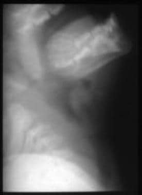
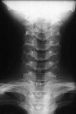 Normal anteroposterior radiograph of the neck. The normal convex borders (shoulders) of the vocal cords are outlined in the larynx.
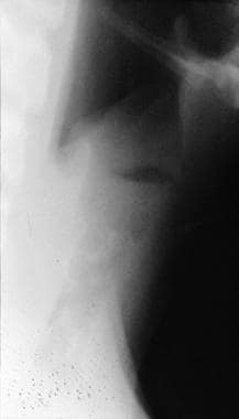
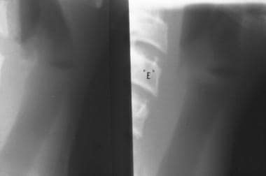 Normal lateral neck radiographs. During inspiration, the undersurface of the vocal cords is wide apart and not visualized. During phonation (saying "e"), the undersurface of the vocal cords are well visualized.
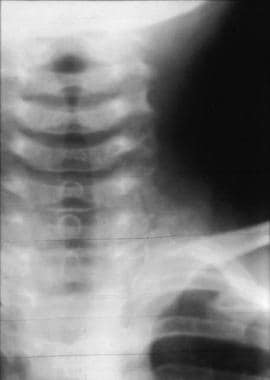 Anteroposterior radiograph in a patient with croup. This image shows the steeple sign, with loss of the normal shoulders of the subglottic larynx.
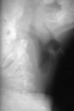 Lateral radiograph in a patient with croup. This image shows the presence of subglottic haziness and narrowing, as well as distention of the hypopharynx. The epiglottis and prevertebral soft tissues are normal.
Preferred examination
Most children with clinical croup require no testing beyond a thorough history and physical examination. Observation and frequent physical examination remain the best ways to monitor affected children. Pulse oximetry is useful if the patient also has bronchiolitis or pneumonia. The oral cavity and oropharynx are examined in the emergency department to exclude other causes of stridor or respiratory distress such as peritonsillar or retropharyngeal abscess or uvulitis. [1, 2, 3]
Laryngoscopy and airway support in a well-controlled environment is required if complete airway obstruction is imminent. Flexible nasopharyngoscopy can be used safely during the acute episode to evaluate the glottic and supraglottic areas. The subglottic area can frequently be visualized by looking through the vocal cords—take care not to pass the scope below the glottis.
Endoscopy has a role in atypical, severe, or recurrent cases of laryngotracheobronchitis. [7, 10] In addition, endoscopy may be used to evaluate children in whom extubation has failed and in whom evidence is seen of severe subglottic trauma, in which case reintubation may not be advisable. [11, 12]
Neck radiographs may be helpful to evaluate the various causes of stridor. [6, 1, 13] A multicenter study demonstrated that pediatric-focused emergency departments are significantly less likely to rely on imaging studies in the work-up of croup. [6]
Initial radiography may have a role in predicting the need for hospitalization in children with croup. A study, by Yang et al, of radiographs of 192 children reported that the severity of croup was significantly correlated with frontal ratio (FR), and lateral ratio (LR). Children with an LR >0.6 and FR >0.65 were found to be at low risk for hospitalizaton, whereas 71% of children with Fan R < 0.23 or and LR < 0.45 required longer hospitalization. [14]
Limitations of techniques
The diagnosis of croup is primarily clinical and requires no further testing. However, AP and lateral soft-tissue technique radiographs of the neck can help differentiate croup from other causes of stridor and respiratory distress, such as foreign body, epiglottitis, and retropharyngeal abscess. Lateral neck radiographs detect croup with up to 93% sensitivity and 92% specificity. The steeple sign on AP radiographs is not specific for croup and may be seen in some children with epiglottitis. The steeple sign can also be absent in some children with croup. A pseudo-steeple sign, which is a normal variant, may be seen at times during the respiratory cycle in some children without croup. [15, 16]
Differential diagnosis and other problems to be considered
Acute epiglottitis and subglottic stenosis are part of the differential diagnosis.
Conditions that cause obstruction in the region of the larynx also include laryngeal foreign body aspiration; acute angioedema (presents with other evidence of swelling of face and neck); retropharyngeal/parapharyngeal abscess; bacterial tracheitis; infectious mononucleosis; laryngeal diphtheria; Paraquat poisoning; burns or thermal injuries; smoke inhalation; neoplasm or hemangioma; acute laryngeal fracture; Chiari I, Chiari II, and Dandy-Walker malformation; laryngomalacia; laryngeal papillomatosis; and extrinsic obstruction by a vascular ring.
Westley score
The Westley score is the most commonly used classification system for measuring the severity of croup. It utilizes a scoring system that ranges from 0 to 17 points based on stridor, retractions, cyanosis, level of consciousness, and air entry, as follows [2, 17] :
-
Inspiratory stridor: 0 (none), 1 (when agitated), 2 (at rest)
-
Retractions: 0 (none), 1 (mild), 2 (moderate), 3 (severe)
-
Air entry: 0 (normal), 1 (decreased); 2 (markedly decreased)
-
Cyanosis: 0 (none), 4 (when crying); 5 (at rest)
-
Level of consciousness: 0 (alert), 5 (disoriented)
A Westley score 2 or less indicates mild croup; 3-5 indicates moderate croup; 6-11 indicates severe croup, and12 or higher indicates impending respiratory failure. Over 85% of affected patients present with mild disease, and severe croup occurs in less than 1%. [2, 17]
Radiography
Perform anteroposterior and lateral radiographs using a high-kilovoltage technique, or perform digital fluoroscopy and rapid-sequence imaging to optimize visualization of the airway. Although high-kilovoltage techniques are preferred, conventional techniques may be used.
The vocal cords, larynx, and lateral walls of the subglottic larynx and trachea are well depicted on the frontal view. The hypopharynx, epiglottis, aryepiglottic folds, prevertebral soft tissues, larynx, and subglottic airway can be evaluated on the lateral projection.
(The 2 images below demonstrate normal lateral and AP neck radiographs.)
Normal anteroposterior radiograph of the neck. The normal convex borders (shoulders) of the vocal cords are outlined in the larynx.
Normal lateral neck radiographs. During inspiration, the undersurface of the vocal cords is wide apart and not visualized. During phonation (saying "e"), the undersurface of the vocal cords are well visualized.
On frontal neck radiographs, the lateral walls of the subglottic larynx are normally convex or shouldered. Wall edema in croup narrows this space, with loss of lateral convexity, and creates a steeple shape below the vocal cords (as shown in the image below). The narrowing may extend for 5-10 mm below the vocal cords.
Anteroposterior radiograph in a patient with croup. This image shows the steeple sign, with loss of the normal shoulders of the subglottic larynx.
On lateral neck radiographs, the hypopharynx is overdistended during inspiration, and the subglottic region is hazy as a result of narrowing of the airway by mucosal edema (as in the following image). The larynx airway is indistinct. The undersurface of the vocal cords that would normally be identified during phonation is not well identified. However, the epiglottis, aryepiglottic folds, and prevertebral spaces appear normal.
Lateral radiograph in a patient with croup. This image shows the presence of subglottic haziness and narrowing, as well as distention of the hypopharynx. The epiglottis and prevertebral soft tissues are normal.
Degree of confidence
Airway radiographs detect croup with up to 93% sensitivity and 92% specificity. Note that subglottic haziness and the steeple sign can also be seen in a small percentage of children who have epiglottitis; however, additional radiographic findings that are specific for epiglottitis are present on the lateral radiograph. Subglottic narrowing from laryngotracheal hemangiomas is typically asymmetric.
A pseudo-steeple sign may be present in children without symptoms of croup. Other radiographic signs of obstruction are absent. Distention of the hypopharynx can be due to any condition that causes upper airway obstruction, such as epiglottitis, foreign body aspiration or ingestion, subglottic hemangioma, or bacterial tracheitis. [18, 19, 20]
Epiglottitis is associated with a distended hypopharynx and subglottic narrowing, but this condition also causes thickening of the epiglottis and aryepiglottic folds (see the image below).
 Lateral radiograph in a 2-year-old child with stridor and fever. This image shows the swelling of the epiglottis and aryepiglottic folds that is typical of epiglottitis. The epiglottis contour resembles a thumb.
The most common nonopaque foreign bodies include foods such as peanuts, candy, and hot dogs. Foreign bodies can cause extrinsic airway obstruction if they lodge in the proximal trachea or esophagus. The most common radiopaque foreign bodies are coins, which can lodge in the esophagus at the level of the cricopharyngeus muscle or aortic arch. Airway obstruction is caused by mechanical compression of the posterior trachea or esophagotracheal edema.
Subglottic hemangioma usually presents in the first 3 months of life. If the subglottic hemangioma extends superiorly to involve the true cords, hoarseness may be present in addition to stridor. Subglottic hemangiomas most commonly cause eccentric narrowing of the subglottic airway. Typically, croup causes symmetric subglottic narrowing.
In membranous croup, inflammation of the larynx, trachea, and bronchi, with an adherent or semi-adherent mucopurulent membrane in the subglottic space and upper trachea, is present. Radiographs of the airway show marked irregularity and edema of the walls of the trachea (see the image below). A detached membrane may be seen in the lumen of the trachea and may be mistaken for a tracheal foreign body. If severe obstruction is present, endoscopic removal of the obstructing membrane may improve the clinical condition of the patient.
 Lateral radiograph in a patient with membranous croup (bacterial tracheitis). This image shows haziness in the subglottic region of the trachea. Soft-tissue defects are identified within the airway. The hypopharynx is overdistended.
-
Normal anteroposterior radiograph of the neck. The normal convex borders (shoulders) of the vocal cords are outlined in the larynx.
-
Normal lateral neck radiographs. During inspiration, the undersurface of the vocal cords is wide apart and not visualized. During phonation (saying "e"), the undersurface of the vocal cords are well visualized.
-
Anteroposterior radiograph in a patient with croup. This image shows the steeple sign, with loss of the normal shoulders of the subglottic larynx.
-
Lateral radiograph in a patient with croup. This image shows the presence of subglottic haziness and narrowing, as well as distention of the hypopharynx. The epiglottis and prevertebral soft tissues are normal.
-
Lateral radiograph in a patient with membranous croup (bacterial tracheitis). This image shows haziness in the subglottic region of the trachea. Soft-tissue defects are identified within the airway. The hypopharynx is overdistended.
-
Lateral radiograph in a 2-year-old child with stridor and fever. This image shows the swelling of the epiglottis and aryepiglottic folds that is typical of epiglottitis. The epiglottis contour resembles a thumb.