Background
Ventricular repair, or cardiorrhaphy, has long been one of the most dramatic and lifesaving procedures performed in the emergency department (ED). Around 3000 BCE, in the Edwin Smith Surgical Papyrus, the first reports of trauma to the thorax were described. [1] The first successful human cardiorrhaphy was performed by the German physician Rehn in 1896 to repair a right ventricular injury sustained during a fencing match. [2]
The first successful cardiorrhaphy in the United States was performed by Hill in 1902; he operated on a teenage stabbing victim on a kitchen table in Montgomery, Alabama. [3] This began the practice of emergency cardiac repair in patients who sustain life-threatening penetrating trauma to the heart.
Cardiac trauma is divided into two mechanisms, as follows:
-
Blunt injuries - Cardiac rupture following blunt thoracic trauma usually causes death at the scene and is not typically encountered clinically [4, 5, 6]
-
Penetrating injuries - These are also commonly lethal but can be salvageable; the most common causes of death from penetrating trauma wounds are cardiac tamponade and exsanguination; in contradistinction to other forms of trauma, preoperative resuscitation plays a limited role, and immediate surgical intervention is a superior determinant of survival [7, 8, 9, 10, 11, 12, 13]
Consider cardiac injury in the differential whenever penetrating injury has occurred in the thorax or upper abdomen. The area of most concern is known as the cardiac box. This is an area of the trunk in which penetrating injuries risk damage to the heart. Anatomically, it is a triangular region bordered by the midclavicular lines laterally, the clavicles superiorly, and the costal margins inferiorly. [7]
Clinical presentations can vary widely, from hemodynamically stable to cardiac arrest. Cardiac tamponade is commonly thought to correlate with the Beck triad (hypotension, elevated jugular venous pressure, and muffled heart sounds), but these symptoms are found only in a minority of patients. Signs of shock (eg, tachycardia, hypotension, diaphoresis, and agitation) are better correlated with tamponade and should, therefore, be assessed. [7]
The chest radiograph does not commonly show an enlarged cardiac silhouette, even in the setting of acute tamponade. In cases of suspected penetrating cardiac injury, radiography should be used to assess for clues of cardiac injury, including retained intracardiac foreign bodies, hemothorax, pneumothorax, or pneumoperitoneum. [7, 14, 15] For more information, see Pneumothorax Imaging. (See the image below.)
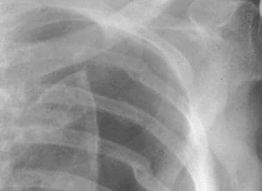 A true pneumothorax line. Note that the visceral pleural line is observed clearly, with the absence of vascular marking beyond the pleural line.
The FAST (Focused Assessment with Sonography in Trauma) examination is increasingly popular as the diagnostic tool of choice in the ED for identifying pericardial effusions. It does have its limitations, however, in that it is both operator- and patient-dependent. It should be used primarily to direct and prioritize management. [16]
An essential part of the management of patients in extremis from cardiac injury is thoracotomy for cardiac resuscitation, [17] and possible cardiorrhaphy. The thoracotomy involves making a large incision to expose the heart, aorta and hilum. It allows for internal defibrillation and cardiac massage. Once the heart is exposed, a pericardial incision can be made to release any tamponade. The heart is then examined to look for any penetrating wounds. For a detailed description of this procedure, see Emergency Bedside Thoracotomy.
Multiple methods are used in the emergency management of cardiac wounds, including the following:
-
Digital pressure - This method is used for small wounds and can be used to control bleeding on the way to the operating room (OR)
-
Foley catheter: For larger wounds, a Foley catheter can be inserted through the wound and inflated, and with gentle traction, tamponade can be accomplished; placing a Foley catheter can also stop hemorrhage so that cardiorrhaphy can be performed or a patient can be transported to the OR for repair; an additional advantage of using the Foley catheter is the ability to administer intracardiac medications, fluids, or blood [18, 19]
-
Immediate cardiorrhaphy (in the ED) - This method has been found to yield a survival rate up to 31% in patients presenting with penetrating cardiac injuries [16]
Indications
The science of surgical resuscitation has advanced tremendously since Rehn performed the first human cardiorrhaphy. The popularity of this procedure has waxed and waned as a result of changes in surgical techniques and differing analyses of patient outcome data. Currently, the indications for emergency thoracotomy, with or without cardiorrhaphy, include the following:
-
Persistent hypotension or signs of cardiac tamponade in the setting of penetrating chest trauma
-
Cardiac arrest in the ED, in the setting of blunt trauma
Contraindications
Contraindications for verntricular repair include the following:
-
Clinically stable patient
-
Blunt trauma in patients with no signs of life upon arrival [22]
-
Patient with evidence of prolonged death (eg, rigor mortis, livor mortis)
-
Patient whose life is unsalvageable (eg, decapitation)
-
Primary incision in the fourth or fifth intercostal space.
-
Open the pleura with scissors.
-
Chest opened with rib spreader.
-
Initial opening of the pericardium with the scalpel.
-
Locate the laceration.
-
Suture threading.
-
Completed mattress stitch.
-
Sufficiently approximated laceration.
-
A true pneumothorax line. Note that the visceral pleural line is observed clearly, with the absence of vascular marking beyond the pleural line.