Background
The extensor tendons of the hand are in a relatively superficial position; consequently, they are highly susceptible to injury from lacerations, bites, burns, or blunt trauma. Extensor tendon injuries are commonly diagnosed in the emergency department (ED). Certain injuries can be repaired in the ED, [1] whereas others should be repaired by a hand surgeon at a later time in an operating room (OR).
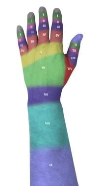
The dorsum of the hand, wrist, and forearm are divided into nine anatomic zones to facilitate classification and treatment of extensor tendon injuries. Odd numbers are used for regions overlying articular structures, with even numbers being assigned to the regions between joints, as follows (see the image below) [2, 3, 4] :
-
Zone 1 (distal interphalangeal [DIP] joint)
-
Zone 2 (middle phalanx)
-
Zone 3 (proximal interphalangeal [PIP] joint)
-
Zone 4 (proximal phalanx)
-
Zone 5 (metacarpophalangeal [MCP] joint)
-
Zone 6 (dorsum of hand)
-
Zone 7 (wrist)
-
Zone 8 (distal forearm)
-
Zone 9 (proximal forearm)
Treatment of extensor tendon injuries may require operative intervention, depending on the complexity of the injury and the zone of the hand involved. [5, 6] Although there is some limited evidence for the view that selected partial extensor tendon lacerations may be treated with splinting and physiotherapy, further study is required. [7] Complete lacerations generally are treated surgically. For treatment of injuries to specific zones of the hand, see Technique.
The goal of repair is to restore tendon continuity and function, with a secondary goal of allowing early motion of the injured digit. Optimal preparation and technique are critical for minimizing adhesions and scar tissue formation and ensuring the best possible outcome.
Extensor tendon injuries are often more difficult to treat than flexor tendon injuries, owing to several issues specific to extensor tendons. The extensor mechanisms of the hand are in a superficial position, not enclosed in tendon sheaths (as flexor tendons are), and often have limited retraction after injury.
Extensor tendons also tend to be thinner and flatter than flexor tendons are, as well as being in very close proximity to bony structures. This leaves them highly susceptible to adhesions and shortening, which can severely impair function and range of motion.
With injuries that are proximal to the anatomic juncturae tendinum, extensor function may still be preserved distally. Additionally, up to 90% of an extensor tendon can be lacerated and still retain preserved function to gravity, necessitating careful physical examination and wound exploration. [8, 4, 9]
Extensor tendon injuries may be iatrogenic as well, occurring either as a consequence of surgical error or as a complication of a previous procedure or medication (eg, a fluoroquinolone). Extensor tendon ruptures following plating of wrist and hand fractures are well documented, including ruptures that have been reported after intramedullary nailing of pediatric forearm fractures. [10, 11]
Indications
Indications for extensor tendon repair include the following:
-
Tendon laceration greater than 50%
-
Tendon laceration less than 50% with significantly decreased strength in comparison with the contralateral finger
-
Tendon laceration associated with significant overlying skin loss, joint-space penetration, or bony fracture
Repair can be accomplished immediately in the ED or after a delay of several (typically, up to 7) days following the injury. [12] Simple lacerations can be repaired in the ED. If repair is delayed or postponement is necessitated by contamination or complexity, then the wound should be irrigated and debrided, the skin should be approximated loosely with interrupted sutures, and the hand should be placed in a volar resting extension splint. [13] Ideally, lesions proximal to zone 6 should be treated in an OR; such injuries tend to require significant exposure of the tissues for appropriate reapproximation of the tendon. [4]
Contraindications
Extensor tendon repair should not be attempted in the ED or acute care setting in any of the following circumstances:
-
Unavailability of an appropriately skilled physician
-
Contaminated injuries, particularly open zone 1, 3, or 5 "fight bite" injuries
-
Presence of associated bony fractures, open joint space, or significant overlying skin loss (which require an orthopedist or hand surgeon for repair)
-
Injuries proximal to zone 6 - Caution is required with these injuries, owing to the frequent necessity of significant surgical exposure to achieve adequate repair
In these cases, the repair should be performed by an experienced hand surgeon, preferably in the OR.
Technical Considerations
Anatomy
Several anatomic structures contribute to the extensor mechanism, including the extrinsic muscles of the forearm, intrinsic muscles such as the interosseous and lumbricals, and fibrous structures.
The extrinsic tendons pass through the extensor retinaculum on the dorsum of the wrist. This structure is divided into six compartments, each of which has tendons that pass through.
The third compartment is of particular importance in injury patterns. Injuries to the distal radius can result in rupture of the extensor pollicis longus (EPL), which passes through this compartment. Of significance, the sixth compartment contains the extensor carpi ulnaris (ECU), which serves as a major stabilizer for the distal radioulnar joint (DRUJ) as well as extends the wrist. [4, 8] Also significant anatomically is the presence of the extensor indicis proprius (EIP) and the extensor digiti minimi or quinti (EDM or EDQ), which extend the index and small fingers, respectively.
The extensor apparatus of the triphalangeal digits has three separate insertion sites and is formed through a complex interplay among the extrinsic extensors, the intrinsic extensors, and the digital ligaments. Proximally, it is attached at the level of the metacarpal heads by a sagittal band, which centers the tendon and prevents hyperextension. The most important insertion site is the insertion of the central slip at the base of the middle phalanx to extend the PIP joint. There is a third insertion point distally to extend the DIP joint, comprising the conjoined lateral bands.
It is important to be aware of the insertion sites of the extensor tendons during repair. (See the image below.)
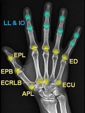 Extensor tendon repair. Extensor tendon insertion sites. LL = musculi lumbricales; IO = musculi interossei; EPL = musculus extensor pollicis longus; EPB = musculus extensor pollicis brevis; ED = musculus extensor digitorum communis; ECRLB = musculus extensor carpi radialis longus et brevis; ECU = musculus extensor carpi ulnaris; APL = musculus abductor pollicis longus. Extrinsic muscles are colored in yellow, intrinsic muscles in blue. Image courtesy of Dr Roberto Schubert, Radiopaedia.org, http://radiopaedia.org/cases/flexor-and-extensor-insertions-at-the-hand-and-wrist.
For more information about the relevant anatomy, see Hand Anatomy and Wrist Joint Anatomy.
Outcomes
A study by Dalton (137 digits; minimum follow-up, 8 wk) compared the effects of early (< 14 d; 110 digits) and delayed (≥14 d; 27 digits) extensor tendon repair on patient outcomes. [14] The two study groups were each further divided by location of injury (zones 1-4 vs zones 5-8). No significant differences between early and delayed repair were found with respect to final total active motion, final extension, return to activity, or surgical complications for either zone 1-4 or zone 5-8 repairs.
-
Povidone-iodine solution, 1% lidocaine, 10-mL syringe, and 25-gauge needle.
-
Irrigation equipment.
-
Equipment for extensor tendon repair.
-
Polypropylene and nylon sutures.
-
Shears, aluminum splint, tape.
-
Mallet finger splint, side view.
-
Mallet finger splint, end view.
-
Boutonnière splint, side view.
-
Boutonnière splint, view from above and to side.
-
Extensor tendon repair. Suture techniques.
-
Extensor tendon repair. Dermatotenodesis.
-
Extensor tendon repair. Video clip of splint application.
-
Division of dorsum of hand and forearm into anatomic zones.
-
Extensor tendon repair. Extensor tendon insertion sites. LL = musculi lumbricales; IO = musculi interossei; EPL = musculus extensor pollicis longus; EPB = musculus extensor pollicis brevis; ED = musculus extensor digitorum communis; ECRLB = musculus extensor carpi radialis longus et brevis; ECU = musculus extensor carpi ulnaris; APL = musculus abductor pollicis longus. Extrinsic muscles are colored in yellow, intrinsic muscles in blue. Image courtesy of Dr Roberto Schubert, Radiopaedia.org, http://radiopaedia.org/cases/flexor-and-extensor-insertions-at-the-hand-and-wrist.


