Overview
Epidural steroid injections (ESIs) have been endorsed by the North American Spine Society and the Agency for Healthcare Research and Quality (formerly, the Agency for Health Care Policy and Research) of the Department of Health and Human Services as an integral part of nonsurgical management of radicular pain from lumbar spine disorders.
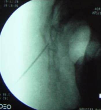
The first documented epidural medication injection, which was performed using the caudal approach (see the image below; see also Approaches for Epidural Injections) was performed in 1901, when cocaine was injected to treat lumbago and sciatica (presumably pain referred from lumbar nerve roots). [1] According to reports, epidurals from the 1920s to 1940s involved using high volumes of normal saline and local anesthetics. Injection of corticosteroids into the epidural space for the management of lumbar radicular pain was first recorded in 1952.
ESIs can provide diagnostic and therapeutic benefits. Diagnostically, ESIs may help to identify the epidural space as the potential pain generator, through pain relief after local anesthetic injection to the site of presumed anatomic pathology. In addition, if the patient receives several weeks or more of pain relief, then it may be reasonable to assume that an element of inflammation was involved in his or her pathophysiology. Since prolonged pain relief is presumed to result from a reduction in an inflammatory process, it is also reasonable to assume that during the period of this analgesia, the afflicted nerve roots were relatively protected from the deleterious effects of inflammation. Chronic inflammation can result in edema, wallerian degeneration, and fibrotic changes to the neural tissues.
Mechanisms of Radicular Low Back Pain
Radicular pain often is the result of nerve root inflammation with or without mechanical irritation. Clinical practice and research demonstrate that mechanical compression alone to the nerves causes only motor deficits and altered sensation but does not necessarily cause pain. Inflammation within the epidural space and nerve roots, as can be provoked by a herniated disk, is a significant factor in causing radicular pain.
Historical evidence of nerve root inflammation has been demonstrated during surgery in patients with radicular low back pain (LBP) from lumbar disk herniation. Animal research in dogs and rats also has revealed severe inflammation locally within the epidural space and nerve root after injection of autologous nuclear material into the epidural space. A high level of phospholipase A2 (PLA2), an enzyme that helps to regulate the initial inflammatory cascade, has been demonstrated in herniated disk material from surgical samples in humans. Leukotriene B4, thromboxane B2, and inflammatory products also have been discovered within herniated human disks after surgery. Animal models have demonstrated that injection of PLA2 into the epidural space induces local demyelination of nerve roots, with resultant ectopic discharges (which is considered the primary pathophysiologic mechanism for sciatica [radicular pain]).
The radicular LBP caused by spinal stenosis is probably related to the inhibition of normal nerve root vascular flow with resultant nerve root nutrition, nerve root edema, and nerve root dysfunction. Chronic nerve root compression can induce axon ischemia, impede venous return, promote plasma protein extravasation, and cause local inflammation. If dorsal root ganglia are chronically compressed and irritated, this theoretically can lead to their sensitization and resultant radicular pain. Similar mechanisms of radicular pain are postulated to occur in the thoracic and cervical spine as well.
In summary, clinical practice and animal research suggest that radicular pain is the result of inflammation of the nerve root in the epidural space provoked by leakage of disk material, compression of the nerve root vasculature, and/or irritation of dorsal root ganglia from spinal stenosis.
Rationale for Use of Steroids in Back Pain
Since lumbar radicular pain may originate from inflammation of the epidural space and the nerve root, analgesic effects of corticosteroids most likely are related to the following mechanisms:
-
Inhibition of PLA2 and inflammation
-
Inhibition of neural transmission in nociceptive C fibers
-
Reduction of capillary permeability
Indications and Contraindications for Epidural Steroid Injections
Although the primary indication for epidural steroid injection (ESI) is radicular pain associated with a herniated nucleus pulposus, a variety of other indications have been reported in the literature.
Lumbar, thoracic, and cervical ESIs may be indicated for lumbar radicular pain associated with any of the following conditions:
-
Lumbosacral disk herniation
-
Spinal stenosis with radicular pain (central canal stenosis, foraminal and lateral recess stenosis)
-
Compression fracture of the lumbar spine with radicular pain
-
Facet or nerve root cyst with radicular pain
-
Postherpetic neuralgia
Absolute contraindications for ESIs include the following:
-
Systemic infection or local infection at the site of a planned injection
-
Bleeding disorder or fully anticoagulated (eg, on a fully therapeutic dose of warfarin, heparin, or other anticoagulant)
-
History of significant allergic reactions to injected solutions (eg, contrast, anesthetic, corticosteroid)
-
Acute spinal cord compression
-
Patient refusal to proceed with the injection procedure
In addition, fluoroscopy should not be used in epidural injections for women who are pregnant, to avoid exposing the fetus to ionizing radiation. Caution should be used when performing injections in patients with poorly controlled diabetes, since the corticosteroid may transiently, but significantly, increase blood glucose levels. Patients with a history of immunosuppression may require additional precautions, such as preprocedure laboratory testing and/or antibiotics. Caution should be exercised when performing injections in individuals who have a history of congestive heart failure because of the potential for steroid-induced fluid retention.
Efficacy of Epidural Injections and Rationale for Fluoroscopy and Contrast
Efficacy of epidural steroid injections (ESIs)
Although numerous articles have supported the benefit of ESIs for LBP, especially if the pain is caused by radiculopathy, other studies have disputed the efficacy of these procedures. Unfortunately, most of the earlier studies (those that failed to show a benefit from the injection) had significant limitations. Aside from using a less-than-desirable research methodology, most of these studies did not use fluoroscopy and radiographic contrast to document accurate placement of the injected substance into the epidural space. Many also failed to demonstrate that injection was performed at a presumed level of pathology, which has been shown to be critical to the success of ESIs.
Studies have reported that without fluoroscopy and radiographic contrast confirmation, incorrect injection placement (ie, placement outside the epidural space) occurs in 30% of cases, on average, even when ESI is performed by experienced injectionists. These methodologic problems most likely were the major factors that led to the mixed assessment of ESIs.
The standard of care in many practices is to obtain a pretreatment MRI prior to performing an epidural injection, since the MRI can help guide the location of the injection. However, in a retrospective study of 367 with back and radicular pain, all patients received a clinical exam and pre-epidural X-rays, but approximately half of the patients also received a pre-epidural MRI. [2] TFESI yielded similar outcomes in both groups, regardless of whether the patient had a pre-treatment MRI. This finding supports that in the setting of clear physical exam and X-ray evidence of the affected spinal level, skipping a pre-epidural MRI may not reduce the efficacy of a TFESI. [2]
Factors affecting the efficacy of ESIs
As with other medical procedures, the efficacy of the ESIs is related to many factors. Aside from the clinician's experience and training, other factors that play an important role include patient selection, symptom duration, underlying pathophysiology, ESI approach, the use of fluoroscopy and contrast enhancement, and the vocational status, as well as the socioeconomic and psychological circumstances, associated with the individual patient.
In general, patients who have had symptoms for less than 3 months have response rates of 90%. When patients have had radiculopathy symptoms for less than 6 months, response decreases to approximately 70%. Response decreases to 50% in patients who have had symptoms for over 1 year. Patients with symptoms of shorter duration have more sustained relief than those with chronic pain. Patients with chronic back pain generally have better response if they develop an acute radiculopathy. Patients with factors favoring the use of ESIs also include those who have not had previous back surgery, who are not on workers’ compensation, who are aged younger than 60 years, and who are nonsmokers.
In a cross-sectional study design at a university spine center, 76 patients with sciatica were followed for a mean of 122 days after receiving transforaminal ESIs. Of these patients, 47% experienced improvement, 28% were unchanged, and 16% worsened. The least favorable outcomes were associated with patients receiving Social Security Disability Insurance (SSDI) or workers’ compensation payments and with those whose work required heavy lifting.
Patient response to ESIs is also related to underlying pathophysiology. In general, acute radicular pain from lumbar disk herniation responds more favorably than does radicular pain from lumbar spinal stenosis. Patients with radicular pain after lumbar spine surgery frequently receive less benefit from ESIs unless the radicular pain is from a recurrent herniated nucleus pulposus. Still, ESIs are often helpful for radicular pain from stenosis.
Lumbar transforaminal epidural injections
The following research demonstrated the efficacy of lumbar transforaminal epidural injections (see Approaches for Epidural Injections) in patients with persistent sciatica from lumbar disk herniation or spinal stenosis:
-
In 2002, Vad and colleagues reported a prospective randomized study comparing transforaminal lumbar epidural injection with lumbar paraspinal trigger-point injection. [3] They randomized 48 patients with sciatica from herniated disk pulposus (confirmed by lumbar spine MRI) into 2 groups. One group received transforaminal lumbar epidural injection, and the other received a lumbar paraspinal intramuscular injection with saline. The average follow-up period was 16 months. The authors used patient satisfaction, the Rolland-Morris scale, and pain reduction extent as indices for efficacy. The success rate in the transforaminal injection group was 84%, compared with 48% in the saline group.
-
Botwin and colleagues demonstrated the efficacy of the transforaminal epidural injection in their retrospective cohort study in patients with sciatica (caused by lumbar spinal stenosis). [4] Thirty-four patients who did not respond to nonsteroidal anti-inflammatory agents and oral analgesics received 1.9 injections (average). At 1 year, 75% of the patients had greater than 50% pain reduction, 64% improved their walking duration, and 57% increased their standing tolerance. An additional benefit of ESIs in many patients is that the injections can potentially obviate the need for hospitalization and surgery in many patients.
-
Riew and colleagues reported results from a prospective, randomized, double-blinded, controlled clinical trial on 55 patients with severe sciatica from spinal stenosis or lumbar disk herniations. [5] These patients had not responded to 6 weeks of conservative treatment and were considered to be surgical candidates. The patients were divided into 2 groups; 1 group received lumbar epidural injection with bupivacaine and steroid, while the other group received only bupivacaine. Up to 4 lumbar epidural injections were delivered if needed. The follow-up period was 2-3 years. The study demonstrated that only 23% of patients in the group that received lumbar ESIs needed surgery, while 67% of patients in the bupivacaine injection group underwent surgery. The difference was statistically significant.
-
A follow-up study at 5 years found that 17 (81%) of 21 patients surveyed still had still not opted for surgery. [6] This report demonstrated a benefit from lumbar ESIs in patients who had been diagnosed with lumbar spinal stenosis or herniated nucleus pulposus, with the injections helping to reduce the need for surgery.
-
A systematic review of 14 randomized and 10 nonrandomized studies on the efficacy of lumbar transforaminal epidural steroid injections for lumbar radicular pain was performed by Manchikanti L et al. [7] The results demonstrated that the efficacy is good for radiculitis secondary to disk herniation with local anesthetics and steroids and fair with local anesthetic only, efficacy is fair for radiculitis secondary to spinal stenosis with local anesthetic and steroids, and efficacy is limited for axial pain and postsurgery syndrome using local anesthetic with or without steroids.
-
Roberts, in another systematic review, reported evidence suggesting transforaminal epidural steroid injections are superior to placebo for radicular symptoms. He also noted good evidence supporting these injections as a surgery-sparing intervention. [8]
-
MacVicar et al. reviewed the literature and found that 70% of patients with radicular pain secondary to disc herniations are at least 50% better at 1 to 2 months following epiduralsteroid injections; 40% of patients reported improvement at 12 months and up to 30% experienced complete pain relief. There were better results in patients with contained disc herniations or low-grade compression. [9]
Lumbar caudal epidural injections
Results from studies on caudal epidural injections are as follows:
-
Barre and colleagues reported that in 35% of patients with symptomatic lumbar spinal stenosis who received caudal ESIs, a visual numeric score improvement of 50% or greater was seen. [10] Long-term treatment success was seen in 35% of patients after a mean follow-up of 32 months.
-
Anwar and coauthors demonstrated that caudal injections could benefit patients with limited straight leg raise and symptoms of radicular pain or spinal stenosis; in this study, 65% of patients were noted to have some improvement at 3 months.
-
A systematic appraisal of the literature on caudal epidural steroid injections included 11 randomized trials and 5 nonrandomized studies. This study concluded that for lumbar disk herniation, the efficacy is good for short- and long-term relief of chronic pain secondary to disk herniation or radiculitis with local anesthetic and steroids and is fair with local anesthetic only. In managing chronic axial or diskogenic pain, spinal stenosis, and postsurgery syndrome, the indicated evidence is fair. [11]
Lumbar interlaminar epidural injections
A systematic review included 15 fluoroscopically guided randomized trials and 11 nonrandomized studies. The efficacy is good for radiculitis secondary to disk herniation with local anesthetics and steroids and fair with local anesthetic only, whereas it is fair for radiculitis secondary to spinal stenosis with local anesthetic and steroids and fair for axial pain without disk herniation with local anesthetic with or without steroids. [12]
A prospective study demonstrated that as compared with conventional lumbar interlaminar epidural injections, the lateral parasagittal interlaminar epidural approach has higher rate of contrast spread into the anterior epidural space. [13]
A recent prospective randomized study compared the efficacy of lumbar ESI using a parasagittal interlaminar (PIL) approach and midline interlaminar (MIL) approach. Thirty-seven patients were randomized to receive injection of 80 mg methylprednisolone either by the PIL (n = 19) or MIL (n = 18). A maximum of 3 injections were performed with 15-day intervals between injections, if necessary. Follow up was 6 months post injection. Patients were evaluated for effective pain relief (≥50% from baseline) by visual analog scale and improvement in disability by the modified Oswestry Disability Questionnaire at intervals of 15 days and 1, 2, 3, and 6 months. The results demonstrated that PIL approach has better ventral epidural spread in contrast (89.7% in the PIL vs 31.7% in the MIL group). At the end of 6 months, the PIL group had a significantly higher percentage of pain relief in the visual analogue scale (PIL 13 [68.4%] of 19) vs MIL 3 [16.7%] of 18)and improvement of disability using Oswestry disabilityquestionnaire score, as well as fewer total injections (29 in PIL vs 41 in MIL, P = .043). [14]
It is important to know that at least 3 cases of lumbar paraplegia have been reported, and each developed after interlaminar lumbar epidural steroid injections. [15] The suspected mechanism is similar to a paraplegia caused by a lumbar transforaminal ESI in which the epidural needle penetrates the radicular medullary artery, and the particulate corticosteroid being injected into this artery inside the spinal canal results in an embolism of spinal cord and subsequent paraplegia.
In fact, the anatomical studies have demonstrated that after the radicular medullary arteries enter the neuroforamen in the anterior aspect of exiting nerve root and dorsal root ganglion, they often travel a distance superiorly and laterally in the lateral epidural space to join the anterior spinal artery supplying the anterior two thirds of the spinal cord. Additionally, in about 63% of cases of cadaver studies, there is a posterior branch of the radicular medullary artery going to the dorsal aspect of the cauda equina. It is conceivable that the epidural needle in the interlaminar lumbar epidural steroid injection will very likely encounter the radicular medullar artery in the lateral aspect of the epidural space or midline posterior epidural space.
As the paraplegia after interlaminar lumbar ESIs is often underreported, the exact frequency of this event cannot be determinted. It is clear that in light of the anatomical positions of these radicular medullary arteries inside the spinal canal as described above, neither midline nor parasagittal interlaminar lumbar ESIs are completely risk free with respect to vascular injury and paraplegia. The alternative approach using the Kambin triangle may be the better choice (see below for description).
Comparison of interlaminar vs transforaminal lumbar epidural injections
More evidence favors the use of transforaminal ESIs in the lumbar spine compared with the cervical spine. Although the interlaminar approach (see Approaches for Epidural Injections) may allow the injectate to flow to the site of pathology by migrating around the thecal sac and into the ventral epidural space, the transforaminal route is presumably more reliable for delivering the steroid to the affected area in cases of disk herniation in which the disk comes into contact with the nerve root.
Rhee and colleagues found a difference in patients undergoing interlaminar and transforaminal ESI. [16] Those patients who underwent transforaminal injections had a 46% reduction in their pain score, and 10% went on to need surgery. In contrast, patients who had interlaminar injections had a 19% reduction in pain, and 25% required surgery.
Recently, a randomized, prospective, blinded, and controlled trial on the 38 patients with lumbar subacute radicular pain was conducted. The study demonstrated that while both groups improved, the transforaminal ESIs provided better pain relief in up to 16 days post injections compared with the interlaminar group. [17]
However, a separate randomized and prospective research study enrolled 32 patients in each group with chronic lumbar radiculopathy and 6 months follow up; the study again revealed the improvement in pain and disability in both groups. However, no significant differences were noted in pain reduction and the Oswestry disability scale between the transforaminal and interlaminar groups at the end of 6 months. [18]
The above discrepancy of efficacy may be due to the lower response to epidural steroid injections in general because of the chronicity of the radiculopathy; alternatively, it may reflect the differences of timing in follow up between the 2 studies. It is generally agreed that ESIs offer short-term (several months) pain relief.
ESIs may avoid unnecessary surgery
The Spine Patient Outcomes Research Trial (SPORT) is a prospective, multicenter study of operative versus nonoperative treatment of lumbar intervertebral disk herniation. The results demonstrated a significant difference in the preference for surgery between groups (19% in the ESI group compared with 56% in the non-ESI group, P< .001). No difference was noted in primary or secondary outcome measures at 4 years between the groups. A higher percentage of patients changed from surgical to nonsurgical treatment in the ESI group (41% vs 12% in non-ESI, P< .001). [19]
No deleterious effects on diskectomy
Buttermann et al conducted a study involving individuals with a lumbar disk herniation of greater than 25% of the cross-sectional area of the spinal canal; the patients were administered ESIs one level above the herniation. [20] The patients received up to 3 injections, with 42-56% of these individuals reporting the treatment to be effective. There was a cross-over of patients who first underwent ESIs and then diskectomy. For those who underwent an initial trial of ESIs, the delay in surgical decompression was not found to be detrimental to neurologic recovery at time of follow-up.
Cervical ESIs
No randomized, controlled trials have been performed to date on the efficacy of ESIs for the cervical spine and treatment of upper limb radicular pain. A prospective study by Rowlingson and Kirschenbaum described significant reduction in upper limb pain after cervical ESIs, and other studies (retrospective and prospective) identified radicular pain relief via interlaminar and transforaminal approaches. [21]
Given the similar mechanisms of radicular pain postulated for the lumbar and cervical regions, compelling evidence regarding the efficacy of lumbar ESIs might be applicable to treatment of upper lumbar interlaminar ESIs. A systematic review included fluoroscopically guided 15 randomized trials and 11 nonrandomized studies. The evidence is good for radiculitis secondary to disk herniation with local anesthetics and steroids and fair with local anesthetic only, whereas it is fair for radiculitis secondary to spinal stenosis with local anesthetic and steroids and fair for axial pain without disk herniation with local anesthetic with or without steroids. [12]
In terms of potential efficacy, transforaminal cervical ESIs are preferred over the interlaminar approach by several authors, because the transforaminal cervical injections allow for the delivery of higher concentrations of medications to isolated nerve roots and neuroforamina where stenosis may be present. [22] Overall, cervical interlaminar studies outnumber investigations of cervical transforaminal injections. [23]
According to House et al, differences in the vasculature and width of the dorsal epidural space compared with the characteristics of these structures in the ventral epidural space result in different risks associated with cervical interlaminar epidural injections in comparison with the transforaminal approach. For example, the dorsal epidural space has a rich venous plexus that increases the risk of an epidural hematoma. This is a potentially serious complication that can lead to permanent neurologic injury if not rapidly decompressed. The risk of dural puncture or direct spinal cord trauma is also increased above the C7-T1 level due to diminished diameter of the epidural space. [23]
A systematic review of cervical interlaminar epidural injections concluded that such injections are effective for relief of cervical radicular pain in the upper limbs; the report strongly recommended the procedure. However, there were no randomized trials identified in this review.
Rationale for fluoroscopy and contrast
Reports suggest that injection without fluoroscopic guidance (ie, blind injection) results in 30-40% of needle misplacements, such as needle tip placement outside the epidural space (including intravascular injection) and placement not at the presumed level of pathologic process. Therefore, it is recommended that ESIs be performed under fluoroscopic guidance and with radiographic contrast documenting appropriate placement in order to improve the safety, accuracy, and potential efficacy of ESIs.
Fluoroscopy in conjunction with contrast is used to improve efficacy and minimize potential complications. Furman and coauthors discovered that for lumbar spine ESIs, using flash or positive blood aspirate to predict intravascular injections was 97.9% specific but only 44.7% sensitive. [24] This suggests that negative aspiration of blood does not ensure a lack of vascular uptake. Similarly, in the cervical spine, vascular uptake injections occurred at a rate of nearly 20% with the use of fluoroscopy (confirmed by contrast injection), via a transforaminal approach. Again, a visible flash of blood in the needle hub or positive aspiration of blood demonstrated similar specificity and sensitivity to the lumbar injection study.
In a prospective study involving 191 patients who underwent single-level lumbar transforaminal epidural injection, simultaneous epidural and vascular injection was found to be 8.9%. Therefore, live fluoroscopy is recommended during contrast injection for confirmation of lumbosacral transforaminal epidural injections. [25]
Rationale and role of CT-guided technique
Fluoroscopy with contrast remains the standard of care for performing transforaminal epidural steroid injection (TFESI), and in many cases is the preferred technique. Results of a study of 38 patients with subacute or chronic low back pain comparingTFESI using CT guidance versus using fluoroscopic guidance showed that both groups had similar therapeutic effectiveness at 3-month followup. Thus, in regions where fluoroscopy is not readily available, this supports an equally effective alternative option of using CT guidance instead. [26]
Rationale and role of ultrasound-guided technique
There may be an emerging role for ultrasound-guided ESIs. Specifically, ultrasound-guided techniques may have a role in certain clinical scenarios and be useful in cervical and caudal ESIs. Ultrasound may provide some potential advantages including no radiation exposure, direct real-time visualization of vessels and nerves, shorter procedure time, and a high success rate for first-attempt injection. This may be useful in several clinical scenarios such as pregnant population, chronic pain requiring multiple injections, and technically challenging anatomy.
One emerging role for ultrasound-guided ESIs is a cervical epidural steroid injection particularly at lower cervical levels. In one study, there was no difference in the treatment effect for 6 months when compared to fluoroscopic-guided cervical ESI. In addition to similar efficacy, ultrasound may avoid intravascular injections and vulnerable structures in the needle trajectory. However, if an intravascular injection does occur ultrasound may not be able to detect it as opposed to fluoroscopy detection with a contrast pattern suggestive of vascular infiltration. Color Doppler and a test anesthetic injection may be used to help detect such intravascular injections. [27] One disadvantage to ultrasound is that it can be technically challenging and requires proper technique and experience in order to safely administer epidural injections. Another disadvantage with using ultrasound alone (without fluoroscopy) is that ultrasound cannot confirm the level the injectate reaches (whereas fluoroscopy does show that information, via showing the contrast flow pattern).
Another specific emerging role for ultrasound-guided ESIs is for ultrasound-guided caudal ESIs. A study by Senkal et al demonstrated ultrasound-guided caudal ESIs had a shorter procedure time and a higher successful first-attempt injection rate compared to the fluoroscopic-guided technique. Ultrasound provides immediate confirmation of the needle location and allows the physician to visualize soft tissue, vessels, and specifically the sacrococcygeal ligament, sacral hiatus, and sacral canal that cannot be visualized using fluoroscopy. The study also found pain relief and improvement were similar in both ultrasound-guided and fluoroscopic-guided techniques. [28]
The fluoroscopic-guided technique remains essential for lumbar transforaminal ESI. Due to shadowing of the foraminal area from bony structures, using ultrasound alone would not allow for visualization of the needle at the desired location. At this time, there are not enough advantages of the ultrasound-guided technique for it to have a role in lumbar transforaminal ESIs. [29]
Safety of Epidural Injections
Potential complications of epidural steroid injections (ESIs)
When performed by a skilled, experienced clinician in an appropriate setting and with carefully selected patients, the chance of significant complication from ESIs is remote. Nonetheless, similar to regional analgesia procedures, there are risks associated with ESIs. The more common risks from lumbar epidural injections are as follows:
-
Backache, postural puncture headache (0.5–1% for lumbar interlaminar injections and 0.6% for caudal epidural injections), nausea, vomiting, dizziness, and vasovagal reaction have been reported.
-
Bleeding along the trajectory of the injection, including in proximity to the nerve roots and/or the spinal cord (epidural hematoma), is a rare but potentially serious complication. Epidural hematoma occurs in 0.01-0.02% of procedures.
-
Infection is a rare complication but may be relatively more common in immunocompromised patients and can include epidural abscess and meningitis.
-
Nerve root injury has been reported.
-
Mild hypothalamic-pituitary-adrenal axis suppression has been reported from 1-3 months after receiving a total of 3 epidural injections (once weekly) with 80 mg of Aristocort in 7 mL of 1% lidocaine.
-
Other rare complications include anterior cord syndrome, presumably resulting from the injection of particulate steroid into the artery of Adamkiewicz, a radiculomedullary artery that supplies the anterior spinal artery feeding the anterior two third of the spinal cord in the thoracolumbar region. (See below for more details.)
-
A report by Arici et al urges caution when approaching transforaminal epidural injections due to the potential presence of anatomical variants in the location of the artery of Adamkiewicz (AKA). The artery is commonly located on the left side at the T8-L1 levels, thus at highest risk for complications when injections are left-sided at the upper lumbar or lower thoracic levels. But there are people who have a right-sided AKA, or an AKA location as superior as T2 or as inferior as S1. Rarely, a patient may even have two AKAs. The AKA is the most important nourishing artery of the thoracolumbar spinal cord and trauma may lead to occlusion and anterior spinal artery syndrome. This is a devastating consequence and presents with bilateral lower limb motor and sensory impairments as well as urinary and/or fecal incontinence. [30]
Cervical ESIs carry similar risks, with the apparent caveat that any damage to the spinal cord at the level of the cervical spine often results in greater impairment than damage at the lumbar levels and may precipitate respiratory arrest at higher cervical levels. There is the risk of spinal cord trauma if the operator performs direct injection into the spinal cord via an interlaminar approach, a risk that is essentially absent at the lower lumbar spine, since the spinal cord terminates at the level of L2.
Cord trauma can also result from compression of the spinal cord from an epidural abscess or an epidural hematoma.
Despite these risks, most agree that complications are minimal when ESIs are performed by a physician with the proper equipment, training, and technique.
Safety of the transforaminal approach
The complications of ESIs can probably be averted by using fluoroscopic guidance, contrast enhancement to avoid vascular uptake, DSA, nonparticulate corticosteroid, and a test dose of local anesthetic before injection of corticosteroid. In the lumbar spine, transforaminal ESI using the Kambin triangle is recommended to avoid needle injury to the radiculomedullary artery or nerve root.
Cervical transforaminal ESI
An anatomic study of 95 cervical intervertebral neuroforamina in 10 embalmed cadavers was conducted. Twenty-one arterial branches were found in the posterior aspect of neuroforamina. Seven of them were potential radicular or segmental medullary vessels to the spinal cord. There were variable anastomoses between vertebral arteries and cervical arteries. The study demonstrated that the critical arteries are found in the posterior aspect of the intervertebral neuroforamina and that they may be vulnerable to injection or injury during transforaminal ESI. [31]
Therefore, aside from the use of live-time fluoroscopy with contrast injection during the cervical transforaminal ESIs to visualize and avoid intravascular injections, the use of smaller-particulate corticosteroid preparations was also recommended to further reduce the risk of central nervous system infarct.
Digital subtraction angiography (DSA) has demonstrated the ability to be able to pick up more venous flow and nonvisible arterial flow during cervical transforaminal ESIs. [32] Therefore, applying DSA during the cervical transforaminal ESIs is recommended to increase safety.
Using smaller-particulate corticosteroid such as dexamethasone may reduce the probability of vascular embolism in case of inadvertent intraarterial needle placement during ESIs. A randomized study comparing the effectiveness of dexamethasone and triamcinolone used in cervical transforaminal epidural injection found that at 4 weeks postinjection, both groups exhibited statistically and clinically significant improvement. Although the dexamethasone was slight less effective than triamcinolone, the difference was neither statistically nor clinically significant. [33] To date, the authors are unaware of any published literature pertaining to central nervous infarcts secondary to the dexamethasone used in the ESIs.
It is advocated injecting a test dose of local anesthetic (nonparticulate medication) after the needle is placed and the correct position is verified with contrast and then waiting for up to 2 minutes to ensure that the patient does not experience adverse central nervous system effects. These adverse effects, including seizures, transient paresis, and respiratory depression, are generally thought to be reversible and an additional indicator of possible vascular uptake.
Additionally, avoidance of heavy sedation during procedures has been recommended to decrease adverse effects.
Lumbar transforaminal ESI
The artery of Adamkiewicz, a critical radiculomedullary artery that supplies the anterior spinal artery, is known to be localized in the superior and anterior aspect of dorsal root ganglion and the exiting nerve root in the lumbar neuroforamen. Therefore, the traditional supraneural approach of transforaminal ESI runs the risk of penetration by the spinal needle and injury to this critical radiculomedullary artery, inducing vascular ischemia or injection of particulate corticosteroid, resulting distal embolism and leading to the paraplegia. Even with the DSA technique, the risk cannot be avoided. [34]
In fact, Glaser and Shah have indicated that the “safe triangle” target of the conventional lumbar transforaminal approach is an “unsafe triangle”. [35] There were more than 10 published cases of lumbar paraplegia after the transforaminal ESIs through traditional or conventional approaches. It is important to note that in each of these cases, use of particulate corticosteroid was involved. Aside from advocating using nonparticulate corticosteroids, to prevent these “black swan” events, using a different approach, Kambin triangle for lumbar transforaminal ESIs, is gaining more and more attention.
Studies have demonstrated that lumbar transforaminal ESIs through the Kambin triangle have the same efficacy as lumbar transforaminal ESIs through traditional approaches, with no report of arterial penetration or paraplegia and less risk of nerve injury post injection. [36]
In fact, surgical studies with endoscopic discectomy through the Kambin triangle also reported fewer nerve and vascular injury events. To these authors’ knowledge, there have been no case reports on the paraplegia after lumbar transforaminal ESIs using the Kambin triangle. A retrospective study on lumbar transforaminal ESIs with the Kambin triangle using live fluoroscope and DSA techniques on 200 patients revealed no arterial uptake of contrast during injections (accepted and to be presented in the 21st annual scientific meeting of International Spine Intervention Society). Thus, we agree with other interventionists. [37] Lumbar transforaminal ESIs should be performed with the Kambin-triangle approach.
Timing, Frequency, Dose, and Volume of Epidural Injections
The optimal timing of epidural injection is unknown. Patients with radicular symptoms often undergo a few weeks of treatment, including relative rest or activity modification, medication, physical therapy, and/or manual therapy, prior to undergoing epidural injections. If the patient does not have success with such a program or if the therapy cannot progress because the patient's pain is too severe (as long as there are no signs of progressive neurologic deficits), epidural injection is indicated for pain control.
In contrast, early use of epidural steroid injections (ESIs) can be considered in patients with severe radicular pain that does not respond even to opioid medication or in whom the pain is severely interfering with sleep habits and daily functioning. Early ESIs also carry the theoretical benefit of controlling inflammation at the early stage and of preventing permanent neural damage, such as nerve fibrosis from the prolonged inflammatory process. Under these circumstances, early administration of ESIs may have a more beneficial effect than would later/delayed use.
The interval between injections varies with the steroid preparation used. Because injected methylprednisolone has been reported to remain in situ for approximately 2 weeks, the clinician should consider waiting approximately 2 weeks after the injection to assess the patient's response and to determine if it would likely be beneficial to administer a repeat injection. However, this 2-week interval may be reduced if a different (short-acting) steroid is used or if the clinical scenario warrants an earlier performance of the repeat epidural. In general, however, routine performance of a predetermined fixed number of epidural injections without a clinical reevaluation in between injection procedures should be discouraged.
The ideal number of epidural injections to be administered for a given clinical scenario is often unclear, because data in the current literature are not clear on the exact number of ESIs to be administered and the timing that should be used. Clinical practice patterns, however, suggest that up to 3-4 injections may be used for acute radicular pain syndromes. Reevaluation by a physician after each injection seems to be indicated to determine the need for additional procedures prior to pursuing a series of 3 epidural injections, regardless of clinical response, since there are no medical outcome studies to clearly support such a regimen.
Studies have suggested that depending on the particular clinical scenario, the total dose of methylprednisolone should probably not exceed approximately 3 mg/kg of body weight, in order to prevent excessive salt and water retention. A study of methylprednisolone dosage in patients with chronic lower back pain found that a 40-mg dose is just as effective as an 80-mg dose in improving disability. The lower dose should be considered for patients who receive repeat injections. [38]
In practice, patients should ideally receive a dose that is high enough to provide the desired therapeutic benefit, but low enough to minimize potential side effects, but there is no gold standard for the ideal dose in a given patient. For interlaminar ESIs, the typical corticosteroid doses are 12-18 mg for betamethasone and 80-120 mg for methylprednisolone. Half of these steroid doses are generally used when performing transforaminal ESIs. The epidural steroid is injected in a diluent, such as lidocaine (1-2%) and/or normal saline.
The volume of the injectate is dictated mainly by the approach used. In cervical and thoracic epidural injections, a total of 3-5 mL may be used for ESIs using the interlaminar approach. However, in cervical and thoracic transforaminal ESIs, clinicians generally use a total volume of only about 1.5-2 mL. The volume used for lumbar ESIs is slightly greater, generally being 6-10 mL for interlaminar ESIs, up to 20 mL for caudal ESIs, and 3-4 mL for transforaminal ESIs.
Approaches for Epidural Injections
A variety of approaches can be used to inject corticosteroids into the epidural space. For purposes of this article, the authors generally refer to all epidural steroid injections as ESIs, only specifying the specific type of approach if needed for a point of distinction or clarification.
Cervical and thoracic epidural injections can be carried out using interlaminar and transforaminal approaches. Lumbar epidural injections can be performed using 3 approaches: transforaminal, interlaminar, and caudal.
Interlaminar epidural injections
Interlaminar epidural injection refers to injection into the epidural space using an interlaminar approach. The injection can be performed through paramedian or midline approaches. (See image below.)
The epidural needle penetrates the following (in order, from superficial to deep):
-
Skin
-
Subcutaneous tissue
-
Paraspinal muscles (paramedian approach) or interspinous ligament (midline approach)
-
Ligamentum flavum
Upon contact with the ligamentum flavum, a “loss of resistance” technique is implemented with saline until proper advancement results in penetration into the epidural space.
Botwin and colleagues used 5 mL of contrast media via a lumbar interlaminar approach to assess for flow. [39] In 36% of the injections, the contrast was noted to spread into the ventral space. Unilateral flow was observed in 84% of the injections. The amounts of cephalad and caudad flow averaged 1.28 and 0.88 levels, respectively.
Transforaminal epidural injections
The transforaminal approach is performed by placing the needle under the pedicle in the neuroforamen, superior and ventral to the dorsal root ganglion and exiting nerve root. The needle is directed in an oblique approach towards a target point on the upper margin of an imaginary triangle (the “safe triangle”), formed by a line tangential to the lower margin of the pedicle, a line tangential to the lateral margin of the pedicle, and the hypotenuse passing obliquely inferiorly and laterally from the inferior medial corner of the pedicle. Advancement is made under lateral and anteroposterior views to provide a 3-dimensional spatial representation. (See image below.)
Caudal epidural injections
Caudal lumbar epidural injections may be performed by inserting a needle through the sacral hiatus into epidural space at the sacral canal. Because of the large area in the caudal space and the distance from the nerve roots, larger volumes of injectate are required to deliver drug to the area of the pain generator. (See image below.)
Medications Used in Epidural Injections
The medication composite for an epidural injection consists of a steroid preparation and, in most cases, an anesthetic.
A contrast medium also is generally used, along with fluoroscopic-guided procedures, to verify placement and distribution of the injectate. Newer formulations, labeled as nonionic radiocontrast agents, are commonly used at present for spinal injection procedures. Although these agents do contain iodine atoms, there is minimal free iodine present, allowing for a formulation with a more physiologic osmolality, as well as less free iodine. These media undergo primarily renal excretion with little, if any, metabolism and deiodination.
For patients with a suspected allergy to contrast medium, consideration should be given to pretreatment orally with antihistamines and corticosteroids prior to the procedure. Acute allergic reactions generally occur in the first few minutes after injection. Ideally, therefore, patients should be observed for at least a few minutes after the procedure.
Local anesthetics bind with intracellular sodium channels to prevent sodium influx, which occurs during depolarization of the neural membrane. The nerve impulse fails to propagate if sufficient sodium channels are blocked. When local anesthetics are used to minimize pain during an epidural procedure, the medication is acting on closed sodium channels. In this case, the duration of the local anesthetic is shorter than it is when pain is present and the sodium channels are open.
A local anesthetic's time to onset of action can be decreased by (1) buffering the anesthetic by adding a base (eg, bicarbonate) and (2) increasing its concentration. Typically, 1% lidocaine is used as a skin anesthesia for epidural steroid injections (ESIs).
Anesthetics are classified as amides or esters. Esters, such as procaine, are metabolized by plasma cholinesterase and are then excreted by the kidney. Amides, such as lidocaine and bupivacaine, are metabolized by the liver and then in the kidney. Because amides are metabolized more slowly than esters, they have a longer duration of action.
Different steroid preparations are used for ESIs. Examples include dexamethasone (Decadron), triamcinolone (Kenalog), betamethasone (Celestone), and methylprednisolone (Solu-Medrol).
The fact that corticosteroids differ significantly in microscopic size has become an important consideration because of an awareness that the larger a particle is, the greater are its chances of occluding a blood vessel should the compound be inadvertently injected intravascularly. A study that analyzed the microscopic size of the aforementioned corticosteroids found the following:
-
Dexamethasone - Particles were 5-10 times smaller than red blood cells, contained few particles, and showed no aggregation.
-
Triamcinolone - Particles varied greatly in size, were densely packed, and formed extensive aggregations.
-
Betamethasone - Particles varied greatly in size, were densely packed, and formed extensive aggregations.
-
Methylprednisolone - Particles were relatively uniform in size, smaller than red blood cells, and densely packed and did not form very many aggregations.
A study that compared dexamethasone and methylprednisone in the treatment of lumbar radiculopathy by lumbar epidural injection found the corticosteroids equivalent in efficacy and adverse effects. [40]
In a randomized, controlled, double-blind trial, caudal epidural injection with local anesthetic with or without steroids was found to be effective in 120 patients with disk herniation or radiculitis, although relief with first and second procedures was significantly higher in the steroid group at 1-year follow-up. [41] Meta-analysis data indicate that either bupivicaine or sodium chloride with corticosteroids is not effective in the treatment of spinal stenosis or lumbar radiculopathy. However, lidocaine with or without corticosteroids is effective. [42]
Most patients take several days to respond to ESIs. Celestone Soluspan provides an extended anti-inflammatory effect, whereas Depo-Medrol and Kenalog provide a more rapid anti-inflammatory effect that is of shorter total duration.
Multidose vials of Depo-Medrol contain benzyl alcohol, which is potentially toxic when administered locally to neural tissue and may increase the risk of arachnoiditis or meningitis. Thus, for epidural injections, many physicians prefer to use steroid preparations without such preservatives. One option is to use only single-dose vials of the corticosteroids, because these generally do not contain benzyl alcohol. However, for some steroid products, even the single-dose vials may contain at least some preservatives or antimicrobial additives, so physicians should be aware of all of these factors when deciding on the steroid to be used for the injection.
Alternative options
One alternative to epidural steroid injection could potentially be epidural injection of platelet-rich plasma (PRP). For a single level lumbar herniated disc, one study compared epidural steroid injection to epidural PRP injection. Epidural PRP injection resulted in no statistically significant difference in outcomes in terms of back pain or disability scores, but PRP showed better outcomes in terms of leg pain. However, this was only a small sample size of 15 patients per group, and the study only compared PRP against one type of corticosteroid (Triamcinolone), thus additional research is needed. [43]
Conclusions
The judicial use of epidural steroid injections (ESIs) in conjunction with a properly designed rehabilitation program may play a very important role in the conservative management of patients with severe radicular pain, improving their quality of life and function.
Questions & Answers
Overview
What are epidural steroid injections (ESIs)?
What are the mechanisms of radicular low back pain relative to epidural steroid injections (ESIs)?
What are the mechanisms of action for epidural steroid injections (ESIs) for back pain?
What are the indications for epidural steroid injections (ESIs)?
What are the contraindications for epidural steroid injections (ESIs)?
What is the efficacy of epidural steroid injections (ESIs) for LBP?
Which factors affect the efficacy of epidural steroid injections (ESIs)?
What is the efficacy of lumbar transforaminal epidural steroid injections (ESIs)?
What is the efficacy of lumbar caudal epidural steroid injections (ESIs)?
What is the efficacy of lumbar interlaminar epidural steroid injections (ESIs)?
What is the rate of spinal surgery following epidural steroid injections (ESIs)?
What are effects of epidural steroid injections (ESIs) on diskectomy outcomes?
What is the efficacy of cervical epidural steroid injections (ESIs)?
What are the possible complications of lumbar epidural steroid injections (ESIs)?
What are the possible complications of cervical epidural steroid injections (ESIs)?
How can the risk of complications from epidural steroid injections (ESIs) be minimized?
How safe are cervical transforaminal epidural steroid injections (ESIs)?
How safe are lumbar transforaminal epidural steroid injections (ESIs)?
What is the optimal timing of epidural steroid injections (ESIs)?
What is the frequency of epidural steroid injections (ESIs)?
What is the dosage used for epidural steroid injections (ESIs)?
What are the approaches used for epidural steroid injections (ESIs)?
How is an interlaminar epidural steroid injection (ESI) administered?
How are transforaminal epidural steroid injections (ESIs) administered?
How are caudal epidural steroid injections (ESIs) administered?
Which medications are used in epidural steroid injections (ESIs)?
What are clinical findings of corticosteroids used in epidural steroid injections (ESIs)?
What is the role of epidural steroid injections (ESIs) in the management of radicular pain?
-
Caudal approach, lateral view.
-
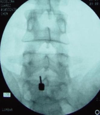
Interlaminar approach at the L5-S1 level on the left, anteroposterior view.
-
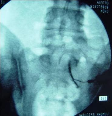
Transforaminal approach at the S1 level on the right, anteroposterior view.
Tables

What would you like to print?
- Overview
- Mechanisms of Radicular Low Back Pain
- Rationale for Use of Steroids in Back Pain
- Indications and Contraindications for Epidural Steroid Injections
- Efficacy of Epidural Injections and Rationale for Fluoroscopy and Contrast
- Safety of Epidural Injections
- Timing, Frequency, Dose, and Volume of Epidural Injections
- Approaches for Epidural Injections
- Medications Used in Epidural Injections
- Conclusions
- Questions & Answers
- Show All
- Media Gallery
- References